Potentiometric Sensors for Pharmaceutical Drug Monitoring: Advances in Solid-Contact Designs, Wearable Integration, and Clinical Applications
This article provides a comprehensive review of the latest advancements in potentiometric sensors for therapeutic drug monitoring (TDM).
Potentiometric Sensors for Pharmaceutical Drug Monitoring: Advances in Solid-Contact Designs, Wearable Integration, and Clinical Applications
Abstract
This article provides a comprehensive review of the latest advancements in potentiometric sensors for therapeutic drug monitoring (TDM). Aimed at researchers, scientists, and drug development professionals, it explores the fundamental principles of ion-selective electrodes (ISEs), the critical shift from liquid-contact to solid-contact designs using novel materials like conducting polymers and carbon nanomaterials, and their application in monitoring drugs with narrow therapeutic indices in biological fluids. The scope extends to emerging trends including 3D-printed, paper-based, and wearable potentiometric sensors for decentralized clinical analysis and point-of-care testing. The discussion includes methodologies for optimizing sensor performance, addressing key challenges in selectivity and stability, and protocols for analytical and clinical validation, positioning potentiometry as a powerful, versatile tool for the future of personalized medicine.
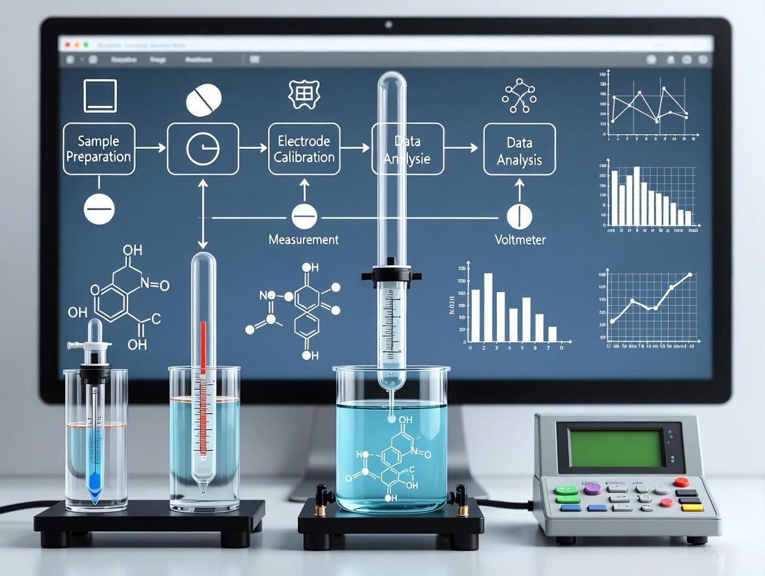
Principles and Evolution of Potentiometric Drug Sensors
Potentiometric sensors have evolved from fundamental principles based on the Nernst equation to sophisticated modern readout systems capable of precise pharmaceutical drug monitoring. This transformation has been propelled by advances in solid-contact electrodes, miniaturization through printing technologies, and enhanced signal processing techniques. These developments have enabled the transition of potentiometric sensing from traditional laboratory settings to point-of-care diagnostic applications, offering rapid, cost-effective analysis of pharmaceutical compounds. This application note details the core principles, fabrication methodologies, and experimental protocols underlying modern potentiometric sensors, with particular emphasis on their application within pharmaceutical research and therapeutic drug monitoring.
Potentiometry represents a cornerstone of electrochemical analysis, enabling the determination of ion activities and concentrations through potential measurement under zero-current conditions [1]. In pharmaceutical sciences, the ability to monitor drug concentrations in complex biological matrices is paramount for therapeutic drug monitoring, pharmacokinetic studies, and quality control processes. Modern potentiometric sensors have undergone significant transformation through the development of solid-contact electrodes and advanced manufacturing techniques, including both 2D and 3D printing technologies [2]. These innovations have addressed critical challenges in sensor miniaturization, reproducibility, and integration, paving the way for their application in wearable devices and electronic skin for continuous physiological monitoring [2]. This document outlines the fundamental principles and practical implementation of potentiometric sensors tailored specifically for pharmaceutical research applications.
Fundamental Principles
The Nernst Equation
The theoretical foundation of potentiometry rests upon the Nernst equation, which describes the relationship between the measured electrochemical potential and the activity of target ions in solution [1]. The equation is expressed as:
E = E° + (RT/nF) ln(a_i)
Where:
- E is the measured electrode potential
- E° is the standard electrode potential
- R is the universal gas constant (8.314 J/(mol·K))
- T is the temperature in Kelvin
- n is the charge number of the ion
- F is Faraday's constant (96,485 C/mol)
- a_i is the activity of the ion of interest [1]
For practical analytical applications where concentration rather than activity is measured, the equation can be adapted to relate potential to the logarithm of the concentration of the target analyte, establishing the fundamental working principle for quantitative analysis using ion-selective electrodes (ISEs) [1].
Potentiometric Cell Architecture
A conventional potentiometric measurement requires a complete electrochemical cell comprising several essential components. The system employs two electrodes: an indicator electrode (or working electrode) whose potential responds to the activity of the target ion, and a reference electrode that maintains a constant, known potential regardless of the solution composition [1]. These electrodes are immersed in the sample solution, which is connected via a salt bridge containing an inert electrolyte to complete the electrical circuit while preventing mixing of solutions [1]. The potential difference between these electrodes is measured under conditions of zero current flow, ensuring the system remains at equilibrium and the composition of the solution remains unchanged during measurement [1].
Diagram 1: Fundamental architecture of a potentiometric cell showing key components and signal flow.
Modern Potentiometric Readouts
Evolution from Conventional to Solid-Contact Sensors
The transition from traditional liquid-contact to solid-contact electrodes represents the most significant advancement in potentiometric sensor design [2]. Conventional sensors utilized internal filling solutions, which imposed limitations on miniaturization, orientation requirements, and maintenance needs. Solid-contact sensors eliminate this liquid component by incorporating an ion-to-electron transducer layer between the ion-selective membrane and the conductive electrode substrate [2]. This architectural innovation has enabled the development of miniaturized, robust, and flexible sensors compatible with point-of-care testing and wearable monitoring devices [2]. The solid-contact configuration has now become the mainstream design for modern potentiometric applications, particularly in pharmaceutical and clinical settings where miniaturization and operational simplicity are paramount.
Advanced Fabrication Methods
Printing technologies have revolutionized potentiometric sensor fabrication, enabling precise, reproducible, and scalable manufacturing. The table below summarizes the primary printing methods employed in modern sensor production:
Table 1: Printing Technologies for Potentiometric Sensor Fabrication
| Method | Type | Key Characteristics | Typical Applications | Considerations |
|---|---|---|---|---|
| Screen Printing | 2D (Stencil) | Simplicity, low cost, high efficiency, wide applicability [2] | Disposable sensors, stretchable electrodes on flexible substrates [2] | Pattern resolution limitations, contact-based process |
| Inkjet Printing | 2D (Non-stencil) | Digital manufacturing, high resolution, non-contact process [2] | Precise deposition of sensing membranes, customized electrode patterns [2] | Ink formulation challenges, potential nozzle clogging |
| Spray/Electrospray Printing | 2D (Stencil) | Uniform thin films, adaptable to various substrates [2] | Fabrication of sensitive membranes using ion-selective cocktails [2] | Material waste concerns, requires masking |
| 3D Printing | Additive | Complex geometries, integrated functional structures [2] | Reproducible sensitive membranes, customized sensor housings [2] | Limited resolution, material compatibility considerations |
Miniaturization and Point-of-Care Adaptation
The development of miniaturized potentiometric devices for point-of-care applications has focused on addressing the ASSURED criteria (Affordable, Sensitive, Specific, User-friendly, Rapid and robust, Equipment-free, and Deliverable to end-users) established by the World Health Organization [3]. Recent innovations in this domain include various sensor configurations:
- Strip-type sensors: Planar devices with co-planar indicator and reference electrodes patterned on a single substrate
- Sandwich-type sensors: Multi-layer architectures that incorporate microfluidic channels for sample handling
- Fully integrated sensors: Systems that incorporate sampling, detection, and readout components in a single device
- Fiber and yarn-based sensors: Textile-integrated sensors for wearable health monitoring applications [3]
Each configuration presents distinct advantages and challenges regarding fabrication complexity, analytical performance, and user operability, with the optimal selection dependent on the specific pharmaceutical monitoring application.
Application in Pharmaceutical Drug Monitoring
Sensor Design for Drug Analysis
The application of potentiometric sensors to pharmaceutical drug monitoring requires careful design considerations to address the complex matrices and specific analytical challenges presented by biological samples. Effective drug monitoring sensors typically incorporate:
- Drug-selective membranes: Polymeric membranes containing ionophores with selective recognition capabilities for target pharmaceutical compounds [2]
- Solid-contact transducers: Intermediate layers that facilitate stable ion-to-electron transduction, often utilizing conductive polymers or nanostructured carbon materials
- Miniaturized reference electrodes: Integrated reference systems that maintain stable potential in small sample volumes
- Sample handling interfaces: Components that enable direct analysis of complex biological samples such as blood, serum, or urine with minimal pretreatment
The development of novel high-performance ionophores remains a key research focus, continually expanding the range of detectable pharmaceutical analytes [2].
Experimental Protocols
Protocol 1: Fabrication of Screen-Printed Potentiometric Sensors
Principle: Create disposable, cost-effective sensors through stencil-based deposition of conductive and sensing inks [2].
Materials:
- Screen printer with appropriate mesh size
- Conductive ink (e.g., carbon, silver/silver chloride)
- Polymer matrix (e.g., PVC, polyurethane)
- Plastic or ceramic substrate
- Ion-selective cocktail (ionophore, ion exchanger, plasticizer)
- Reference membrane components
Procedure:
- Substrate Preparation: Clean and dry the substrate material (typically plastic or ceramic) to ensure proper adhesion of printed layers.
- Electrode Printing: Align the stencil pattern and sequentially print conductive tracks, working electrode, and reference electrode using appropriate inks.
- Curing: Thermally cure the printed electrodes according to ink manufacturer specifications (typically 60-80°C for 30-60 minutes).
- Ion-Selective Membrane Application: Prepare the ion-selective cocktail by dissolving membrane components (1-2% ionophore, 0.5-1% ion exchanger, 30-33% polymer matrix, and balance plasticizer) in tetrahydrofuran.
- Membrane Deposition: Apply the ion-selective cocktail solution via drop-casting or spraying onto the working electrode area.
- Solvent Evaporation: Allow the solvent to evaporate slowly under ambient conditions for 24 hours to form a homogeneous membrane.
- Conditioning: Condition the completed sensors in a solution containing the target ion (typically 10⁻³ M) for 12-24 hours before use.
Quality Control:
- Verify electrode conductivity using impedance measurements
- Confirm membrane adhesion through visual inspection and stability testing
- Test batch-to-batch reproducibility using standard solutions
Diagram 2: Workflow for fabricating screen-printed potentiometric sensors for drug monitoring.
Protocol 2: Calibration and Measurement Procedure for Drug Analysis
Principle: Establish quantitative relationship between sensor potential and drug concentration using standard solutions [1].
Materials:
- Potentiometric sensor (fabricated as in Protocol 1)
- High-impedance potentiometer or pH/mV meter
- Standard solutions of target drug (e.g., 10⁻² to 10⁻⁶ M)
- Stirring platform
- Temperature control system
- Data recording system
Procedure:
- Sensor Preparation: Remove sensors from conditioning solution and rinse gently with deionized water.
- Standard Preparation: Prepare at least five standard solutions spanning the expected concentration range (typically 10⁻² to 10⁻⁶ M) in appropriate background electrolyte.
- Measurement Sequence: Immerse sensors in standard solutions from lowest to highest concentration under constant stirring.
- Potential Recording: Record the stable potential reading for each standard solution (typically achieved within 30-180 seconds depending on sensor design).
- Calibration Curve: Plot potential (mV) versus logarithm of drug concentration and perform linear regression analysis.
- Sample Measurement: Immerse sensors in unknown samples and record stable potential values.
- Concentration Determination: Calculate sample concentrations from the calibration curve using the measured potential values.
Quality Assurance:
- Perform daily calibration with fresh standard solutions
- Monitor slope and intercept consistency for sensor performance tracking
- Include quality control samples with known concentrations to verify accuracy
- Maintain constant temperature during measurements (±1°C)
Research Reagent Solutions
Table 2: Essential Materials for Potentiometric Pharmaceutical Analysis
| Reagent/Material | Function | Typical Composition/Examples | Application Notes |
|---|---|---|---|
| Ion-Selective Cocktail | Recognition element for target analyte | Ionophore (1-2%), Polymer matrix (30-33%), Plasticizer (balance), Additives (0.5-1%) [2] | Composition must be optimized for each specific drug target |
| Solid-Contact Material | Ion-to-electron transduction | Conducting polymers (PEDOT, polypyrrole), nanostructured carbons (graphene, CNTs) [2] | Critical for potential stability; prevents water layer formation |
| Conductive Inks | Electrode fabrication | Carbon, silver, silver/silver chloride pastes [2] | Compatibility with substrate and membrane materials is essential |
| Reference Membrane | Stable reference potential | PVC matrix with salt additives (KCl, NaCl) [3] | Must demonstrate low drift in biological samples |
| Conditioning Solution | Sensor activation and storage | Solution containing target ion (10⁻³ M) in appropriate background | Conditioning time affects response stability and reproducibility |
Data Analysis and Interpretation
Analytical Performance Metrics
The quantitative performance of potentiometric sensors for pharmaceutical applications is evaluated using several key metrics:
- Response Slope: Determined from the calibration curve, with theoretical Nernstian slope being 59.16 mV/decade at 25°C for monovalent ions [1]. Significant deviations may indicate non-ideal behavior or membrane formulation issues.
- Limit of Detection (LOD): Calculated by extrapolation from the linear response region, typically defined as the concentration where the response curve intersects the baseline potential plus three standard deviations [3].
- Selectivity Coefficients: Quantified using the Separate Solution Method or Fixed Interference Method to evaluate sensor performance in the presence of potentially interfering ions endemic to pharmaceutical or biological samples.
- Response Time: Typically measured as the time required to reach 95% of the final steady-state potential after sample introduction, critical for high-throughput applications and point-of-care testing.
Troubleshooting Common Issues
Diagram 3: Diagnostic and troubleshooting pathway for common potentiometric sensor performance issues.
The evolution from the fundamental Nernst equation to modern potentiometric readouts has transformed pharmaceutical drug monitoring capabilities, enabling precise, rapid, and cost-effective analysis of therapeutic compounds. Advances in solid-contact electrodes, printing technologies, and miniaturized designs have addressed previous limitations while opening new applications in point-of-care testing and continuous monitoring. The experimental protocols and analytical frameworks presented in this application note provide researchers with practical methodologies for implementing potentiometric sensing in pharmaceutical research. Future developments will likely focus on further miniaturization, multi-analyte detection capabilities, and enhanced integration with digital health platforms, continuing to expand the role of potentiometry in pharmaceutical sciences and personalized medicine.
Therapeutic Drug Monitoring (TDM) is a critical component of precision medicine, enabling the optimization of drug dosage to maximize efficacy while minimizing toxicity. This practice is especially vital for drugs with a narrow therapeutic index (NTI), where the difference between the minimum effective concentration and the minimum toxic concentration is small [4] [5]. Traditional TDM methods, including chromatography and immunoassays, often require skilled operators, involve complex sample preparation, and typically capture drug concentrations only at a single time point, failing to monitor dynamic changes [6].
Potentiometry, an electrochemical technique that measures the potential difference between two electrodes under conditions of negligible current flow, presents a powerful alternative [4] [7]. The emergence of advanced potentiometric sensors, particularly solid-contact ion-selective electrodes (SC-ISEs), offers a compelling solution for TDM. These sensors are characterized by their ease of design, rapid response, high selectivity, and suitability for miniaturization and continuous monitoring [4] [7] [8]. This application note details the intrinsic advantages of potentiometric sensors for NTI drug monitoring and provides a validated experimental protocol for their application.
Advantages of Potentiometry for NTI Drug TDM
The following table summarizes the key challenges in NTI drug monitoring and how potentiometric sensors address them.
Table 1: Addressing NTI Drug Monitoring Challenges with Potentiometry
| Monitoring Challenge for NTI Drugs | Potentiometric Solution | Impact on TDM |
|---|---|---|
| Need for rapid, frequent measurement | Rapid response time (e.g., ~15 seconds [9]) and direct readout | Enables near-real-time dose adjustment and high-throughput analysis. |
| Risk of toxic side effects from small concentration fluctuations | High selectivity via ionophores [7] [8] and low detection limits (e.g., 10-8 mol L-1 [9]) | Accurately measures clinically relevant concentration ranges, minimizing false results. |
| Requirement for simple, cost-effective analysis | Ease of design, fabrication, and modification [4] [5]; minimal sample pre-treatment | Reduces operational complexity and cost, suitable for point-of-care testing. |
| Desire for continuous monitoring | Compatibility with wearable platforms [4] [6] and solid-contact designs [7] [8] | Allows for tracking dynamic pharmacokinetic profiles, moving beyond single time-point data. |
The Solid-Contact Advantage
A significant advancement in potentiometry is the transition from traditional liquid-contact ISEs to solid-contact ISEs (SC-ISEs). SC-ISEs replace the inner filling solution with a solid-contact layer that acts as an ion-to-electron transducer, overcoming issues of evaporation, mechanical instability, and difficult miniaturization associated with their liquid-contact counterparts [7] [8]. Key materials used as solid contacts include:
- Conducting Polymers (CPs): Such as poly(3,4-ethylenedioxythiophene) (PEDOT) and polyaniline (PANI), which operate via a redox capacitance mechanism [8].
- Carbon-based nanomaterials: Including graphene, carbon nanotubes, and colloid-imprinted mesoporous carbon, which function based on a high electric-double-layer capacitance [7] [8].
- Nanocomposites: Materials like MoS2 nanoflowers filled with Fe3O4 or tubular gold nanoparticles, which provide synergistic effects, enhancing capacitance and signal stability [7].
These materials are pivotal for developing the next generation of robust, miniaturized, and wearable potentiometric sensors for continuous TDM [8].
Experimental Protocol: Potentiometric Determination of an NTI Drug
This protocol provides a generalized methodology for determining drug concentration in a pharmaceutical formulation using a solid-contact potentiometric sensor. The example can be adapted for various NTI drugs by selecting an appropriate ionophore.
Research Reagent Solutions
Table 2: Essential Materials and Reagents
| Item | Function/Brief Description |
|---|---|
| Ionophore | A selective molecular recognition element (e.g., Schiff base, macrocyclic compound) that binds the target drug ion. |
| Polymer Matrix | (e.g., PVC) Forms the bulk of the ion-selective membrane. |
| Plasticizer | (e.g., o-NPOE, DBP) Provides mobility for membrane components and influences dielectric constant. |
| Ionic Additive | (e.g., Lipophilic salt) Ensures ionic conductivity and reduces membrane resistance. |
| Solid-Contact Material | (e.g., PEDOT, Carbon nanotubes) Ion-to-electron transducer layer on the electrode substrate. |
| Graphite Powder | Conductive substrate for carbon paste electrodes [9]. |
| Standard Drug Solution | High-purity reference standard for sensor calibration and validation. |
Sensor Fabrication Workflow
The following diagram illustrates the multi-step process for fabricating a solid-contact potentiometric sensor.
Step-by-Step Procedure
Preparation of the Solid-Contact Layer
- Select an electrode substrate (e.g., glassy carbon, screen-printed carbon). Deposit the solid-contact material. For conducting polymers like PEDOT, this can be achieved via electrochemical polymerization or drop-casting a ready-made dispersion. For carbon nanomaterials, prepare a homogeneous ink and drop-cast it onto the substrate, allowing it to dry [8].
Preparation of the Ion-Selective Membrane (ISM)
- Combine the following components in a glass vial:
- Polymer matrix (e.g., PVC): 30-33 mg (wt. 33%)
- Plasticizer (e.g., o-NPOE): 60-66 mg (wt. 66%)
- Ionophore (specific to the target drug): 0.5-2 mg (wt. 1-2%)
- Lipophilic ionic additive (e.g., NaTPB or KTFPB): ~0.5 mg (wt. 1%)
- Add ~1 mL of tetrahydrofuran (THF) to the vial and cap it. Vortex the mixture until all components are completely dissolved, forming a homogeneous ISM cocktail [9].
- Combine the following components in a glass vial:
Sensor Assembly and Conditioning
- Using a micropipette, deposit a precise volume (e.g., 50-100 µL) of the ISM cocktail directly onto the solid-contact layer.
- Allow the THF to evaporate slowly at room temperature for at least 12 hours, forming a uniform polymeric membrane.
- Condition the assembled sensor by soaking it in a standard solution of the target drug (e.g., 1.0 × 10⁻³ mol L-1) for 24 hours to establish a stable potential [9].
Calibration, Measurement, and Data Analysis
Calibration
- Prepare a series of standard solutions of the drug across a concentration range (e.g., 1 × 10⁻⁷ to 1 × 10⁻² mol L-1) using a constant ionic strength background.
- Immerse the conditioned sensor and a reference electrode (e.g., Ag/AgCl) in the standard solutions from the lowest to the highest concentration.
- Measure the equilibrium potential (in mV) for each solution under stirring. Rinse the sensor gently with deionized water between measurements.
- Plot the measured potential (E) vs. the logarithm of the drug concentration (log C). The plot should yield a linear Nernstian response (slope of ~59.2/z mV/decade for cations at 25°C) [7] [9].
Sample Analysis
- For pharmaceutical formulations (e.g., tablets), weigh and powder a representative number of tablets. Dissolve an accurately weighed portion of the powder in the appropriate solvent and dilute to volume.
- Measure the potential of the sample solution using the calibrated sensor.
- Determine the drug concentration in the sample from the calibration curve.
Method Validation
- Assess the sensor's performance according to the following criteria, ideally over multiple days to establish intermediate precision [10]:
- Linearity: Coefficient of determination (R²) of the calibration curve.
- Accuracy: Recovery studies (e.g., 95-105%) by standard addition or comparison with a reference method.
- Precision: Repeatability (intra-day) and intermediate precision (inter-day), expressed as Relative Standard Deviation (RSD %).
- Selectivity: Determine potentiometric selectivity coefficients (KpotA,B) against common interfering ions using the Separate Solution Method (SSM) or Fixed Interference Method (FIM) [9].
- Assess the sensor's performance according to the following criteria, ideally over multiple days to establish intermediate precision [10]:
Table 3: Exemplary Performance Characteristics of a Potentiometric Sensor
| Performance Parameter | Exemplary Value | Reference |
|---|---|---|
| Linear Range (mol L-1) | 1 × 10⁻⁷ – 1 × 10⁻¹ | [9] |
| Nernstian Slope (mV/decade) | 29.571 ± 0.8 (for a divalent ion) | [9] |
| Detection Limit (mol L-1) | 5.0 × 10⁻⁸ | [9] |
| Response Time | ~15 seconds | [9] |
| Working pH Range | 3.5 – 6.5 | [9] |
| Lifespan | > 2 months | [9] |
Future Perspectives: Wearable Potentiometric Sensors
The future of TDM lies in continuous, real-time monitoring, and potentiometry is perfectly positioned to enable this through wearable sensors [4] [6]. These devices can be integrated into patches or textiles to measure drug concentrations non-invasively in biofluids like sweat, thereby providing a comprehensive pharmacokinetic profile [8] [6].
The logical pathway from a laboratory sensor to a personalized dosing recommendation is outlined below.
Potentiometric sensors offer a critical advantage for the TDM of NTI drugs. Their simplicity, speed, low cost, and high selectivity directly address the limitations of conventional analytical techniques. The advent of solid-contact and wearable sensors further extends their applicability towards continuous, real-time monitoring, paving the way for a new era of truly personalized pharmacotherapy. The protocol provided herein serves as a robust foundation for researchers and scientists to harness this powerful technology in drug development and clinical monitoring.
Ion-Selective Electrodes (ISEs) are potentiometric sensors that quantitatively measure the activity of specific ions in solution, forming a critical technology platform for pharmaceutical research and therapeutic drug monitoring [11] [7]. The core function of an ISE relies on an ion-selective membrane (ISM) that generates a membrane potential by selectively interacting with target ions [12]. The configuration of the interface behind this membrane fundamentally differentiates ISE designs, primarily into liquid-contact (LC-ISE) and solid-contact (SC-ISE) configurations [12] [7]. This application note details the anatomical structure, working principles, and performance characteristics of both configurations within the specific context of pharmaceutical drug analysis, providing validated experimental protocols for their implementation.
Structural Configurations and Operational Principles
Liquid-Contact ISE (LC-ISE) Anatomy
The traditional LC-ISE employs an internal filling solution as a stable ionic bridge between the ion-selective membrane and the internal reference electrode [7]. Its structure consists of:
- Ion-Selective Membrane (ISM): A polymer membrane (typically PVC or acrylic) containing an ionophore (selective ion carrier), ion exchanger, plasticizer, and polymer matrix [12].
- Internal Filling Solution: An aqueous solution containing a fixed concentration of the target ion [7].
- Internal Reference Electrode: Typically an Ag/AgCl wire immersed in the internal solution, providing a stable potential reference [12] [7].
The potential difference (EMF) measured between the ISE and an external reference electrode follows the Nernst equation: E = E⁰ + (RT/zF)ln(a), where S = RT/zF represents the theoretical Nernstian slope (approximately 59.16 mV/decade for a monovalent ion at 23°C) [13]. The primary function of the internal solution is to establish a stable potential at the interface between the inner reference electrode and the back side of the ISM [12].
Solid-Contact ISE (SC-ISE) Anatomy
SC-ISEs eliminate the internal filling solution, replacing it with a solid-contact (SC) layer that acts as an ion-to-electron transducer [12] [7]. This design revolutionizes the electrode by enabling miniaturization, portability, and simplified manufacturing [13]. The three core components are:
- Conductive Substrate: An electron-conducting material such as glassy carbon, platinum, or screen-printed electrodes [12].
- Solid-Contact (SC) Layer: A material with both ionic and electronic conductivity that facilitates the transduction of ionic currents from the membrane to electronic currents in the substrate [14].
- Ion-Selective Membrane (ISM): Similar in composition to that used in LC-ISEs [12].
Two primary mechanisms govern the transduction at the SC layer [12] [14]:
- Redox Capacitance Mechanism: Utilizes conducting polymers (e.g., PEDOT, PANI, POT) that undergo reversible oxidation/reduction reactions, providing a stable potential through their high redox capacitance [12] [14].
- Electric Double-Layer (EDL) Capacitance Mechanism: Employs capacitive materials like carbon nanotubes (MWCNTs), graphene, or nanocomposites, where charge separation at the ISM/SC interface creates a stable double-layer capacitance [12] [14].
Figure 1: Anatomical comparison of Liquid-Contact and Solid-Contact Ion-Selective Electrodes.
Comparative Analysis: LC-ISE vs. SC-ISE
The structural differences between the two configurations lead to distinct performance characteristics, particularly relevant to pharmaceutical applications such as drug dissolution testing and therapeutic drug monitoring (TDM) [11] [15].
Table 1: Performance Comparison of LC-ISE and SC-ISE Configurations
| Parameter | Liquid-Contact ISE (LC-ISE) | Solid-Contact ISE (SC-ISE) |
|---|---|---|
| Key Structural Feature | Internal filling solution [7] | Solid-contact transduction layer [12] |
| Miniaturization Potential | Limited by internal solution volume [12] | Excellent, ideal for wearable sensors [11] [7] |
| Potential Stability | Generally stable, but sensitive to filling solution changes [12] | Can achieve high stability with optimal SC layer [13] [14] |
| Maintenance Requirements | High (refill solution, membrane maintenance) [12] | Low (no liquid components) [13] |
| Response Time | Seconds to minutes | Often < 10-30 seconds [15] [16] |
| Lifetime | Months with maintenance | Weeks to months (single-use possible) [11] |
| Primary Limitations | Solution evaporation/leakage, pressure/temperature sensitivity, difficult miniaturization [12] | Potential water layer formation, signal drift with poor SC layer [13] [17] |
| Ideal Pharmaceutical Use Case | Benchtop dissolution testing, quality control labs [15] | Portable analysis, wearable monitors, in-field testing [11] [7] |
Experimental Protocol: Fabrication of a Solid-Contact ISE for Drug Analysis
This protocol details the construction of a SC-ISE with a conductive polymer solid-contact layer, suitable for the determination of cationic drugs (e.g., Venlafaxine, Lidocaine, Propranolol) [15] [14].
Materials and Reagents
Table 2: Essential Research Reagents for SC-ISE Fabrication
| Reagent/Material | Function | Example Specifications |
|---|---|---|
| Polyvinyl Chloride (PVC) | Polymer matrix for the ISM, provides structural integrity [12] [14] | High molecular weight, Selectophore grade [14] |
| Plasticizer (e.g., o-NPOE, DOS) | Imparts plasticity and mobility to membrane components, influences dielectric constant [12] | 2-Nitrophenyl octyl ether (o-NPOE), Bis(2-ethylhexyl) sebacate (DOS) [12] [14] |
| Ionophore | Selectively binds target ion (drug molecule) [12] | Drug-specific (e.g., ion-pair complex, macrocyclic host) [16] [17] |
| Ion Exchanger (e.g., NaTFPB, KTpClPB) | Introduces ionic sites into membrane, crucial for proper operation with neutral ionophores [12] [15] | Potassium tetrakis(4-chlorophenyl) borate (KTpClPB) [15] |
| Solid-Contact Material | Ion-to-electron transducer (e.g., Conducting polymer, MWCNTs) [14] | Poly(3-octylthiophene-2,5-diyl) (POT), Multi-Walled Carbon Nanotubes (MWCNTs) [13] [14] |
| Conductive Substrate | Electron-conducting support [12] | Glassy Carbon Electrode (GCE), Screen-Printed Electrode (SPE) [13] [17] |
| Tetrahydrofuran (THF) | Solvent for membrane casting [15] [14] | Analytical grade, anhydrous |
Step-by-Step Procedure
- Substrate Preparation: Polish a glassy carbon electrode (GCE) successively with fine alumina slurries (e.g., 1.0, 0.3, and 0.05 µm) to a mirror finish. Ricate thoroughly with distilled water and dry [13].
- Solid-Contact Layer Deposition:
- Option A (Conducting Polymer): Prepare a 1-5 mg/mL solution of the conducting polymer (e.g., POT) in a suitable solvent (e.g., tetrahydrofuran or acetonitrile). Deposit 10-50 µL of this solution onto the polished GCE surface and allow it to dry under ambient conditions to form a thin film [13] [14].
- Option B (Nanomaterial): Disperse 1-2 mg of MWCNTs in 1 mL of organic solvent (e.g., xylene/THF mixture) via sonication. Drop-cast 10-50 µL of the dispersion onto the GCE and dry [13] [17].
- Ion-Selective Membrane (ISM) Cocktail Preparation: In a glass vial, accurately weigh and combine the following components to make approximately 200 mg of total membrane mass [15] [14]:
- 1.0 wt% Ionophore (e.g., drug-TPB⁻ ion-pair)
- 0.5-1.0 wt% Ion Exchanger (e.g., NaTFPB)
- 32-65 wt% Plasticizer (e.g., o-NPOE)
- 33-66 wt% Polymer Matrix (e.g., PVC) Add 1-2 mL of THF and stir until the components are completely dissolved, forming a homogeneous, viscous cocktail.
- Membrane Deposition: Drop-cast 50-100 µL of the ISM cocktail directly onto the prepared solid-contact layer. Carefully cover the vial and allow the THF to evaporate slowly over 24-48 hours at room temperature to form a uniform, dry membrane with a thickness of 100-300 µm [15] [14].
- Electrode Conditioning: Soak the newly fabricated SC-ISE in a stirring solution of the target drug (e.g., 1.0 × 10⁻³ M Venlafaxine HCl or Lidocaine HCl) for 12-24 hours (or overnight) to establish a stable equilibrium at the membrane-sample interface [15] [14].
- Calibration and Use: Calibrate the conditioned electrode in a series of standard solutions of the target drug (e.g., from 1 × 10⁻⁷ M to 1 × 10⁻² M). Measure the potential versus a commercial Ag/AgCl reference electrode under stirring. A stable, Nernstian response (slope of ~59 mV/decade for monovalent cation) confirms successful fabrication [14] [16].
Figure 2: Solid-contact ISE fabrication workflow.
Application in Pharmaceutical Analysis: Drug Release Monitoring
SC-ISEs are ideally suited for monitoring drug release from solid dosage forms due to their rapid response, minimal sample preparation, and ability to analyze colored/turbid solutions [11] [15].
Experimental Workflow for Drug Release Profiling:
- Setup: Use the fabricated drug-selective SC-ISE and a reference electrode in a standard dissolution vessel (e.g., 100 mL volume, 300 rpm, 37°C) containing the dissolution medium [15].
- Measurement: Introduce the drug-loaded dosage form (e.g., a polymer film or a coated porous substrate) into the medium.
- Data Acquisition: Continuously record the potential output of the SC-ISE at short intervals (e.g., every 10 seconds) using a high-impedance data acquisition system [15].
- Data Conversion: Convert the measured potential (mV) to drug concentration (mol/L) in real-time using the pre-established calibration curve [15].
- Validation: Compare the resulting release profile with data obtained from a standard technique like UV spectrophotometry to validate the method [15].
This potentiometric method offers significant advantages over UV spectroscopy, as it is unaffected by sample turbidity, air bubbles, or the presence of other UV-absorbing excipients, providing a more robust and direct measurement of drug activity [15].
The evolution from liquid-contact to solid-contact configurations represents a significant advancement in ISE technology, directly addressing the needs of modern pharmaceutical research. While LC-ISEs remain robust for controlled laboratory environments, SC-ISEs offer superior advantages in miniaturization, portability, and operational simplicity, enabling their application in wearable sensors and point-of-care diagnostic devices [11] [7]. The critical design choice lies in selecting and optimizing the solid-contact material—whether based on redox capacitance (conducting polymers) or double-layer capacitance (nanocarbons)—to ensure potential stability and prevent the formation of a detrimental water layer [13] [14] [17]. By providing a detailed anatomical understanding and a validated fabrication protocol, this application note equips researchers to effectively utilize ISEs for advanced pharmaceutical analysis, including therapeutic drug monitoring and real-time dissolution testing.
Potentiometric sensors, specifically ion-selective electrodes (ISEs), are established tools in electrochemical analysis for determining ion concentrations in diverse samples. Traditional liquid-contact ISEs (LC-ISEs) contain an internal solution that facilitates ion-to-electron transduction. While effective, this design suffers from fundamental limitations including mechanical instability, evaporation or leakage of the internal solution, challenges in miniaturization, and a short shelf-life, restricting their use in miniaturized, portable, or wearable applications [7] [18].
The "solid-contact revolution" addresses these limitations by replacing the internal solution with a solid-contact (SC) layer, also known as an ion-to-electron transducer. This innovation has given rise to solid-contact ISEs (SC-ISEs), which offer superior mechanical robustness, ease of miniaturization and integration, enhanced potential stability, and the prevention of a detrimental water layer between the membrane and the substrate [7] [18]. This transition is particularly impactful for pharmaceutical drug monitoring, enabling the development of point-of-care devices, wearable sensors for real-time therapeutic drug monitoring (TDM), and highly reproducible, automated production [19] [20]. This document details the critical materials, experimental protocols, and applications underpinning this technological shift.
Key Transducer Materials and Performance
The solid-contact layer is the core of an SC-ISE, responsible for efficient ion-to-electron transduction and potential stability. Various classes of materials have been explored, each with distinct properties and transduction mechanisms.
Table 1: Critical Assessment of Common Ion-to-Electron Transducer Materials
| Material Class | Example Materials | Reported Performance Metrics | Advantages | Disadvantages |
|---|---|---|---|---|
| Conducting Polymers | Poly(3,4-ethylenedioxythiophene) (PEDOT), Polypyrrole (PPy) | Capacitance: N/AShort-term drift: N/AMechanism: Redox Capacitance [18] | High redox capacitance, good electrical conductivity, well-established deposition methods. | Susceptible to interferences from O₂, CO₂, and light; Swelling in aqueous solutions can affect stability. |
| Carbon Nanomaterials | Multi-Walled Carbon Nanotubes (MWCNTs) | Capacitance: N/AShort-term drift: N/AMechanism: EDL Capacitance [21] [18] | Very high surface area, hydrophobicity, excellent electrical conductivity. | Potential for agglomeration; Batch-to-batch variability. |
| Graphene-based Materials | Graphene, Graphene Oxide, Reduced Graphene Oxide | Capacitance: 383.4 ± 36.0 µFShort-term drift: 2.6 ± 0.3 µV s⁻¹Total Resistance: 216.1 ± 27.4 kΩ [21] | Highest reported capacitance, very hydrophobic, high electroactive surface area, low potential drift. | Cost and complexity of production for some forms. |
| Nanocomposites | Fe₃O₄/MoS₂, Tubular Gold Nanoparticles (Au-TFF) | Capacitance: HighShort-term drift: N/AMechanism: Synergistic [7] | Tailored properties, enhanced stability and capacitance, improved electron transfer kinetics. | More complex synthesis and fabrication process. |
| 3D-Printed Carbon-Composites | Polylactic acid-Carbon Black (PLA-CB) | Cost: ~€0.32/sensor [20]Reproducibility (E⁰ RSD): ± 3 mV [20] | Extreme low cost, automated fabrication, high reproducibility, custom designs. | Lower conductivity compared to pure carbon materials; Requires optimization of print parameters. |
*N/A: Specific values not provided in the cited search results, but the mechanism is confirmed.
Experimental Protocols
Protocol 1: Fabrication of Graphene-Based SC-ISEs for Lithium Sensing
This protocol outlines the procedure for creating high-performance SC-ISEs using graphene as a transducer, adapted from a critical assessment study [21].
Principle: A commercially available screen-printed electrode (SPE) modified with graphene provides the solid-contact transducer substrate. A lithium-ion selective membrane (ISM) is then drop-cast onto this substrate to create the final sensor.
The Scientist's Toolkit: Table 2: Essential Materials for Graphene-Based Lithium SC-ISE
| Item | Function |
|---|---|
| Graphene-modified SPE | Serves as the ion-to-electron transducer and conductive substrate. |
| Lithium Ionophore | The selective recognition element within the ISM that complexes with Li⁺ ions. |
| Ion Exchanger | (e.g., NaTFPB) Provides ionic sites in the membrane for proper potentiometric response. |
| Plasticizer | (e.g., DOS) Creates a fluid matrix for the ISM, enabling ion mobility. |
| Polymer Matrix | (e.g., PVC or PU) Provides structural integrity to the ISM. |
| Tetrahydrofuran (THF) | Volatile solvent used to dissolve the ISM components for drop-casting. |
Procedure:
- ISM Cocktail Preparation: In a glass vial, prepare the ISM cocktail by dissolving the following components in THF (e.g., 1 mL total volume):
- Lithium Ionophore (e.g., 1% by weight)
- Ion Exchanger (e.g., Sodium Tetrakis[3,5-bis(trifluoromethyl)phenyl]borate (NaTFPB), 0.5% by weight)
- Plasticizer (e.g., Dioctyl Sebacate (DOS), 65% by weight)
- Polymer Matrix (e.g., Poly(Vinyl Chloride) (PVC), 32.5% by weight)
- Membrane Deposition: Using a micropipette, apply 5 aliquots of 10 µL of the ISM cocktail onto the working electrode surface of the graphene-modified SPE. Allow each aliquot to dry completely for 20 minutes at room temperature before applying the next.
- Final Conditioning: After the final layer is deposited, allow the sensor to dry for an additional 1 hour. Condition the completed SC-ISE overnight in a solution of 10 mM LiCl to equilibrate the membrane.
- Potentiometric Measurement: Connect the SC-ISE and a reference electrode (e.g., Ag/AgCl) to a high-input impedance potentiometer. Measure the potential while immersing the electrodes in a series of standard Li⁺ solutions with stirring. Construct a calibration curve by plotting the measured potential (mV) vs. the logarithm of Li⁺ activity.
Protocol 2: Automated Fabrication of 3D-Printed Solid-Contact ISEs
This protocol describes a highly reproducible and automated method for producing SC-ISEs using multimaterial fused filament fabrication (FFF) 3D-printing [20].
Principle: A 3D printer is used to fabricate an electrode body with an integrated well, using an insulating filament (PETg) and a conductive carbon-composite filament (PLA-CB) that functions as both the electrode and the ion-to-electron transducer.
Procedure:
- Electrode Design: Design a 3D model of the electrode (e.g., dimensions 30 × 10 × 1.2 mm) featuring a recessed well (e.g., 5 mm diameter) to contain the ISM. The design should include a base insulator layer, a conductive CB-PLA electrode layer, and a top insulator layer.
- Slicing and Setup: Export the design as an STL file and import it into a slicer program (e.g., PrusaSlicer). Use the following key print settings:
- Infill: 100%
- Extrusion Multiplier: 1.1 (to prevent void formation)
- Nozzle Temperature: 240 °C (for CB-PLA)
- Bed Temperature: 90 °C
- Print Speed: 25 mm/s
- Multimaterial Printing: Execute the print using a single-nozzle printer with a modified gcode to perform filament swaps. This will produce a complete, insulated electrode with a CB-PLA working electrode at the bottom of the well.
- ISM Application and Conditioning: Prepare a potassium-ISM cocktail (e.g., 1% valinomycin, 0.5% NaTFPB, 65% DOS, 33.5% PVC in THF). Apply 5 × 10 µL aliquots of the cocktail into the well of the 3D-printed electrode, allowing each to dry for 20 minutes. Condition the finished 3DP-SC-ISE overnight in 10 mM KCl.
Response Mechanisms of Solid-Contact Transducers
The potential stability in SC-ISEs is governed by the interfacial capacitance at the substrate/ISM junction. Two primary mechanisms have been experimentally verified, depending on the transducer material [18].
Redox Capacitance Mechanism: This mechanism is characteristic of conducting polymers (e.g., PEDOT) that exhibit reversible redox behavior. The ion-to-electron transduction is achieved via a reversible Faradaic process. The potential is thermodynamically defined and highly stable because the redox couple's concentrations are fixed within the polymer layer [18]. For a PEDOT-based K⁺-ISE, the overall reaction can be summarized as:
PEDOT⁺Y⁻ (SC) + K⁺ (aq) + e⁻ (GC) ⇌ PEDOT (SC) + Y⁻ (ISM) + K⁺ (ISM)Electric-Double-Layer (EDL) Capacitance Mechanism: This non-Faradaic mechanism is typical for carbon-based materials like graphene, CNTs, and CB. The transduction occurs through the electrostatic separation of charges at the interface between the electronic conductor and the ionic conductor (ISM), forming an EDL. The high stability of these transducers stems from their exceptionally high surface area and hydrophobicity, which lead to a large capacitance and effectively prevent the formation of a water layer [21] [18].
Applications in Pharmaceutical Drug Monitoring
The solid-contact revolution has directly enabled advanced applications in pharmaceutical research and clinical monitoring.
Wearable Sensors for Therapeutic Drug Monitoring (TDM): SC-ISEs are ideal for wearable, non-invasive monitoring of pharmaceutical drugs in biofluids like sweat. For example, an enzyme-based wearable sensor was developed for real-time detection of the anti-Parkinson's drug L-Dopa in sweat, demonstrating a strong correlation with blood pharmacokinetic profiles [19]. This allows for personalized dosing and minimizes side effects.
Rapid Purity and Potency Analysis: SC-ISEs can be designed for specific drugs to rapidly assess purity during industrial production. A quality-by-design (QbD) approach was used to develop a potentiometric sensor for Hydroxychloroquine (HCQ) that is highly selective against its toxic starting materials (DCQ and HND). This enables at-line monitoring of reaction kinetics and final product purity without complex instrumentation [22].
High-Reproducibility Production for Clinical Use: Automated fabrication methods like 3D printing are critical for producing highly reproducible SC-ISEs suitable for clinical TDM. The consistency in standard potential (E⁰) offered by these methods moves the field closer to "calibration-free" sensors, simplifying their use by end-users in decentralized healthcare settings [20] [23].
Biomarker Comparison Across Biological Fluids
The selection of an appropriate biological matrix is fundamental to the success of any drug monitoring protocol. Blood, urine, and saliva each offer distinct advantages and limitations for quantifying pharmaceutical compounds and their metabolites.
Table 1: Comparative Analysis of Biological Matrices for Drug Monitoring
| Feature | Blood/Plasma/Serum | Urine | Saliva |
|---|---|---|---|
| Invasiveness | Invasive (venipuncture) [24] | Non-invasive | Non-invasive [25] [24] [26] |
| Collection Ease | Requires trained personnel [27] | Simple, but requires restroom facilities | Simple, no special training needed [25] [26] |
| Matrix Complexity | High (proteins, cells, lipids) | Moderate to High | Moderate (proteins, enzymes, microbes) [26] |
| Biomarker Concentration | Represents systemic circulation | Often concentrated; suitable for metabolite profiling | Generally lower, correlates with free, unbound drug fraction [27] |
| Primary Applications | Gold standard for pharmacokinetics (TDM) [27] | Compliance testing, metabolite identification, occupational exposure | Therapeutic Drug Monitoring (TDM), especially for drugs with narrow therapeutic index [28] [27] |
| Correlation to Blood Levels | Reference standard | Variable, often qualitative or semi-quantitative | Strong correlation for specific drugs (e.g., paracetamol) [27] |
| Key Analytical Challenges | Sample preprocessing (centrifugation), hemolysis | Variable dilution (requires creatinine correction), analyte stability | Contamination (food debris, oral hygiene), lower analyte concentration, requires sensitive sensors [26] [27] |
Saliva is particularly advantageous for therapeutic drug monitoring (TDM) as it often contains the free, pharmacologically active fraction of a drug [27]. Its non-invasive nature facilitates frequent sampling, improving patient compliance and enabling real-time pharmacokinetic profiling [25]. For instance, paracetamol concentrations in saliva show a strong correlation with plasma levels, making it a viable alternative for monitoring [27].
Advanced Analytical Methodologies
Potentiometric Sensor Platforms
Potentiometric sensors are a cornerstone of modern analytical chemistry for ion concentration determination. The core principle involves measuring the potential difference between an Ion-Selective Electrode (ISE) and a reference electrode under conditions of negligible current flow [7].
Solid-Contact Ion-Selective Electrodes (SC-ISEs) represent a significant advancement over traditional liquid-contact ISEs. They eliminate the inner filling solution, which enhances mechanical stability, prevents solution evaporation, and allows for easier miniaturization and integration into wearable formats [7] [8]. A key component of SC-ISEs is the solid-contact layer, which acts as an ion-to-electron transducer.
Table 2: Common Solid-Contact Materials Used in Potentiometric Sensors
| Material Category | Examples | Key Properties & Functions |
|---|---|---|
| Conducting Polymers | Poly(3,4-ethylenedioxythiophene) (PEDOT), Polypyrrole (PPy), Polyaniline (PANI) | Function primarily via a redox capacitance mechanism, providing stable potential and efficient transduction [8]. |
| Carbon-Based Nanomaterials | Carbon nanotubes, Graphene, Mesoporous carbon | Offer a high double-layer capacitance due to their large surface area, contributing to signal stability [8]. |
| Nanocomposites | MoS2 nanoflowers with Fe3O4; Tubular gold nanoparticles with Tetrathiafulvalene | Combine materials for a synergistic effect, enhancing capacitance, stability, and electron transfer kinetics [8]. |
The mechanism of solid-contact ISEs can follow one of two primary pathways, depending on the transducer material. The redox capacitance mechanism is typical for conducting polymers, where the polymer's oxidation/reduction provides the charge transfer. In contrast, carbon-based materials often operate via an electric-double-layer (EDL) capacitance mechanism, storing charge at the electrode-electrolyte interface [8].
Protocol: Fabrication of a Solid-Contact Potentiometric Sensor
This protocol outlines the steps for creating a generalized solid-contact ion-selective electrode for drug monitoring [8].
- Reagents & Materials: Conducting substrate (e.g., glassy carbon, gold, screen-printed electrode); Solid-contact material (e.g., PEDOT:PSS dispersion, carbon nanotube solution); Ion-selective membrane components: Ionic receptor (ionophore), Ion-exchanger, Polymer matrix (e.g., PVC), Plasticizer; Solvent (e.g., Tetrahydrofuran - THF); Standard solutions of the target drug for calibration.
- Equipment: Potentiostat/Galvanostat; Electrochemical cell; Micropipettes; Ultrasonic bath; Spin coater (optional); Fume hood.
Procedure:
- Substrate Pretreatment: Clean the conducting substrate mechanically (e.g., with alumina slurry) and/or electrochemically (e.g., by cycling in sulfuric acid) to ensure a pristine surface.
- Solid-Contact Layer Deposition: Deposit the transducer material onto the substrate.
- For conducting polymers: This can be done via drop-casting of a polymer solution or by electrochemical polymerization (e.g., chronocoulometry) for more controlled film growth [8].
- For nanomaterials: Drop-cast a homogenous dispersion of the nanomaterial (e.g., carbon nanotubes) and allow the solvent to evaporate.
- Ion-Selective Membrane (ISM) Cocktail Preparation: In a glass vial, dissolve the polymer matrix (e.g., ~30 mg PVC), plasticizer (e.g., ~60-65 mg), ionophore (e.g., ~1-5 mg), and ion-exchanger (e.g., ~0.5-2 mg) in a suitable volatile solvent (e.g., 1-2 mL THF).
- Membrane Deposition: Using a micropipette, drop-cast a precise volume (e.g., 50-100 µL) of the ISM cocktail directly onto the solid-contact layer. Allow the solvent to evaporate slowly under a glass beaker to form a homogeneous, tacky film.
- Conditioning & Calibration: Condition the newly fabricated sensor in a solution containing the target ion (e.g., 1 mM drug solution) for several hours or overnight to establish a stable equilibrium potential. Calibrate the sensor by measuring its potential in a series of standard solutions with known concentrations of the target drug.
Protocol: Quantification of Paracetamol in Saliva Using a Smartphone-Based Electrochemical Biosensor
This protocol details a specific application for monitoring paracetamol (acetaminophen) in artificial saliva, leveraging smartphone technology for point-of-care testing [27].
- Reagents & Materials: Artificial saliva; Paracetamol standards; Phosphate buffer saline (PBS, pH 7.4); Electrochemical cell; Screen-printed carbon electrodes (SPCEs) or a custom low-cost potentiostat (e.g., KickStat); Smartphone with dedicated app (e.g., "MediMeter").
- Equipment: Low-cost potentiostat (e.g., KickStat) compatible with a smartphone [27]; Micropipettes; Vortex mixer.
Procedure:
- Sample Collection & Preparation: Collect saliva sample via passive drool or using a standardized collection device. Centrifuge the sample (e.g., at 10,000 × g for 5 min) to remove particulates and obtain a clear supernatant. For initial method development, use artificial saliva spiked with paracetamol [27].
- Sensor System Setup: Connect the electrochemical sensor (e.g., SPCE) to the potentiostat, which is interfaced with a smartphone running the analytical application.
- Electrochemical Measurement: Transfer a fixed volume (e.g., 50 µL) of the prepared sample or standard onto the sensor. Initiate the measurement through the smartphone app. An optimized electrochemical technique (e.g., chronoamperometry or differential pulse voltammetry) is applied to quantify paracetamol.
- Data Analysis: The smartphone application automatically records the electrochemical signal (e.g., current) and correlates it to a pre-established calibration curve (R² = 0.988 reported for paracetamol [27]). The result, displaying the paracetamol concentration, is presented on the smartphone screen within approximately one minute [27].
The Scientist's Toolkit: Essential Research Reagents and Materials
Table 3: Key Reagent Solutions for Sensor Fabrication and Drug Analysis
| Item | Function/Application |
|---|---|
| Ionophores | Molecular recognition elements within the Ion-Selective Membrane that selectively bind to the target drug ion [8]. |
| Polymer Matrices (e.g., PVC) | Form the bulk of the sensing membrane, providing a supportive matrix for the ionophore and other components [8]. |
| Plasticizers (e.g., DOS, o-NPOE) | Impart flexibility and mobility to the polymer membrane, influencing ionophore dynamics and sensor lifespan [8]. |
| Ion-Exchangers | Introduce ionic sites into the membrane to ensure permselectivity and a stable Nernstian response [8]. |
| Conducting Polymers (e.g., PEDOT:PSS) | Serve as the solid-contact layer, transducing the ionic signal from the membrane into an electronic signal for measurement [8]. |
| Carbon Nanomaterials (e.g., MWCNTs) | Used as high-surface-area solid-contact materials or electrode modifiers to enhance signal stability and sensitivity [8]. |
| Artificial Saliva | A simulated biological fluid used for method development, optimization, and calibration to mimic the matrix of human saliva [27]. |
| Enzymes (for enzymatic assays) | Biological recognition elements used in biosensors to impart high specificity for the target analyte (e.g., enzyme-based paracetamol sensors) [27]. |
Sensor Fabrication, Material Innovations, and Real-World Deployment
The evolution of solid-contact ion-selective electrodes (SC-ISEs) represents a significant advancement in potentiometric sensing for pharmaceutical drug monitoring. Unlike traditional liquid-contact ISEs, SC-ISEs eliminate the internal filling solution, enabling miniaturization, better portability, and more robust detection limits [14]. The critical component in these sensors is the transducer layer, which facilitates the conversion of an ionic signal from the recognition event into an electronic signal that can be measured by the underlying electrode [8]. This ion-to-electron transduction is vital for creating stable, reliable, and sensitive sensors suitable for pharmaceutical applications such as therapeutic drug monitoring and quality control [5].
The selection of transducer material directly governs key sensor performance parameters, including potential stability, sensitivity, detection limit, and operational lifespan. Ideal transducer materials must exhibit high capacitance, excellent hydrophobicity to prevent the formation of water layers, and both electronic and ionic conductivity [8] [14]. Within this context, conducting polymers like poly(3,4-ethylenedioxythiophene) (PEDOT) and polyaniline (PANI), alongside carbon-based nanomaterials such as multi-walled carbon nanotubes (MWCNTs) and graphene, have emerged as the most promising and extensively researched materials. This application note provides a comparative analysis of these materials and details standardized protocols for their implementation in potentiometric sensors for pharmaceutical analysis.
Transduction Mechanisms and Material Properties
The mechanism of ion-to-electron transduction varies fundamentally between conducting polymers and carbon-based nanomaterials, directly influencing sensor design and performance.
Mechanism of Conducting Polymers (PEDOT, PANI)
Conducting polymers function primarily through a redox capacitance mechanism [8] [14]. These polymers possess a conjugated backbone that can be switched between oxidized and reduced states. When used as a transducer in a cation-selective sensor, the overall reaction can be summarized as follows [8]: CP⁺ + B⁻(SC) + L(ISM) + M⁺(aq) + e⁻(C) ⇌ CP⁰(SC) + B⁻(ISM) + LM⁺(ISM) Here, CP⁺/CP⁰ represents the oxidized/reduced state of the conducting polymer (e.g., PEDOT, PANI), B⁻ is the doping anion, L is the ionophore in the ion-selective membrane (ISM), M⁺ is the target cation, and C is the underlying conductor. This reversible redox reaction provides a high thermodynamic capacitance, stabilizing the potential at the interface between the electron-conducting substrate and the ion-conducting membrane [14].
Mechanism of Carbon-Based Nanomaterials (MWCNTs, Graphene)
In contrast, carbon-based nanomaterials like MWCNTs and graphene rely on a double-layer capacitance mechanism [14]. These materials feature exceptionally high specific surface areas. When employed as a transducer, they form an electrical double layer at the interface with the ion-selective membrane. The capacitance of this double layer ((C{dl})) is a crucial parameter, as it dictates the potential stability; a higher (C{dl}) results in lower potential drift [14]. The extensive surface area of MWCNTs and graphene maximizes this double-layer capacitance, leading to highly stable sensor outputs.
The diagram below illustrates and contrasts these two primary transduction mechanisms.
Comparative Performance of Transducer Materials
The choice of transducer material has a direct and measurable impact on the electrochemical characteristics of the resulting sensor. A rational study comparing MWCNTs, PANi, and ferrocene demonstrated distinct performance outcomes [14].
Table 1: Comparative Electrochemical Performance of Different Transducer Materials for VEN-TPB Ion-Pair Based SC-ISEs [14]
| Transducer Material | Slope (mV/decade) | Detection Limit (mol/L) | Linear Range (mol/L) | Potential Drift (ΔE/Δt, µV/s) | Double-Layer Capacitance (C~dl~, µF) |
|---|---|---|---|---|---|
| MWCNTs | 56.1 ± 0.8 | 3.8 × 10⁻⁶ | 1.0 × 10⁻² – 6.3 × 10⁻⁶ | 34.6 | 850 |
| PANi | 55.6 ± 0.7 | 5.0 × 10⁻⁶ | 1.0 × 10⁻² – 8.0 × 10⁻⁶ | 39.8 | 620 |
| Ferrocene | 54.8 ± 0.5 | 7.9 × 10⁻⁶ | 1.0 × 10⁻² – 1.3 × 10⁻⁵ | 47.2 | 390 |
Table 2: Key Characteristics and Applications of Primary Transducer Materials
| Material | Primary Mechanism | Key Advantages | Reported Application Example |
|---|---|---|---|
| PEDOT | Redox Capacitance | High conductivity, good stability, commercial availability | Widely used as a stable solid contact in various SC-ISEs [8]. |
| PANi | Redox Capacitance | Easy synthesis, good environmental stability, acid-doping capability | Used as a transducer for Venlafaxine HCl sensors [14]. |
| MWCNTs | Double-Layer Capacitance | Very high surface area, excellent electrical conductivity, mechanical strength | Demonstrated the best overall performance (capacitance, drift) for Venlafaxine HCl detection [14]. |
| Graphene/Graphene Nanoplatelets | Double-Layer Capacitance | Ultra-high surface area, superior hydrophobicity prevents water layer formation | Used as a transducer layer to stabilize potential response and prevent water layer formation in Donepezil and Memantine sensors [29]. |
Detailed Experimental Protocols
Protocol 1: Sensor Fabrication and Modification
This protocol describes the functionalization of a glassy carbon electrode (GCE) with graphene nanoplatelets and a molecularly imprinted polymer (MIP)-based membrane for the selective detection of pharmaceutical drugs like Donepezil (DON) and Memantine (MEM) [29].
Workflow Overview:
Materials:
- Glassy Carbon Electrode (GCE) (e.g., OD: 10 mm, ID: 5 mm)
- Graphene Nanoplatelets (6–8 nm thick, 5 microns wide) [29]
- Molecularly Imprinted Polymer (MIP) for the target drug (see Protocol 2 for synthesis)
- Poly(Vinyl Chloride) (PVC), high molecular weight
- Plasticizer (e.g., 2-Nitrophenyl octyl ether - o-NPOE)
- Ionic exchanger (e.g., Potassium tetrakis(4-chlorophenyl)borate - K-TCPB)
- Tetrahydrofuran (THF), analytical grade
- Ultrasonication bath
Step-by-Step Procedure:
- Electrode Pretreatment: Polish the GCE surface with successive grades of alumina slurry (e.g., 1.0, 0.3, and 0.05 µm) on a microcloth. Rinse thoroughly with deionized water and then with ethanol. Dry at room temperature [29].
- Graphene Transducer Layer Deposition: Disperse 1.0 mg of graphene nanoplatelets in 1.0 mL of a suitable solvent (e.g., DMF) via ultrasonication for 30-60 minutes to form a homogeneous suspension. Deposit a known volume (e.g., 5-10 µL) of this suspension onto the polished GCE surface and allow it to dry under ambient conditions, forming a uniform solid-contact layer [29].
- Ion-Selective Membrane (ISM) Preparation: Weigh the following components into a glass vial:
- 100 mg of PVC
- 200 mg of plasticizer (o-NPOE)
- 1-5 mg of the synthesized MIP (as ionophore)
- 0.5-2 mg of ionic exchanger (K-TCPB)
- Dissolve the mixture in 2 mL of THF and stir until a clear, homogeneous solution is obtained [29].
- Membrane Casting: Drop-cast a precise volume (e.g., 50-100 µL) of the membrane cocktail directly onto the graphene-modified GCE. Allow the THF to evaporate slowly, preferably by covering the vial loosely, to form a uniform, tacky film over the transducer layer.
- Sensor Conditioning: Before the first measurement, condition the fabricated sensor by soaking in a stirring solution of the target drug (e.g., 1.0 × 10⁻³ M Donepezil HCl) for 12-24 hours. For daily use, store the sensor dry and re-condition in a standard drug solution for 30-60 minutes [29].
Protocol 2: Synthesis of Molecularly Imprinted Polymers (MIPs)
This protocol details the synthesis of MIPs via precipitation polymerization, which can be incorporated into the ISM to provide superior selectivity against interfering ions and the co-formulated drug [29].
Materials:
- Template Molecule: Target drug (e.g., Donepezil or Memantine)
- Functional Monomer: Methacrylic acid (MAA)
- Cross-linker: Ethylene glycol dimethacrylate (EGDMA)
- Initiator: Azobisisobutyronitrile (AIBN)
- Porogenic Solvent: Dimethylsulfoxide (DMSO)
Step-by-Step Procedure:
- Pre-polymerization Mixture: In a glass-capped bottle, dissolve 0.5 mmol of the target drug (template) in 40.0 mL of DMSO. Add 2.0 mmol of MAA and sonicate the mixture for 15 minutes to allow pre-complex formation.
- Polymerization Initiation: To the mixture, add 8.0 mmol of EGDMA (cross-linker) and 0.6 mmol of AIBN (initiator). Sonicate briefly for 1 minute to ensure complete dissolution and mixing.
- Oxygen Removal: Purge the solution with nitrogen gas for 15 minutes to remove dissolved oxygen, which can inhibit the free-radical polymerization.
- Polymerization Reaction: Place the sealed bottle in a thermostatic oil bath at 60 °C for 24 hours to complete the polymerization process.
- Template Removal: After polymerization, wash the resulting polymer particles repeatedly with a solvent (e.g., methanol:acetic acid, 9:1 v/v) to remove the template molecule completely. This leaves behind specific recognition cavities. Finally, dry the MIP under vacuum at 60 °C until a constant weight is achieved [29].
Protocol 3: Electrochemical Characterization of Transducer Layers
Characterizing the transducer layer is crucial for predicting sensor performance. Key parameters include double-layer capacitance ((C_{dl})) and potential drift.
Materials:
- Fabricated SC-ISEs
- Potentiostat/Galvanostat
- Electrochemical cell with reference electrode (e.g., Ag/AgCl) and counter electrode (e.g., Pt wire)
- Aqueous solution of 0.1 M KCl (or other suitable electrolyte)
Part A: Capacitance Measurement via Electrochemical Impedance Spectroscopy (EIS)
- Setup: Immerse the fabricated SC-ISE, a reference electrode, and a counter electrode in a 0.1 M KCl solution.
- Measurement: Run an EIS spectrum at the open-circuit potential over a frequency range of 0.1 Hz to 100 kHz with a small amplitude AC voltage (e.g., 10 mV).
- Analysis: Fit the obtained impedance spectrum to a suitable equivalent circuit. The double-layer capacitance ((C{dl})) can be extracted from the constant phase element (CPE) values in the low-frequency region of the spectrum. A higher (C{dl}) indicates better potential stability [14].
Part B: Potential Drift Assessment via Chronopotentiometry (CP)
- Setup: Use the same three-electrode setup as in Part A.
- Measurement: Apply a constant current (e.g., ±1 nA) for a set duration (e.g., 60 seconds) and record the potential change over time.
- Analysis: Calculate the potential drift (ΔE/Δt) from the slope of the potential-time curve. A lower drift value signifies a more stable sensor [14].
The Scientist's Toolkit: Essential Research Reagent Solutions
Table 3: Key Reagents for SC-ISE Fabrication and Characterization
| Reagent / Material | Function / Role | Example & Notes |
|---|---|---|
| Glassy Carbon Electrode (GCE) | Electron-conducting substrate | Provides a stable, polished surface for transducer layer deposition. |
| Graphene Nanoplatelets | Double-layer capacitance transducer | Hydrophobic material that prevents water layer formation; enhances signal stability [29]. |
| Poly(3,4-ethylenedioxythiophene) (PEDOT) | Redox capacitance transducer | Conducting polymer known for high stability and conductivity; often used doped with poly(styrene sulfonate) (PSS) [8]. |
| Multi-Walled Carbon Nanotubes (MWCNTs) | Double-layer capacitance transducer | Provides very high surface area, leading to high capacitance and low potential drift [14]. |
| Molecularly Imprinted Polymer (MIP) | Selective recognition element in ISM | Synthetic receptor that creates shape-specific cavities for the target drug, drastically improving selectivity [29]. |
| Poly(Vinyl Chloride) (PVC) | Matrix polymer for ISM | Forms the bulk of the ion-selective membrane, providing mechanical stability [29] [14]. |
| Plasticizer (e.g., o-NPOE) | ISM component | Dissolves ionophores, provides mobility for ions within the membrane, and influences selectivity [29]. |
| Ionic Exchanger (e.g., K-TCPB) | ISM component | Introduces permselectivity and facilitates ion exchange at the membrane-sample interface [29]. |
| Tetrahydrofuran (THF) | Solvent | Used to dissolve PVC, plasticizer, and ionophores to create a homogeneous membrane cocktail for casting. |
Potentiometric sensors have emerged as powerful electroanalytical tools in pharmaceutical research, particularly for therapeutic drug monitoring (TDM). These sensors combine the simplicity, portability, and cost-effectiveness of potentiometry with the molecular-level design of sensing membranes capable of selective drug recognition. The core component of such sensors is the ion-selective membrane (ISM), a meticulously formulated layer whose composition directly dictates analytical performance. This application note details the strategic design of ISMs, focusing on the critical triad of components: ionophores (molecular recognition elements), plasticizers (membrane media and solvents), and polymer matrices (structural scaffolds). Framed within a broader thesis on potentiometric sensors for pharmaceutical drug monitoring, this guide provides researchers and drug development professionals with detailed protocols and foundational knowledge to develop robust, selective, and biocompatible sensors for precise drug quantification.
Core Components of the Sensing Membrane
The performance, selectivity, and stability of a potentiometric sensor are governed by the careful selection and proportioning of its membrane components. The table below summarizes the function and key considerations for each critical component.
Table 1: Critical Components of a Potentiometric Sensing Membrane
| Component | Primary Function | Key Considerations & Examples |
|---|---|---|
| Ionophore | Selective recognition and binding of the target ion/drug. | Selectivity, binding constant, lipophilicity. E.g., valinomycin (K⁺), calix[8]arene (Palonosetron) [30], Molecularly Imprinted Polymers (MIPs) (Safinamide) [31]. |
| Polymer Matrix | Provides structural integrity to the membrane. | Biocompatibility, mechanical stability, film-forming ability. E.g., Poly(vinyl chloride) (PVC), polyurethane (PU), poly(methyl methacrylate-co-decyl methacrylate) [32]. |
| Plasticizer | Imparts flexibility and governs the membrane's dielectric constant. | Biocompatibility & leaching potential, lipophilicity, viscosity. E.g., Dioctyl phthalate (DOP), 2-nitrophenyl octyl ether (o-NPOE), bis(2-ethylhexyl sebacate) (DOS) [33]. |
| Ion Exchanger | Ensures ionic conductivity and electroneutrality within the membrane. | Compatibility with the ionophore-target complex. E.g., Sodium tetraphenylborate (Na-TPB) for cations [34] [30]. |
| Additives | Enhance performance characteristics like conductivity or hydrophobicity. | Function-specific. E.g., Multi-walled carbon nanotubes (MWCNTs) to prevent water layer formation [31]. |
The Scientist's Toolkit: Essential Research Reagent Solutions
The following table lists essential materials required for the formulation of potentiometric sensing membranes as described in the cited research.
Table 2: Research Reagent Solutions for Membrane Fabrication
| Reagent / Material | Typical Function | Brief Explanation of Role |
|---|---|---|
| High MW Poly(Vinyl Chloride) (PVC) | Polymer Matrix | Serves as an inert, structural backbone for the membrane [34] [35]. |
| Tetrahydrofuran (THF) | Solvent | Dissolves membrane components for homogeneous film casting [34]. |
| Dioctyl Phthalate (DOP) / o-NPOE | Plasticizer | Solvates ionophore and ion exchanger, imparts membrane flexibility, and modulates permittivity [34] [30]. |
| Sodium Tetraphenylborate (Na-TPB) | Ion Exchanger | Provides lipophilic counter-ions to maintain membrane electroneutrality [34] [30]. |
| Molecularly Imprinted Polymer (MIP) | Ionophore | Synthetic, biomimetic receptor with tailored cavities for highly selective target binding [31]. |
| Multi-Walled Carbon Nanotubes (MWCNTs) | Solid-Contact Transducer | Hydrophobic layer that prevents water formation and enhances signal transduction in all-solid-state sensors [31]. |
Experimental Protocols for Membrane Fabrication and Drug Analysis
This section provides a detailed, step-by-step methodology for fabricating a coated graphite all-solid-state ion-selective electrode (ASS-ISE) and applying it to pharmaceutical analysis, based on validated research [34].
Protocol: Fabrication of a Coated Graphite All-Solid-State Ion-Selective Electrode (ASS-ISE)
Aim: To construct a robust, disposable sensor for the determination of benzydamine hydrochloride (BNZ·HCl) in pharmaceutical cream and biological fluids [34].
Workflow Overview:
Step 1: Preparation of the Ion-Pair Complex
- Procedure: Mix 50 mL of a 10⁻² M solution of the target drug (cationic, e.g., BNZ·HCl) with 50 mL of a 10⁻² M solution of sodium tetraphenylborate (Na-TPB) as the lipophilic anion.
- Precipitation: Allow the resulting solid precipitate (drug-TPB ion-pair) to equilibrate with the supernatant for 6 hours.
- Isolation: Collect the solid by filtration, wash thoroughly with bi-distilled water, and air-dry the powdered ion-pair complex at ambient temperature for 24 hours [34].
Step 2: Formulation of the Sensing Membrane Cocktail
- Weighing: Accurately weigh the following components into a glass petri dish:
- 10 mg of the synthesized ion-pair complex.
- 45 mg of plasticizer (e.g., Dioctyl phthalate, DOP).
- 45 mg of high molecular weight PVC.
- Dissolution: Add 7 mL of tetrahydrofuran (THF) and mix thoroughly until a homogeneous solution is obtained [34].
Step 3: Membrane Casting on Graphite Substrate
- Substrate Preparation: Use a graphite rod electrode (e.g., 4 mm diameter) housed in a Teflon holder. Polish the exposed surface to a shiny finish and clean.
- Coating: Dip the polished surface of the graphite electrode into the membrane cocktail solution.
- Solvent Evaporation: Allow the THF to evaporate completely at room temperature, leaving a thin, uniform PVC film adhered to the graphite surface [34] [35].
Step 4: Sensor Conditioning and Calibration
- Conditioning: Condition the assembled sensor by immersing it in a 10⁻² M solution of the target drug for 4 hours to establish a stable equilibrium at the membrane-sample interface.
- Calibration: Measure the electromotive force (EMF) of the sensor in a series of standard drug solutions across a concentration range (e.g., 10⁻⁶ M to 10⁻² M). Use a pH/mV meter with a double-junction Ag/AgCl reference electrode.
- Data Plotting: Construct a calibration curve by plotting the measured potential (mV) against the logarithm of the drug activity (concentration). A Nernstian slope of approximately 59.2 mV/decade at 25°C for a monovalent ion confirms proper sensor function [34].
Protocol: Enhancing Selectivity with Molecularly Imprinted Polymers (MIPs)
Aim: To incorporate a MIP as a highly selective ionophore for the determination of Safinamide (SAF) in dosage forms and biological fluids [31].
Procedure:
- MIP Synthesis: Prepare the MIP via precipitation polymerization using the target drug (SAF) as a template, methacrylic acid (MAA) as a functional monomer, and ethyleneglycoldimethacrylate (EGDMA) as a cross-linker.
- Template Removal: After polymerization, leach the template molecules from the polymer matrix using a suitable solvent to create specific recognition cavities.
- Membrane Incorporation: Disperse the obtained MIP particles into the standard PVC membrane cocktail (replacing the ion-pair complex) and proceed with the sensor fabrication as described in Protocol 3.1 [31].
Performance Gain: The MIP-based sensor for Safinamide demonstrated a Nernstian slope of 59.30 mV/decade, a low detection limit of 8.0 × 10⁻⁷ M, and excellent selectivity over interfering ions and the drug's degradation products [31].
Advanced Material Strategies for Enhanced Performance
Biocompatibility and Safety
For sensors intended for wearable or implantable use (e.g., continuous monitoring), the biocompatibility of every membrane component is critical. Traditional components like PVC, oNPOE, and DOS plasticizers, along with some ionophores, can exhibit cytotoxicity and may leach into biological fluids [33].
- Strategies: Employ covalent bonding of ionophores to the polymer matrix, use green solvents (e.g., cyclohexanone) during fabrication, and explore biopolymers or graft copolymers as alternative matrices to improve sensor safety and long-term stability [33].
Computational Design of Ionophores
The development of highly selective ionophores is being transformed by Quantitative Structure-Property Relationship (QSPR) modeling. This in-silico approach predicts the potentiometric sensitivity and selectivity of an ionophore based on its molecular structure, significantly reducing time and resource consumption in the development cycle [36].
The strategic design of the sensing membrane is the cornerstone of developing high-performance potentiometric sensors for pharmaceutical applications. The selection of ionophores (from classical to advanced MIPs), polymer matrices, and plasticizers directly controls the critical parameters of selectivity, sensitivity, and stability. The protocols outlined herein, covering fabrication, calibration, and advanced imprinting techniques, provide a robust foundation for research. By integrating these principles with emerging trends in biocompatibility and computational design, researchers can advance the field of therapeutic drug monitoring, paving the way for more precise, personalized, and point-of-care medical treatments.
The integration of additive manufacturing into electrochemical sensor design represents a paradigm shift in pharmaceutical research, particularly for developing personalized therapeutic drug monitoring (TDM) systems [7]. Three-dimensional (3D) printing technologies enable the rapid fabrication of customizable potentiometric sensors with complex geometries, miniaturized features, and integrated functionalities that are difficult to achieve through traditional manufacturing methods [37] [2]. These capabilities are especially valuable for TDM of drugs with narrow therapeutic indices, where precise, patient-specific dosing is critical for treatment efficacy and safety [7] [19].
This protocol details the application of 3D printing for creating solid-contact ion-selective electrodes (SC-ISEs) tailored to pharmaceutical analysis, providing researchers with methodologies to fabricate low-cost, reproducible sensors for decentralized drug monitoring applications [20].
3D Printing Technologies for Potentiometric Sensors
Multiple 3D printing technologies have been adapted for fabricating potentiometric sensors, each offering distinct advantages for specific sensor components and applications. The table below compares the primary techniques used in sensor development:
Table 1: Comparison of 3D Printing Technologies for Potentiometric Sensors
| Printing Technology | Materials Compatible | Best For | Resolution | Key Advantages | Limitations |
|---|---|---|---|---|---|
| Fused Deposition Modeling (FDM) | Thermoplastic polymers (PLA, PETg, conductive PLA-CB, ABS) [20] [38] | Electrode housings, structural components, conductive tracks [37] [39] | ~50-200 μm [39] | Low cost, multi-material capability, wide material selection [20] [39] | Limited resolution, layer adhesion issues, may require post-processing [39] |
| Stereolithography (SLA) | Photopolymer resins | High-resolution features, microfluidics [37] [39] | ~25-100 μm [39] | High resolution, smooth surface finish [37] | Limited material options, poorer mechanical properties [37] |
| Digital Light Processing (DLP) | Photopolymer resins | Integrated lab-on-a-chip systems [39] | ~10-50 μm [39] | Fast printing speed, high resolution [39] | Limited material options, potential cytotoxicity [39] |
For pharmaceutical TDM applications, FDM printing has emerged as the most accessible and versatile technology due to its multi-material capability, which allows simultaneous printing of conductive and insulating elements in a single automated process [20]. This is particularly advantageous for creating integrated sensor systems that combine electrode structures, housings, and microfluidic channels for sample handling [37] [39].
Research Reagent Solutions and Materials
The successful development of 3D-printed potentiometric sensors requires specific materials for both the printing process and the functional components of the electrode system.
Table 2: Essential Materials for 3D-Printed Potentiometric Sensors in Pharmaceutical Applications
| Material Category | Specific Materials | Function/Purpose | Application Notes |
|---|---|---|---|
| Conductive Filaments | PLA-Carbon Black (PLA-CB) [20], Polylactic acid-based composites [38] | Ion-to-electron transducer, working electrode substrate [20] | Provides solid contact; requires 100% infill to prevent voids [20] |
| Insulating/Structural Polymers | Polyethylene terephthalate glycol (PETg) [20], Polylactic acid (PLA) [38] | Electrode housing, insulation layers, microfluidic channels [37] [20] | PETg offers chemical resistance; PLA is biodegradable [38] |
| Ion-Selective Membrane Components | Polyvinyl chloride (PVC) [37], Polyurethane [20], Polylactic acid-based matrix [38] | Membrane polymer matrix | Determines membrane stability and compatibility [37] |
| Plasticizers | Dioctyl sebacate (DOS) [20], Polyethylene glycol derivatives (PEG-400, PEG-1500) [38] | Controls membrane viscosity and flexibility [37] [38] | PEG-400 offers biodegradability [38] |
| Ionophores | Valinomycin (for K+) [20], Acridono-18-crown-6 ether (for Hg2+) [38], drug-selective ionophores | Molecular recognition element for selective target binding [37] | Critical for sensor selectivity; must be compatible with polymer matrix [37] |
| Ion-Exchangers | Sodium tetrakis[3,5-bis(trifluoromethyl)phenyl] borate (NaTFPB) [20] | Imparts permselectivity to the membrane [20] | Lipophilic salts prevent interference from counter-ions [20] |
Experimental Protocol: Fabrication of a 3D-Printed Solid-Contact Ion-Selective Electrode
This protocol describes the complete fabrication process for a multimaterial 3D-printed solid-contact ion-selective electrode optimized for pharmaceutical drug monitoring applications.
Equipment and Software Requirements
- FDM 3D Printer: Capable of multimaterial printing (e.g., Prusa i3 MK3S+)
- Design Software: Fusion 360 (Autodesk) or similar CAD package
- Slicing Software: PrusaSlicer or Ultimaker Cura
- Curing Oven: For drying filament (80°C capability)
- Potentiometer: High-impedance voltmeter (>1 GΩ) for potential measurements
- Chemical Fume Hood: For safe handling of membrane cocktail components
Electrode Design and 3D Printing Process
Sensor Fabrication Workflow
Step 1: CAD Model Design
- Design the electrode using CAD software with a three-layer structure: (1) PETg base (0.8 mm thickness) with groove for electrode, (2) CB-PLA conductive composite (0.4 mm thickness) as working electrode, and (3) PETg insulating cover (0.4 mm thickness) with a 5-mm diameter well for membrane deposition [20].
- Include a recessed electrode geometry with surrounding walls to contain the ion-selective membrane during deposition and prevent spreading [20].
- Export the final design as an STL file for slicing.
Step 2: Slicing and Printer Setup
- Use slicing software with the following parameters for CB-PLA: 100% infill percentage, 1.1 extrusion multiplier, 240°C nozzle temperature, 90°C bed temperature, and 25 mm/s printing speed [20].
- For PETg components, use manufacturer-recommended settings (typically 230-250°C nozzle, 70-80°C bed).
- Implement a multimaterial printing approach using a single-nozzle printer by modifying the gcode to enable filament changes at appropriate layers [20].
Step 3: Printing Process
- Pre-dry all filaments at 80°C for 24 hours to remove moisture [20].
- Execute the print job in a stable environment without drafts.
- A typical batch of 75 electrodes can be printed in approximately 3.5 hours using an optimized printing strategy [20].
Step 4: Post-Printing Inspection and Validation
- Visually inspect electrodes under magnification to ensure proper layer adhesion and absence of defects.
- Validate the electroactivity of the CB-PLA surface using cyclic voltammetry in 1 mM FcMeOH/0.1 M KCl solution [20].
- Electrodes maintain electroactivity for at least three months when stored properly [20].
Ion-Selective Membrane Preparation and Deposition
Step 1: Membrane Cocktail Formulation
- For a potassium-selective membrane (as a model system), prepare a cocktail containing:
- 1.0% (w/w) ionophore (e.g., valinomycin for K+ sensing)
- 0.5% (w/w) ionic additive (NaTFPB)
- 65.0% (w/w) plasticizer (DOS)
- 33.5% (w/w) polymer matrix (PVC or polyurethane) [20]
- Dissolve 100 mg of the total mixture in 1 mL of tetrahydrofuran (THF) and mix thoroughly until a homogeneous solution is obtained.
- For pharmaceutical applications, incorporate drug-selective ionophores (e.g., for lithium, antibiotics, or antipsychotics) using the same proportions.
Step 2: Membrane Deposition
- Using a micropipette, deposit 5 aliquots of 10 μL of the membrane cocktail into the electrode well.
- Allow each layer to dry for 20 minutes at room temperature before adding the next aliquot.
- After the final deposition, cure the membrane for 1 hour at room temperature followed by overnight conditioning in a 10 mM solution of the target ion [20].
Sensor Characterization and Performance Evaluation
Analytical Performance Metrics
Table 3: Performance Characteristics of 3D-Printed Potentiometric Sensors
| Performance Parameter | Typical Results | Measurement Protocol | Importance for Pharmaceutical TDM |
|---|---|---|---|
| Slope (Sensitivity) | 50-59 mV/decade for monovalent ions [37] | Calibration with standard solutions in logarithmic concentration series | Ensures accurate concentration measurements in therapeutic range |
| Detection Limit | 10^-5 to 10^-6 M [37] | Intersection of extrapolated linear regions of calibration curve | Must be sufficient for drug concentrations in biofluids |
| Response Time | 10-30 seconds for 95% response [37] | Time to reach stable potential after sample introduction | Enables rapid analysis for point-of-care applications |
| Working Range | 10^-5 to 10^-1 M [37] | Linear portion of calibration curve | Must cover therapeutic and toxic concentration ranges |
| Reproducibility (E⁰) | RSD ± 0.5-3% [20] | Standard potential variation between sensors from same batch | Critical for manufacturing consistency |
| Selectivity Coefficient (log K) | -2 to -4 for common interferents [37] | Separate solution method or fixed interference method | Ensures minimal interference from endogenous compounds |
| Lifetime | Several weeks to months [37] | Periodic calibration over time | Determands practical usability and storage requirements |
Validation in Pharmaceutical Applications
Drug Monitoring Application Process
For pharmaceutical applications, validate sensor performance with the following protocol:
- Calibration in Relevant Matrices: Perform calibrations in both simple buffers and complex matrices (e.g., artificial serum, sweat, or interstitial fluid) to evaluate matrix effects [19].
- Cross-Selectivity Assessment: Test against common endogenous compounds (Na+, K+, Ca²⁺, Mg²⁺, urea, lactate) and co-administered medications likely to be present in samples [7].
- Correlation with Reference Methods: Validate sensor results against established techniques (HPLC, LC-MS) for drug concentration measurements using appropriate statistical methods (Bland-Altman analysis, linear regression) [19].
- Stability Testing: Evaluate sensor performance over time (potential drift, sensitivity changes) under realistic storage and usage conditions.
Applications in Pharmaceutical Drug Monitoring
3D-printed potentiometric sensors enable several advanced applications in pharmaceutical research and clinical monitoring:
Personalized Therapeutic Drug Monitoring: Create patient-specific sensors for drugs with narrow therapeutic indices (e.g., antiepileptics, immunosuppressants, antibiotics) [7] [19]. The miniaturization and customizability of 3D-printed sensors facilitate development of wearable devices for continuous monitoring [2] [19].
Point-of-Care Drug Level Testing: Deploy low-cost, disposable sensors in clinical settings for rapid drug concentration assessment, enabling immediate dosage adjustments [7]. The automated fabrication of 75 ready-to-use electrodes in under 3.5 hours at approximately €0.32 per sensor makes this approach economically viable [20].
Pharmacokinetic Studies: Employ sensor arrays for high-temporal-resolution monitoring of drug concentration profiles in preclinical studies [19]. The design flexibility of 3D printing allows creation of specialized form factors for specific experimental setups.
Quality Control in Pharmaceutical Manufacturing: Implement printed sensors for in-process monitoring during drug production, particularly in decentralized manufacturing scenarios [40].
Troubleshooting and Optimization Guidelines
Table 4: Troubleshooting Common Issues in 3D-Printed Sensor Fabrication
| Problem | Potential Causes | Solutions | Preventive Measures |
|---|---|---|---|
| Poor reproducibility between sensors | Inconsistent membrane thickness, variable conductive surface | Implement recessed electrode design with containment walls [20] | Standardize deposition protocol, automate membrane application |
| High electrical noise | Voids in conductive material, poor layer adhesion | Increase extrusion multiplier to 1.1, ensure 100% infill [20] | Use fresh, dry filament; optimize printing temperature |
| Short sensor lifetime | Membrane delamination, component leaching | Optimize membrane composition, use compatible materials | Test different polymer matrices (PVC vs. polyurethane) [20] |
| Reduced sensitivity | Contaminated surface, inadequate conditioning | Implement chemical/electrochemical surface treatments [39] | Establish standardized preconditioning protocol |
| Slow response time | Thick membrane, poor ion mobility | Optimize plasticizer type and content [38] | Adjust membrane thickness via deposition volume control |
The protocols outlined herein provide a comprehensive framework for leveraging 3D printing technologies to advance potentiometric sensor development for pharmaceutical drug monitoring applications. The multimaterial fused filament fabrication approach enables rapid production of highly reproducible, low-cost solid-contact ion-selective electrodes with performance characteristics suitable for therapeutic drug monitoring [20].
The integration of 3D printing with potentiometric sensing represents a significant advancement toward truly personalized medicine, enabling the development of patient-specific monitoring systems that can be produced on-demand in decentralized healthcare settings [41]. As materials and printing technologies continue to evolve, 3D-printed sensors are poised to become essential tools for optimizing pharmacotherapy through precise, continuous drug monitoring.
The precise monitoring of pharmaceutical drugs, particularly those with a narrow therapeutic index, is a critical challenge in modern healthcare. Potentiometric sensors, which measure the potential difference across ion-selective membranes to determine ion activities, have emerged as powerful tools for therapeutic drug monitoring (TDM) [7] [42]. These sensors are increasingly being developed on flexible and wearable platforms to enable non-invasive, continuous monitoring outside traditional clinical settings [8]. The transition from conventional liquid-contact ion-selective electrodes (ISEs) to solid-contact ISEs (SC-ISEs) has been pivotal for wearable applications, eliminating internal filling solutions and enabling miniaturization, flexibility, and maintenance-free operation [7] [8].
This article explores the platform diversification of potentiometric sensors for point-of-care pharmaceutical applications, focusing on three principal substrates: paper, textile, and planar platforms. Each platform offers distinct advantages for specific TDM scenarios, from single-use diagnostic strips to fully integrated wearable monitors. We provide detailed application notes and experimental protocols to facilitate the development and implementation of these sensor technologies in pharmaceutical research and clinical practice.
Paper-Based Potentiometric Sensors
Paper-based sensors leverage the intrinsic properties of paper—porosity, hydrophilicity, capillary action, and modifiable surface chemistry—to create low-cost, disposable analytical devices [43] [44]. The cellulose fiber network facilitates fluid transport without external pumps, making paper an ideal substrate for single-use point-of-care tests. Paper types commonly used in sensor fabrication include chromatography paper (e.g., Whatman) for its superior wicking ability, filter paper for larger pore size and higher retention, and nitrocellulose membranes for high protein-binding capacity [43] [44].
In pharmaceutical applications, paper-based potentiometric sensors have been developed for detecting various ionic drugs and biomarkers. Their disposability eliminates cross-contamination risks, while their low cost enables widespread deployment in resource-limited settings. Recent advancements have integrated paper-based sensors with smartphone readouts and artificial intelligence for automated data interpretation [44].
Fabrication Techniques
Table 1: Common Fabrication Techniques for Paper-Based Sensors
| Technique | Resolution | Cost/Device | Key Advantages | Limitations |
|---|---|---|---|---|
| Wax Printing | ~200-500 μm | $0.05 | Simple, low-cost, accessible | Low resolution, poor thermal stability |
| Photolithography | ~20 μm | Higher | High precision, scalable | Expensive equipment required |
| Inkjet Printing | ~50 μm | Moderate | Rapid deposition of functional inks | Potential bio-ink leaching |
| Screen Printing | ~100 μm | Low to moderate | High throughput, compatible with various inks | Limited resolution compared to photolithography |
| Laser Cutting | ~50-100 μm | Moderate | Precise patterning, no chemicals required | Specialized equipment needed |
Protocol: Fabrication of Paper-Based Potentiometric Sensor via Wax Printing
Objective: To create a paper-based solid-contact ion-selective electrode for pharmaceutical TDM applications.
Materials:
- Whatman No. 1 chromatography paper
- Solid wax printer (e.g., Xerox ColorQube)
- Hot plate or oven (maintained at 100°C)
- Carbon conductive ink (e.g., Cabot PE771)
- Ion-selective membrane components: ionophore, ionic sites, polymer matrix (e.g., PVC), plasticizer
- Tetrahydrofuran (THF) or other suitable solvent
- Reference electrode components: Ag/AgCl ink, NaCl, polyvinyl alcohol (PVA) hydrogel
Procedure:
- Design Hydrophobic Barriers: Create a sensor design with defined hydrophilic zones for working and reference electrodes using CAD software. The working electrode area should be 3-5 mm in diameter.
- Wax Printing: Print the design onto the chromatography paper using a solid wax printer.
- Barrier Formation: Heat the printed paper on a hot plate at 100°C for 60-120 seconds to allow wax to penetrate through the paper thickness, forming complete hydrophobic barriers.
- Working Electrode Preparation: a. Apply carbon conductive ink to the designated working electrode area via screen printing or drop-casting. b. Dry at 60°C for 30 minutes to form the solid-contact transducer layer. c. Prepare ion-selective membrane cocktail: Dissolve 1-2% ionophore, 0.5-1% ionic sites, 30-33% PVC, and 65-68% plasticizer in THF. d. Drop-cast 20-50 μL of the membrane cocktail onto the carbon layer and allow solvent evaporation overnight.
- Reference Electrode Preparation: a. Apply Ag/AgCl ink to the designated reference electrode area. b. Dry at 60°C for 30 minutes. c. Apply NaCl-saturated PVA hydrogel over the Ag/AgCl layer to create a stable reference interface.
- Sensor Conditioning: Condition the completed sensor in a solution containing the target ion (0.1-1.0 mM) for 12-24 hours before use.
Quality Control: Verify electrode conductivity and membrane integrity using impedance spectroscopy. Test sensor response in standard solutions before sample analysis.
Signaling Mechanism
The working principle of paper-based potentiometric sensors involves the ion-to-electron transduction mechanism at the solid-contact layer [8]. For cation-selective electrodes, the ionophore in the ion-selective membrane selectively complexes with the target drug cation. This creates a phase boundary potential at the membrane-sample interface proportional to the ion activity. The solid-contact layer (e.g., conducting polymer or carbon nanomaterial) transduces this ionic signal into an electronic signal measurable by the underlying conductor.
Textile-Based Potentiometric Sensors
Textile-based sensors represent the convergence of textiles and electronics, creating wearable platforms that seamlessly integrate with clothing [45] [46]. These sensors offer superior comfort, flexibility, and continuous monitoring capability compared to rigid or semi-flexible alternatives. The global e-textile market is expected to grow from USD 2.82 billion in 2022 to USD 17.94 billion by 2031, reflecting the significant interest in this technology [45].
Textile-based potentiometric sensors are particularly valuable for TDM of drugs like lithium, which require frequent monitoring to maintain concentrations within a narrow therapeutic window (0.6-1.2 mM) [47]. These sensors can be integrated into garments that directly contact skin, enabling non-invasive monitoring of drugs in sweat or interstitial fluid extracted via reverse iontophoresis [47].
Fabrication Techniques
Protocol: Development of Textile-Based Lithium Sensor for Wearable TDM
Adapted from Sweilam et al. (2020) [47]
Objective: To fabricate a cotton-based potentiometric lithium sensor for non-invasive drug monitoring.
Materials:
- Bleached cotton fabric (pre-washed to remove sizing agents)
- Carbon fiber thread (7-10 μm diameter)
- Lithium ionophore VI (e.g., L1,6-Dimethyl-1,10-phenanthroline)
- Poly(vinyl chloride) (PVC)
- 2-Nitrophenyl octyl ether (o-NPOE)
- Potassium tetrakis(4-chlorophenyl)borate (KTpClPB)
- Tetrahydrofuran (THF)
- Polyurethane coating solution
- Ag/AgCl paste or ink
- NaCl
Procedure:
- Substrate Preparation: a. Cut cotton fabric into 1×1 cm squares. b. Clean by soaking in ethanol for 30 minutes, then rinse with deionized water. c. Dry at 60°C for 1 hour.
Working Electrode Fabrication: a. Weave carbon fiber thread through the cotton substrate to create a conductive network. b. Apply carbon ink over the woven thread to enhance conductivity and stability. c. Cure at 80°C for 1 hour. d. Prepare lithium-selective membrane cocktail: 1.0% lithium ionophore VI, 0.5% KTpClPB, 32.5% PVC, and 66% o-NPOE in THF. e. Drop-cast 30-50 μL of the membrane cocktail onto the carbonized area. f. Allow THF evaporation for 24 hours at room temperature.
Reference Electrode Fabrication: a. Weave carbon fiber thread through a separate area of the cotton substrate. b. Apply Ag/AgCl ink to create the Ag/AgCl reference element. c. Cure at 80°C for 1 hour. d. Prepare a reference membrane: 1.0 M NaCl in polyvinyl alcohol hydrogel. e. Apply the hydrogel over the Ag/AgCl layer.
Device Integration: a. Connect working and reference electrodes to lightweight, flexible wires using conductive epoxy. b. Apply polyurethane coating to insulated areas while leaving sensing areas exposed. c. Cure at 60°C for 2 hours.
Sensor Conditioning: Condition the sensor in 0.1 M LiCl solution for 12 hours before use.
Performance Validation:
- Linear range: 0.1-10 mM Li+
- Detection limit: ~0.05 mM
- Response time: <30 seconds
- Stability: >2 weeks with proper storage
Research Reagent Solutions
Table 2: Key Research Reagents for Textile-Based Lithium Sensors
| Reagent | Function | Typical Concentration | Notes |
|---|---|---|---|
| Lithium Ionophore VI | Selective recognition of Li+ ions | 1.0% (w/w in membrane) | Critical for sensor selectivity against Na+ and K+ |
| KTpClPB | Ionic sites in the membrane | 0.5% (w/w in membrane) | Establishes permselectivity and lowers membrane resistance |
| o-NPOE | Plasticizer for membrane | 66% (w/w in membrane) | Provides optimal polarity for Li+ ionophore function |
| PVC | Polymer matrix for membrane | 32.5% (w/w in membrane) | Forms stable, flexible membrane on textile substrate |
| Carbon fiber thread | Solid-contact transducer | N/A | Provides flexible conduction pathway on textile |
| Polyurethane | Protective coating | 5-10% (w/v in solution) | Enhances wash fastness while maintaining breathability |
Planar and Flexible Potentiometric Sensors
Planar sensors fabricated on flexible polymeric substrates (e.g., PET, PI) represent an intermediate platform between rigid commercial sensors and fully textile-integrated systems [8]. These sensors benefit from established microfabrication techniques while offering mechanical flexibility suitable for wearable applications. Recent innovations include integration with microfluidic systems for sample handling and the use of novel solid-contact materials such as conducting polymers and carbon nanomaterials [7] [8].
In pharmaceutical TDM, planar sensors are particularly valuable for their potential to create multi-analyte arrays on a single chip, enabling simultaneous monitoring of a drug and relevant metabolites or electrolytes. This capability is crucial for drugs whose effects are influenced by physiological conditions, such as pH or competing ions.
Fabrication Techniques
Protocol: Fabrication of Flexible Planar Potentiometric Sensor Array
Objective: To create a flexible, multi-analyte potentiometric sensor array for simultaneous monitoring of pharmaceutical compounds and physiological ions.
Materials:
- Flexible polyester (PET) or polyimide substrate (100-200 μm thickness)
- Photolithography supplies: photoresist (SU-8), developer, mask aligner
- Metal evaporation system (for Au or Pt deposition)
- Conducting polymer (e.g., PEDOT:PSS) or carbon nanotube dispersion
- Ion-selective membrane components for target analytes
- Screen printing equipment (optional for larger-scale production)
- Lamination film for device encapsulation
Procedure:
- Substrate Preparation: a. Clean flexible substrate with ethanol and oxygen plasma treatment (5 minutes, 100 W). b. Enhance adhesion through silanization or application of adhesion promoter.
Electrode Patterning: a. Deposit 10-20 nm Cr adhesion layer followed by 100-200 nm Au layer via thermal or e-beam evaporation. b. Pattern electrode structures using photolithography and wet etching. c. Alternatively, use screen printing to deposit conductive patterns for lower-resolution applications.
Solid-Contact Formation: a. Electropolymerize conducting polymer (e.g., PEDOT) on working electrode sites using chronoamperometry at 0.9-1.0 V vs. Ag/AgCl for 30-60 seconds. b. Alternatively, drop-cast 10-20 μL of carbon nanotube dispersion and dry at 60°C for 1 hour.
Ion-Selective Membrane Deposition: a. Prepare ion-selective membrane cocktails for each target analyte (drug, metabolites, electrolytes). b. Use micro-dispensing or inkjet printing to deposit specific membranes on designated working electrodes. c. Allow solvent evaporation for 24 hours.
Reference Electrode Formation: a. Apply Ag/AgCl ink on the reference electrode area. b. Cure at 80°C for 1 hour. c. Apply reference membrane (e.g., polyvinyl acetate with NaCl) if creating all-solid-state reference.
Device Encapsulation: a. Laminate with protective film, leaving sensing areas and contact pads exposed. b. Verify insulation integrity through impedance testing in buffer solution.
Experimental Workflow
Comparative Analysis and Performance Metrics
Table 3: Performance Comparison of Sensor Platforms for Pharmaceutical TDM
| Parameter | Paper-Based | Textile-Based | Planar/Flexible |
|---|---|---|---|
| Cost per Device | $0.05-$0.50 | $1-$10 | $5-$50 |
| Fabrication Complexity | Low | Moderate | High |
| Mechanical Flexibility | Moderate (foldable) | High (stretchable) | Moderate (bendable) |
| Multi-analyte Capability | Limited | Moderate | High |
| Sample Volume Requirement | 5-50 μL | Contact-based (sweat) | 10-100 μL |
| Detection Limit | 0.1-1 μM | 1-10 μM | 0.01-0.1 μM |
| Response Time | 30-60 seconds | 30-120 seconds | 10-30 seconds |
| Operational Lifetime | Single-use | Days to weeks | Weeks to months |
| Washability/Durability | Not applicable | Moderate to high | Moderate |
The field of potentiometric sensors for pharmaceutical TDM is rapidly evolving, with several emerging trends shaping its future. The integration of artificial intelligence and machine learning for data analysis is enhancing the accuracy and predictive capabilities of these sensors [44]. Simultaneously, advances in materials science are yielding novel solid-contact materials with higher capacitance and better stability, such as MXenes and metal-organic frameworks (MOFs) [7] [8].
Sustainability considerations are driving research toward eco-friendly alternatives, including the replacement of synthetic fibers with bioderived materials and the development of biodegradable sensor components [45]. Additionally, the convergence of microfluidics with sensor technology enables sophisticated sample handling and preprocessing on-chip, expanding the range of analyzable biological samples [46] [48].
For pharmaceutical applications specifically, the future lies in closed-loop systems where drug monitoring sensors are integrated with drug delivery devices for automated dosage adjustment. This approach is particularly promising for drugs with narrow therapeutic windows, such as antiepileptics, immunosuppressants, and chemotherapeutic agents [42].
In conclusion, the platform diversification of potentiometric sensors—spanning paper, textile, and planar technologies—provides researchers and clinicians with a versatile toolkit for therapeutic drug monitoring. Each platform offers distinct advantages for specific application scenarios, from low-cost screening to continuous wearable monitoring. As these technologies continue to mature, they hold significant promise for personalized pharmacotherapy optimized through real-time drug concentration monitoring.
Wearable potentiometric sensors represent a transformative technological advancement for the decentralized monitoring of pharmaceutical drugs, enabling real-time, non-invasive tracking of patient compliance and pharmacokinetic profiles. These sensors are fundamentally based on the principle of ion-selective electrodes (ISEs), which translate the activity of a target ion (such as a drug molecule) into a measurable potentiometric signal (electromotive force, EMF) [49]. The core component is an ion-selective membrane (ISM) that hosts an ionophore—a molecular recognition element capable of forming a selective complex with the target drug [50]. The resulting potential difference across the membrane follows a Nernstian relationship with the logarithmic activity of the target ion, allowing for quantitative determination [49]. The transition of these sensors from conventional laboratory setups to wearable, all-solid-state configurations has unlocked their potential for continuous on-body analysis, moving beyond traditional biomarkers to include therapeutic drugs [49] [50]. This paradigm shift is driven by the need for personalized medicine, where understanding individual drug metabolism and ensuring adherence to treatment regimens are critical for therapeutic success, particularly in chronic conditions.
Application Notes: Gemifloxacin as a Model Analyte
Quantitative Performance of Gemifloxacin Sensors
The application of wearable potentiometric sensors for drug monitoring is exemplified by recent research into the detection of gemifloxacin (GF), a broad-spectrum fluoroquinolone antibacterial agent [50]. Supramolecular chemistry principles have been employed to develop three distinct polyvinylchloride (PVC) membrane sensors for GF, utilizing β-cyclodextrin (β-CD), γ-cyclodextrin (γ-CD), and 4-tert-butylcalix[8]arene (calixarene) as ionophores. The performance characteristics of these sensors are summarized in Table 1.
Table 1: Performance Characteristics of Gemifloxacin Potentiometric Sensors [50]
| Sensor | Ionophore | Slope (mV/decade) | Linear Range (mol L⁻¹) | Detection Limit (mol L⁻¹) | Response Time (s) |
|---|---|---|---|---|---|
| Sensor 1 | β-Cyclodextrin | 55 ± 0.3 | 2.4 × 10⁻⁶ to 1.0 × 10⁻² | 8.0 × 10⁻⁷ | <15 |
| Sensor 2 | γ-Cyclodextrin | 56 ± 0.4 | 2.7 × 10⁻⁶ to 1.0 × 10⁻² | 8.5 × 10⁻⁷ | <15 |
| Sensor 3 | Calix[8]arene | 60 ± 0.3 | 2.42 × 10⁻⁶ to 1.0 × 10⁻² | 7.5 × 10⁻⁷ | <15 |
The data demonstrates near-Nernstian responses for all three configurations, with Sensor 3 (calixarene-based) exhibiting the highest sensitivity. The rapid response time and low detection limits confirm the feasibility of these sensors for the therapeutic monitoring of GF in pharmaceutical formulations and biological fluids [50].
Membrane Composition and Optimization
The performance of potentiometric sensors is critically dependent on the composition of the ion-selective membrane. A typical membrane comprises a polymer matrix, an ionophore, a plasticizer, and a lipophilic additive.
Table 2: Optimized Membrane Composition for Gemifloxacin Sensors [50]
| Membrane Component | Function | Optimal Mass (mg) | Notes |
|---|---|---|---|
| Polyvinyl Chloride (PVC) | Polymeric matrix providing mechanical stability. | 30.0 | High molecular weight PVC is standard. |
| Ionophore | Molecular recognition element for target drug. | 25.0 | β-CD, γ-CD, or Calix[8]arene for GF. |
| o-NPOE Plasticizer | Imparts mobility and lowers glass transition temperature. | 65.5 | Higher dielectric constant preferred for cationic drugs. |
| KTpClPB Additive | Lipophilic ion-exchanger; improves selectivity and response. | 5.0 | Neutralizes charge in host-guest complexes. |
The optimization process involves systematically varying the ratios of these components. For GF sensors, a mass ratio of ionophore to lipophilic ion (KTpClPB) of 5:1 was found to be optimal [50]. The choice of plasticizer is also crucial; for GF, o-nitrophenyl octyl ether (o-NPOE) with its higher dielectric constant yielded a superior Nernstian response compared to dibutyl phthalate (DBP) or dioctyl phthalate (DOP) [50] [51].
Experimental Protocols
Protocol 1: Fabrication of a Wearable Potentiometric Drug Sensor
Principle: This protocol describes the fabrication of an all-solid-state potentiometric sensor integrated into a flexible, wearable platform for continuous drug monitoring, using gemifloxacin as a model analyte [50] [49].
The Scientist's Toolkit: Research Reagent Solutions
- Polyvinyl Chloride (PVC): Serves as the structural polymer matrix for the ion-selective membrane.
- Ionophore (e.g., β-CD, γ-CD, Calixarene): The key recognition element that selectively binds to the target drug molecule.
- Plasticizer (e.g., o-NPOE): Provides membrane fluidity, ensuring proper ion mobility and rapid response.
- Lipophilic Additive (e.g., KTpClPB): Acts as an ion-exchanger, enhances selectivity, and reduces membrane impedance.
- Tetrahydrofuran (THF): A volatile solvent used to dissolve membrane components for casting.
- Flexible Conductive Substrate (e.g., Carbon/PEDOT:PSS-based ink): Forms the solid-contact transducer layer between the membrane and the electrode.
Procedure:
- Membrane Cocktail Preparation: Precisely weigh 30.0 mg PVC, 25.0 mg ionophore (e.g., calix[8]arene), 65.5 mg o-NPOE plasticizer, and 5.0 mg KTpClPB lipophilic additive into a glass vial [50].
- Dissolution: Add 3 mL of tetrahydrofuran (THF) to the vial and cap it securely. Mix on a vortex mixer until all components are completely dissolved, forming a homogeneous, viscous solution.
- Substrate Preparation: A flexible substrate (e.g., polyester, textile) with a pre-patterned conductive track (e.g., screen-printed carbon) is used. The conductive surface may be modified with a solid-contact layer (e.g., PEDOT:PSS) to enhance potential stability [49].
- Membrane Casting: Using a micropipette, deposit a precise volume (e.g., 50-100 µL) of the membrane cocktail onto the active area of the conductive substrate.
- Solvent Evaporation: Allow the THF to evaporate slowly at room temperature for 24 hours, covered to prevent dust contamination. This process forms a uniform, dry polymeric membrane with a thickness of approximately 0.3 mm [50] [51].
- Conditioning & Validation: Prior to use, condition the fabricated sensor in a solution of the target drug (e.g., 1.0 × 10⁻³ M GF) for several hours. Validate the sensor's performance by measuring its potential response in a series of standard solutions to construct a calibration curve [49].
Protocol 2: Analytical and On-Body Validation
Principle: This protocol outlines the procedure for validating the analytical performance of the wearable drug sensor according to IUPAC guidelines, including its application in on-body monitoring scenarios [50] [49].
Procedure:
- Calibration Curve: Immerse the conditioned sensor and a reference electrode (e.g., Ag/AgCl) in a series of standard gemifloxacin solutions with concentrations ranging from 1.0 × 10⁻⁷ M to 1.0 × 10⁻² M. Record the stable potential (EMF) reading for each solution under stirring at a constant temperature (e.g., 25 °C) [50].
- Data Analysis: Plot the recorded EMF (mV) versus the logarithm of the GF activity (log a_GF). Perform linear regression analysis. The slope of the curve (ideally 59.16 mV/decade for a monovalent cation at 25°C), linear range, and detection limit should be reported [49].
- Selectivity Assessment: Determine the potentiometric selectivity coefficient (KPotGF, J) for common interfering ions (e.g., Na⁺, K⁺, Ca²⁺) using the Separate Solution Method (SSM) or Fixed Interference Method (FIM) [50] [51].
- On-Body Testing (Sweat Analysis): a. Integration: Integrate the validated sensor into a wearable platform, such as an epidermal patch or a sweatband, that ensures intimate contact with the skin [49]. b. Sample Handling: Incorporate a fluidic cell for continuous sweat sampling during physical activity or use iontophoresis to induce sweat for spot measurements in clinical settings [49]. c. Data Collection: Connect the sensor to a portable, miniaturized potentiometer to record potential changes continuously or at set intervals. d. Reference Analysis: Collect sweat samples at defined time points for subsequent validation using a reference method, such as high-performance liquid chromatography (HPLC) [49].
Wearable potentiometric sensors present a robust, low-cost, and highly adaptable platform for the continuous, non-invasive monitoring of pharmaceutical drugs, as demonstrated by the sensitive detection of gemifloxacin. The successful implementation of these sensors hinges on the rational design of the ion-selective membrane and rigorous analytical validation. Future development in this field will focus on expanding the library of drug-selective ionophores, improving the long-term stability and biocompatibility of wearable interfaces, and integrating sensors with wireless communication modules and data analytics (AI/ML) for closed-loop therapeutic systems. This will ultimately pave the way for truly personalized pharmacotherapy, where drug dosing is dynamically adjusted based on real-time feedback from the patient.
Overcoming Key Challenges: Selectivity, Stability, and Biofouling
The pursuit of high-fidelity analytical measurements in complex biological matrices represents a central challenge in pharmaceutical drug monitoring. Potentiometric sensors, despite their advantages of portability, rapid response, and cost-effectiveness, often face compromised specificity and accuracy due to interference from endogenous ions and metabolites. This application note delineates strategic approaches to mitigate such interference, drawing on recent advancements in sensor design and metabolomic profiling. We detail protocols for employing quality-by-design (QbD) principles in sensor development, incorporating selective ionophores, and optimizing membrane composition to enhance selectivity for target pharmaceutical compounds. Furthermore, we discuss the critical role of metabolomics in identifying and characterizing common endogenous interferents, such as proline-containing dipeptides found in urine, enabling proactive countermeasures. The synthesized strategies and detailed protocols herein provide a structured framework for researchers and drug development professionals to develop robust potentiometric sensors capable of reliable operation in biologically complex environments.
Potentiometric sensors based on ion-selective electrodes (ISEs) have garnered significant attention for therapeutic drug monitoring and pharmaceutical analysis due to their simple design, rapid response, and applicability to colored and turbid solutions [5]. The ability to monitor drug levels in biological matrices such as plasma, urine, and milk is crucial, particularly for pharmaceuticals with a narrow therapeutic index [5]. However, the biological milieu is replete with endogenous ions and small-molecule metabolites (<1500 Da), including amino acids, organic acids, lipids, and nucleotides, which can obstruct accurate measurement [52]. These metabolites represent the final downstream products of cellular processes, and their concentrations can vary widely, creating a dynamic background of potential interferents [52].
A prominent example of such interference is found in human urine, where broad obscuring signals in liquid chromatography-mass spectrometry (LC-MS) metabolic profiles have been unambiguously identified as proline-containing dipeptides, specifically N,N,N-trimethyl-l-alanine-l-proline betaine (l,l-TMAP) and N,N-dimethyl-l-proline-l-proline betaine (l,l-DMPP) [53]. These metabolites exhibit slow interconversion between cis and trans isomers, leading to unusually broad elution profiles that can obscure the signals of co-eluting analytes [53]. In potentiometry, such interferents can compete with the target ion at the sensing membrane, leading to a diminished selectivity coefficient and inaccurate concentration readings. Overcoming these challenges is paramount for developing sensors that are fit-for-purpose in industrial at-line monitoring and clinical settings.
Strategic Approaches to Mitigation
A multi-faceted approach is essential to ensure the specificity of potentiometric sensors. The following strategies, summarized in the table below, provide a comprehensive path forward.
Table 1: Strategic Approaches to Mitigate Interference
| Strategy | Core Principle | Key Implementation | Effect on Specificity |
|---|---|---|---|
| QbD Sensor Optimization | A systematic, statistically driven approach to sensor design and development. | Use custom experimental designs (e.g., DOE) to optimize membrane composition [22]. | Maximizes selectivity against known impurities and modulates general ion interference. |
| Advanced Membrane Engineering | Employ materials that selectively recognize the target ion over structurally similar interferents. | Incorporate selective ionophores (e.g., calix[8]arene, β-cyclodextrin) and optimize plasticizer/ion-exchanger combinations [22]. | Directly improves the sensor's discrimination capability via molecular recognition. |
| Metabolomic Profiling | Proactively identify and characterize common endogenous interferents in the target biofluid. | Use LC-MS/MS and NMR to isolate and identify interfering metabolites like urinary dipeptides [53]. | Informs the sensor design process by defining the real-world interference landscape. |
| Chromatographic Mitigation | (For LC-MS workflows) Modify separation conditions to resolve the target from interferents. | Adjust column temperature and mobile-phase pH to reduce the chromatographic footprint of interferents [53]. | Reduces ionization suppression and mitigates the obscuring effect on the metabolic profile. |
The following diagram illustrates the logical workflow for developing an interference-resistant potentiometric sensor, integrating these core strategies.
Detailed Experimental Protocols
Protocol 1: Quality-by-Design Sensor Optimization for Hydroxychloroquine
This protocol, adapted from a recent study, details the QbD approach for fabricating a potentiometric sensor selective for Hydroxychloroquine (HCQ) in the presence of its toxic impurities, 4,7-Dichloroquinoline (DCQ) and hydroxynovaldiamine (HND) [22].
3.1.1 Research Reagent Solutions Table 2: Essential Materials for Sensor Fabrication
| Reagent/Material | Function in Sensor Membrane |
|---|---|
| Polyvinyl Chloride (PVC) | High molecular weight polymer that forms the structural matrix of the sensing membrane. |
| Plasticizer (e.g., NPOE, DBP) | Imparts plasticity to the PVC membrane and influences the dielectric constant, affecting ionophore selectivity and ion exchanger dynamics. |
| Ion Exchanger (e.g., TPB, PT) | Provides initial ion sensitivity and facilitates ion-to-electron transduction at the membrane interface. |
| Selective Ionophore (e.g., Calix[8]arene, β-Cyclodextrin) | Key component for selectivity; molecular recognition element that preferentially binds the target ion over interferents. |
| Tetrahydrofuran (THF) | Solvent used to dissolve the membrane components and create a uniform cocktail for electrode coating. |
3.1.2 Step-by-Step Procedure
- Experimental Design: Utilize software (e.g., Design Expert) to create a custom experimental design. Define independent variables, typically:
- Factor A: Ion exchanger type (e.g., TPB vs. PT).
- Factor B: Plasticizer type (e.g., NPOE vs. DBP).
- Factor C: Ionophore type (e.g., β-Cyclodextrin vs. Calix[8]arene). This generates a set of membrane recipes (e.g., 16 compositions) for systematic testing [22].
- Membrane Cocktail Preparation: For each recipe, quantitatively mix the components in a 5-mL volumetric flask. A standard mass composition is 32% w/w PVC, 65% w/w plasticizer, 1% w/w cation exchanger, and 2% w/w ionophore. Use THF as the solvent to achieve a homogeneous cocktail [22].
- Sensor Fabrication: a. Pre-treat the glassy carbon electrode (GCE) by polishing and electrochemically coating it with a conductive polymer like polyaniline. b. Apply 60 µL of the membrane cocktail onto the modified, dry GCE surface. c. Allow the THF to evaporate completely, leaving a solid PVC sensing membrane. d. Condition the fabricated sensor by immersing it in a 1 × 10⁻² M standard solution of the target drug (e.g., HCQ) for one hour before use [22].
- Sensor Validation & Optimization: a. Calibration: Immerse the sensor and a reference electrode (e.g., Ag/AgCl) in a series of standard solutions (e.g., 1 × 10⁻⁷ – 1 × 10⁻² M). Record the potential reading for each concentration and plot the calibration curve (Potential vs. log[Concentration]). b. Response Characterization: From the calibration curve, determine the Nernstian slope (mV/decade), linear range, limit of detection (LOD), and response time. c. Selectivity Assessment: Using the Separate Solution Method, measure the potential developed by the primary ion (HCQ) and the interferents (DCQ, HND, and common endogenous ions). Calculate the potentiometric selectivity coefficient (( K_{HCQ,Int}^{Pot} )) using the following equation [22]:
Protocol 2: Identification and Characterization of Endogenous Interferents
This protocol outlines the process of isolating and identifying endogenous metabolites that cause broad spectral interference, as demonstrated for human urine [53].
3.2.1 Step-by-Step Procedure
- Sample Preparation: Collect and prepare the biofluid of interest (e.g., urine). A simple "dilute-and-shoot" approach may be used initially.
- Chromatographic Isolation: Use liquid chromatography (LC) to separate the components of the biofluid. When broad, obscuring peaks are observed, collect fractions corresponding to these peaks across multiple runs.
- Structure Elucidation: a. Mass Spectrometry (LC-MS/MS): Analyze the isolated fractions using tandem mass spectrometry to obtain structural information and a preliminary molecular formula. b. Nuclear Magnetic Resonance (NMR) Spectroscopy: Subject the purified interferent to NMR analysis (e.g., ¹H, ¹³C) for definitive chemical structure confirmation and to identify features like isomerism (e.g., cis/trans isomers of dipeptides) [53].
- Mitigation via LC-MS Method Development: If developing an LC-MS method, mitigate the identified interference by experimentally adjusting chromatographic parameters such as column temperature and mobile-phase pH, which can shrink the broad elution profile of the interferent and reduce its obscuring effect [53].
The experimental workflow for this protocol is visualized below.
Data Presentation and Analysis
The following table synthesizes quantitative data from the referenced HCQ sensor optimization study, demonstrating the impact of different membrane compositions on sensor performance [22].
Table 3: Performance of Selected Hydroxychloroquine Sensor Membranes [22]
| Membrane Recipe | Slope (mV/decade) | Correlation Coefficient (r) | LOQ (M) | Selectivity Log K (vs. HND) | Response Time (s) |
|---|---|---|---|---|---|
| PVC-NPOE-TPB-CX8 | 30.57 | 0.9931 | 1.07 × 10⁻⁶ | -2.65 | 6.5 |
| PVC-DBP-PT-CX8 | 29.12 | 0.9990 | 1.41 × 10⁻⁶ | -2.10 | 4.0 |
| PVC-NPOE-PT-BCD | 26.51 | 0.9998 | 2.04 × 10⁻⁶ | -1.92 | 4.5 |
| PVC-DBP-TPB-BCD | 28.11 | 0.9996 | 1.86 × 10⁻⁶ | -1.75 | 3.5 |
Abbreviations: NPOE (2-nitrophenyl octyl ether), DBP (dibutyl phthalate), TPB (tetraphenylborate), PT (phosphotungstic acid), CX8 (calix[8]arene), BCD (β-cyclodextrin).
The optimized sensor (PVC-NPOE-TPB-CX8) demonstrated a near-Nernstian slope, a low quantification limit, and most importantly, superior selectivity against the hydroxynovaldiamine (HND) impurity, as evidenced by the highly negative log selectivity coefficient [22]. This quantitative output validates the success of the QbD optimization process in mitigating interference.
Ensuring the specificity of potentiometric sensors in the presence of endogenous interferents is a critical and achievable goal. A reactive approach is insufficient; a proactive strategy that integrates knowledge of the biofluid's metabolome with rational sensor design is paramount. The combination of metabolomic profiling to identify common interferents and Quality-by-Design optimization of the sensor membrane provides a powerful, systematic methodology to this complex problem. By employing selective ionophores, optimizing membrane physico-chemical properties, and rigorously validating sensor selectivity against identified interferents, researchers can develop robust analytical tools. These protocols and strategies provide a clear roadmap for advancing the field of pharmaceutical drug monitoring, enabling the development of reliable sensors for at-line process control and precise therapeutic drug monitoring in biological fluids.
In the field of pharmaceutical drug monitoring, the long-term stability of potentiometric sensors is paramount for obtaining reliable, calibration-free data. Signal drift—the undesired slow change in sensor output over time—poses a significant challenge for continuous monitoring and quality control applications. Two primary interconnected phenomena drive this instability: the formation of an aqueous layer at the solid-contact interface and poorly controlled redox potentials within the sensor architecture. The aqueous layer, a thin film of water that accumulates between the ion-selective membrane and the underlying electron conductor, creates an ill-defined ionic environment that compromises potential stability. Simultaneously, the lack of a well-defined redox couple at the back-side of the sensing membrane leads to potential variations that manifest as signal drift. This application note examines the synergistic role of hydrophobic materials and redox buffers in combating these issues, with a specific focus on applications in pharmaceutical analysis, including the monitoring of drugs such as metoprolol, felodipine, and hydroxychloroquine.
Mechanisms of Signal Drift and Stabilization
The Aqueous Layer Problem
In solid-contact ion-selective electrodes (SC-ISEs), the unintended formation of a water layer between the ion-selective membrane and the solid contact material is a primary cause of signal drift [8] [54]. This aqueous layer creates an unstable secondary ion pathway and allows for the establishment of variable phase boundary potentials. When the composition of the sample changes, ions can accumulate or deplete within this water layer, leading to slow potential drifts as the system struggles to re-establish equilibrium. For pharmaceutical applications where sensors may be used for continuous process monitoring or implanted for therapeutic drug monitoring, this effect can severely compromise measurement accuracy over extended periods.
Redox Buffer Stabilization
Redox buffers function by establishing a well-defined, stable redox potential at the interface between the ion-selective membrane and the solid contact. They typically consist of a conjugate redox pair (both oxidized and reduced forms) present at approximately equal concentrations [55]. This system creates a thermodynamic "sink" that buffers against potential variations caused by minor redox-active impurities or environmental fluctuations. The recent development of hydrophilic redox buffers like cobalt(II/III) bis(terpyridine) has shown particular promise, improving the standard deviation of the standard potential (E⁰) for chloride sensors from 2.7 mV to 0.3 mV [56]. Such precision is essential for pharmaceutical applications where small concentration changes may have significant therapeutic implications.
Hydrophobic Material Protection
Hydrophobic materials combat the aqueous layer problem through physical exclusion of water. Their low surface energy and water-repellent properties prevent the nucleation and growth of water films at critical interfaces. Multi-walled carbon nanotubes (MWCNTs) exemplify this approach, with their inherent hydrophobicity helping to minimize water layer formation [54]. When integrated into the solid contact layer, MWCNTs enhance both the electrical conductivity and hydrophobicity of the interface, leading to more stable potential readings over time, as demonstrated in sensors for metoprolol and felodipine [54].
Table 1: Materials for Combating Signal Drift in Potentiometric Sensors
| Material Category | Specific Examples | Primary Mechanism | Performance Benefits |
|---|---|---|---|
| Redox Buffers | Cobalt(II/III) bis(terpyridine) | Establishes well-defined redox potential | Improves E⁰ reproducibility to ±0.3 mV [56] |
| Hydrophobic Nanomaterials | Multi-walled Carbon Nanotubes (MWCNTs) | Prevents water layer formation via hydrophobicity | Enhances stability & response time [54] |
| Conducting Polymers | PEDOT/PSS, Poly(3-octylthiophene) | Redox capacitance & semi-hydrophobic properties | Potential drift as low as 10 µV/h [8] |
| Hydrophobic Additives | Tetrakis(4-chlorophenyl)borate derivatives | Increases membrane hydrophobicity | Reduces ion exchange & water uptake [57] |
Research Reagent Solutions Toolkit
Table 2: Essential Materials for Stabilized Potentiometric Sensor Development
| Reagent/Material | Function/Application | Key Characteristics |
|---|---|---|
| Cobalt(II/III) bis(terpyridine) | Hydrophilic redox buffer for inner filling solutions | Provides stable redox potential; compatible with anion-sensing membranes [56] |
| Multi-walled Carbon Nanotubes (MWCNTs) | Hydrophobic solid-contact material | High conductivity, large surface area, inherent hydrophobicity minimizes water layer [54] |
| PEDOT/PSS | Conducting polymer solid contact | Dual ion-electron conductor; high redox capacitance; semi-hydrophobic properties [58] [8] |
| Tetrakis(4-chlorophenyl)borate derivatives | Lipophilic ionic additives | Enhance membrane hydrophobicity; reduce water uptake [57] |
| 2-Nitrophenyl octyl ether (o-NPOE) | Plasticizer for polymeric membranes | Creates hydrophobic environment; determines membrane dielectric constant [57] |
| Poly(vinyl chloride) (PVC) | Polymer matrix for sensing membranes | Provides mechanical stability; hosts ion-selective components [22] [57] |
| Valinomycin | Potassium-selective ionophore | Model ionophore for sensor development and testing [57] |
Experimental Protocols
Protocol: Incorporating Redox Buffers in Solid-Contact ISEs
This protocol describes the integration of cobalt(II/III) bis(terpyridine) redox buffer into solid-contact ion-selective electrodes to enhance potential reproducibility, based on the work reported for textile-based sensors [56].
Materials Required:
- Cobalt(II/III) bis(terpyridine) complex
- Appropriate solvent (e.g., deionized water or ethanol)
- Solid-contact ISEs with traditional inner filling solution
- Reference electrodes (e.g., Ag/AgCl)
Procedure:
- Prepare a 10-100 mM stock solution of cobalt(II/III) bis(terpyridine) in suitable solvent.
- Replace the conventional inner filling solution of the ISE with the redox buffer solution.
- For solid-contact electrodes without inner solution, incorporate the redox buffer directly into the solid contact layer during fabrication.
- Condition the modified electrodes in a solution containing the target analyte for 1-2 hours before initial use.
- Validate performance by measuring the standard potential (E⁰) reproducibility across multiple electrode batches (target: standard deviation ≤ 0.5 mV).
Technical Notes:
- The optimal concentration of the redox buffer may vary depending on electrode design and target analyte.
- For anion-sensing applications, ensure compatibility between the redox buffer and the sensing membrane.
- Performance can be assessed by comparing potential drift before and after redox buffer incorporation.
Protocol: Fabricating MWCNT-Modified Solid-Contact ISEs
This protocol details the preparation of hydrophobic solid-contact layers using multi-walled carbon nanotubes, based on the successful stabilization of metoprolol and felodipine sensors [54].
Materials Required:
- Multi-walled carbon nanotubes (MWCNTs)
- Poly(vinyl chloride) (PVC)
- 2-Nitrophenyl octyl ether (o-NPOE)
- Tetrahydrofuran (THF)
- Graphite powder (optional)
- Ion-selective membrane components specific to target analyte
Procedure:
- Prepare MWCNT dispersion by sonicating 2-5 mg MWCNTs in 10 mL THF for 30 minutes.
- Mix the MWCNT dispersion with PVC powder (32% w/w), plasticizer (65% w/w o-NPOE), and any additional membrane components.
- Apply the MWCNT-containing cocktail to the cleaned electrode surface (e.g., glassy carbon) using drop-casting or spin-coating.
- Allow THF to evaporate completely, forming a uniform solid-contact layer.
- Apply the ion-selective membrane cocktail over the MWCNT layer and allow to dry.
- Condition the finished sensor in a solution of the target analyte (e.g., 10⁻² M) for at least 1 hour.
Technical Notes:
- The MWCNT layer thickness can be controlled by varying the volume of applied dispersion.
- For enhanced stability, incorporate lipophilic salts (e.g., tetrakis(4-chlorophenyl)borate derivatives) into the ion-selective membrane.
- Performance validation should include chronopotentiometric measurements to assess potential drift.
Protocol: Performance Validation for Drift Assessment
This protocol standardizes the evaluation of long-term stability for hydrophobic material and redox buffer-modified potentiometric sensors.
Materials Required:
- Potentiostat or high-impedance voltmeter
- Reference electrode
- Constant concentration of target analyte solution
- Data acquisition system
Procedure:
- Immerse the modified sensor and reference electrode in a continuously stirred solution of fixed analyte concentration.
- Record the potential at regular intervals (e.g., every 10 seconds) over an extended period (minimum 2 hours, ideally 24+ hours).
- Calculate potential drift as the slope of the potential versus time plot (µV/h or mV/h).
- Perform constant-current chronopotentiometry by applying a small current pulse (±1 nA) and monitoring the potential transient.
- Calculate the capacitance of the solid-contact layer using the formula: C = i × (dt/dE), where i is current, and dt/dE is the slope of the potential transient.
- Compare the calculated capacitance with unmodified sensors to confirm enhanced charge storage capacity.
Technical Notes:
- Higher capacitance values generally correlate with improved potential stability.
- Testing should be performed at constant temperature to eliminate thermal artifacts.
- For pharmaceutical applications, validate sensor performance in relevant matrices (e.g., simulated biological fluids).
Signaling Pathways and Stabilization Mechanisms
The following diagram illustrates the mechanisms by which hydrophobic materials and redox buffers stabilize the potentiometric signal, highlighting the critical interfaces and processes involved.
Stabilization Mechanisms in Potentiometric Sensors
The diagram illustrates the critical pathways for signal stabilization in potentiometric sensors. The ion-to-electron transduction pathway (shown in blue) represents the desired signal pathway, where ions from the sample interact with the ion-selective membrane, and the signal is transduced through the solid contact to the electrode. The destabilizing pathway (shown in red) occurs when water penetrates the membrane, creating an aqueous layer that establishes an unwanted ion path and causes signal drift. Hydrophobic materials (shown with green dashed lines) prevent this by excluding water at the membrane-solid contact interface. Simultaneously, redox buffers (shown with blue dashed lines) stabilize the thermodynamic potential at the solid contact interface, providing a reference that minimizes drift. The synergistic combination of these approaches addresses both the thermodynamic and physical causes of signal instability.
Application in Pharmaceutical Monitoring
The integration of hydrophobic materials and redox buffers has enabled significant advances in pharmaceutical monitoring applications. For instance, a stability-indicating potentiometric platform incorporating MWCNTs successfully assayed metoprolol and felodipine in combined dosage forms and human plasma, demonstrating the effectiveness of this approach in complex matrices [54]. The sensors showed Nernstian slopes of 55.23 mV/decade for metoprolol and 56.089 mV/decade for felodipine across wide linear ranges (10⁻⁷ to 10⁻² M), with detection limits below 8.0 × 10⁻⁸ M. Similarly, a quality-by-design ecofriendly potentiometric sensor for hydroxychloroquine purity monitoring in the presence of toxic impurities achieved a slope of 30.57 mV/decade with a fast response of 6.5 seconds and excellent selectivity [22]. These performances highlight the practical benefits of the stabilization strategies outlined in this application note.
For researchers and drug development professionals, implementing these material solutions can significantly enhance the reliability of potentiometric sensors for critical applications including active pharmaceutical ingredient (API) purity testing, reaction kinetic studies, and therapeutic drug monitoring in biological fluids. The protocols provided offer a practical starting point for integrating these stabilization approaches into sensor development workflows.
The transition from conventional liquid-contact ion-selective electrodes (LC-ISEs) to solid-contact ISEs (SC-ISEs) represents a critical advancement for the integration of potentiometric sensors into pharmaceutical drug monitoring research. This evolution enables the miniaturization and portability required for point-of-care diagnostics and wearable sensors [7] [59]. However, a significant challenge impeding the reliable application of SC-ISEs is the formation of an undesirable aqueous layer at the buried interface between the solid-contact (SC) material and the ion-selective membrane (ISM) [60] [61]. This water layer acts as an uncontrolled electrolyte reservoir that re-equilibrates with changing sample composition, leading to unstable potentiometric signals, signal drift, and ultimately, inaccurate measurements of drug concentrations [61] [59]. For pharmaceutical applications, where monitoring drugs with narrow therapeutic indices is common, such inaccuracies are unacceptable [7]. This Application Note details the mechanisms of this problem and provides validated protocols to overcome it, with a specific focus on applications in therapeutic drug monitoring (TDM).
The Aqueous Layer Problem: Underlying Mechanisms
In SC-ISEs, the measured potential is the sum of all interfacial potentials within the system. Ideally, only the potential at the sample-ISM interface should change with the activity of the target ion [60]. The formation of a water layer at the SC-ISM interface introduces an additional, unstable phase boundary potential that is highly sensitive to changes in the sample composition, such as variations in pH or the concentration of interfering ions [61] [59].
This aqueous layer originates from the passive uptake of water from the sample solution through the polymeric ISM. The composition of the membrane itself significantly influences this process. For instance, traditional plasticized poly(vinyl chloride) (PVC) membranes are more susceptible to water uptake compared to more hydrophobic alternatives like polymethyl methacrylate/polydecyl methacrylate (PMMA/PDMA) copolymers [61]. Once formed, this water layer facilitates uncontrolled ion fluxes, disrupting the thermodynamic equilibrium and leading to a drifting standard potential (E⁰) [60]. This phenomenon directly compromises the long-term stability and reproducibility of measurements, which are paramount for the continuous monitoring of pharmaceuticals in biological fluids [62].
The following diagram illustrates the structural differences between stable and compromised SC-ISEs and the primary consequences of water layer formation.
Mechanisms for Enhanced Potential Stability
Two primary, non-mutually-exclusive strategies have been developed to counteract the aqueous layer problem: the use of hydrophobic solid-contact materials and the implementation of water-repellent membrane matrices.
Hydrophobic Solid-Contact Materials
The solid-contact layer serves as the ion-to-electron transducer. Its properties are crucial for interfacial stability. Using materials with high hydrophobicity (i.e., high water contact angle) creates a thermodynamic barrier that inhibits water accumulation.
- Conducting Polymers (CPs): CPs like poly(3-octylthiophene) (POT) and polyaniline (PANI) can be engineered for extreme hydrophobicity. Recent research demonstrates that a superhydrophobic PANI solid contact, fabricated via electrodeposition with perfluorooctanoic acid (PFOA) as a co-dopant, resulted in a water contact angle >150° [60]. This SC-ISE exhibited a dramatically lower potential drift of 13.6 ± 3.2 µV/h over 12 hours, which is 55.7 times lower than that of an unmodified SC-ISE [60].
- Carbon-Based Materials & Nanocomposites: Carbon materials such as graphene and carbon nanotubes provide a high double-layer capacitance for ion-to-electron transduction [7] [59]. Their performance is enhanced by forming hydrophobic nanocomposites. For example, a composite of POT and Molybdenum disulfide (MoS₂) has been used to create a highly hydrophobic SC layer [62]. Incorporating lipophilic additives like silver tetrakis [3,5-bis(trifluoromethyl)phenyl]borate (AgTFPB) into graphene-based SCs has been shown to increase the water contact angle from 102.5° to 119.5°, thereby reducing the water layer and improving potential stability [60].
Water-Repellent Membrane Matrices
The composition of the ion-selective membrane itself dictates the rate of water permeation. Replacing standard plasticized PVC with more hydrophobic polymers can significantly slow water uptake.
- PMMA/PDMA Copolymers: Research using neutron reflectometry and electrochemical impedance spectroscopy has proven that PMMA/PDMA copolymer membranes are highly effective at mitigating water layer formation [61]. A study showed that the time for a water layer to form in a PMMA/PDMA-based ISE was nearly twenty times longer than in a plasticized PVC ISE. When combined with a POT solid contact, the water layer was completely eliminated [61].
The synergistic application of these mechanisms is summarized in the table below, which compares the performance of different material strategies.
Table 1: Performance Comparison of Material Strategies for Mitigating the Aqueous Layer
| Material Strategy | Reported Performance Metric | Key Advantage | Exemplary Reference |
|---|---|---|---|
| Superhydrophobic PANI SC | Potential drift: 13.6 µV/h (55.7x improvement); Standard E⁰ deviation: 0.96 mV | Extreme water repellence (contact angle >150°) inhibits water layer formation. | [60] |
| POT/MoS₂ Nanocomposite SC | Applied in nitrate sensor with superior stability after dry storage. | Nanocomposite structure provides high capacitance and hydrophobicity. | [62] |
| PMMA/PDMA Copolymer Membrane | Water layer formation time 20x longer than PVC; elimination with POT SC. | Inherently water-repellent polymer matrix reduces water uptake. | [61] |
| Lipophilic Additive in SC | Water contact angle increased from 102.5° to 119.5°; potential drift reduced to 180 µV/h. | Additives like AgTFPB enhance SC hydrophobicity post-synthesis. | [60] |
Experimental Protocols
The following protocols provide detailed methodologies for fabricating and characterizing a stable SC-ISE, with a focus on mitigating the aqueous layer.
Protocol: Fabrication of a Superhydrophobic Polyaniline Solid-Contact ISE
This protocol is adapted from research demonstrating significant stability improvement for an NH₄⁺-ISE, a principle applicable to cation-selective pharmaceutical sensors [60].
Research Reagent Solutions
| Item | Function / Specification |
|---|---|
| Aniline Monomer | Primary polymer for the solid-contact layer. |
| Perchloric Acid (HClO₄) | Primary dopant acid for PANI electrodeposition. |
| Perfluorooctanoic Acid (PFOA) | Co-dopant to impart superhydrophobicity. |
| Glassy Carbon Electrode | Substrate for electrodeposition (e.g., 3 mm diameter). |
| Ion-Selective Membrane Cocktail | Components: PVC, plasticizer (e.g., DOS), ionophore (e.g., Nonactin for NH₄⁺), ionic additive (e.g., NaTFPB). |
Step-by-Step Procedure:
- Electrode Substrate Preparation: Polish the glassy carbon electrode sequentially with alumina nanoparticles (e.g., 300 nm, 100 nm, 50 nm) and rinse thoroughly with Milli-Q water and acetone. Perform electrochemical cleaning in a supporting electrolyte (e.g., 0.5 M H₂SO₄) via cyclic voltammetry until a stable voltammogram is obtained [61].
- Electropolymerization of PANI SC: Prepare an electrolytic solution containing 0.1 M aniline, 0.1 M HClO₄, and 0.1 M PFOA in a suitable solvent (e.g., water). Use a standard three-electrode system (Glassy Carbon as Working Electrode, Ag/AgCl reference, Pt counter). Perform electrodeposition using chronopotentiometry (CP). Applying a constant current density of 0.1 mA/cm² for 600 seconds has been shown to produce a superhydrophobic PANI layer with the desired nanofiber morphology [60].
- ISM Membrane Casting: Prepare the ion-selective membrane cocktail by dissolving the components (e.g., 1 wt% ionophore, 0.5 wt% ionic additive, 32.5 wt% PVC, 66 wt% plasticizer) in tetrahydrofuran (THF). Mix thoroughly. Drop-cast a defined volume (e.g., 50 µL) of the cocktail onto the PANI-modified electrode. Allow the THF to evaporate slowly under ambient conditions for at least 24 hours to form a homogeneous, defect-free membrane.
- Conditioning and Storage: Condition the fabricated SC-ISE in a solution of the primary ion (e.g., 0.01 M NH₄Cl) for a minimum of 12-24 hours before use. For storage, keep the electrode dry in a desiccator. Studies show that properly fabricated SC-ISEs can retain functionality even after one-month periods of dry storage, provided a sufficient re-conditioning period is applied [62].
Protocol: Water Layer Test via Current-Reversal Chronopotentiometry
This electrochemical method is a standard tool for assessing the presence of a detrimental water layer and the resulting potential stability [60].
Step-by-Step Procedure:
- Setup: Place the conditioned SC-ISE and a reference electrode in a stirred, dilute solution of the primary ion (e.g., 0.001 M).
- Measurement: Apply a constant current pulse (e.g., ±1 nA) for a fixed duration (e.g., 60 s), immediately followed by a current pulse of the same magnitude but opposite polarity for the same duration. Record the potential transient throughout the sequence.
- Analysis: Measure the potential drift (ΔE/Δt) during the last 10% of each current pulse. A stable SC-ISE without a significant water layer will show a very low drift (e.g., tens of µV/h). A high drift value indicates the presence of a conductive water layer that is continuously re-equilibrating. The superhydrophobic PANI SC-ISE, for example, demonstrated a drift of only 13.6 ± 3.2 µV/h using this method [60].
The workflow for developing and validating a stable SC-ISE, integrating the protocols above, is outlined below.
Application in Pharmaceutical Monitoring
The stability of SC-ISEs is a prerequisite for their use in pharmaceutical research, particularly in Therapeutic Drug Monitoring (TDM). TDM is crucial for drugs with a narrow therapeutic index, where the margin between effective and toxic concentrations is small [7]. Potential drift caused by an aqueous layer would render continuous monitoring unreliable.
Ionophore-doped SC-ISEs have been successfully developed for specific pharmaceuticals. For instance, a sensor for Palonosetron HCl incorporated calix[8]arene as an ionophore, which improved selectivity against degradation products and structurally similar drugs [30]. The stability of the underlying SC-ISE platform ensures that such selective molecular recognition is accurately transduced into a stable potentiometric signal. The move towards wearable potentiometric sensors for real-time, non-invasive analysis of ions in biological fluids like sweat further underscores the necessity of solving the aqueous layer problem to achieve commercial and clinical viability [59] [8].
The formation of a water layer at the solid-contact/ISM interface is a fundamental challenge that has long limited the reliability of SC-ISEs. This Application Note has detailed that the most effective mechanism for achieving improved potential stability is the combined use of hydrophobic materials for both the solid-contact layer and the ion-selective membrane. Protocols for fabricating superhydrophobic conducting polymer SCs and for rigorously testing potential stability provide a clear path forward. For researchers in pharmaceutical drug development, adopting these strategies is essential for developing next-generation potentiometric sensors capable of accurate, stable, and reliable drug monitoring in clinical and point-of-care settings.
Potentiometric sensors have become indispensable tools in pharmaceutical drug monitoring due to their cost-effectiveness, rapid response, and capability for real-time analysis [7] [5]. For researchers and drug development professionals, optimizing these sensors' key performance parameters—detection limits, response time, and operational lifespan—is crucial for obtaining reliable data, especially when monitoring drugs with narrow therapeutic indices or in complex biological matrices [7] [5]. This application note provides a detailed examination of evidence-based strategies and practical protocols to enhance these critical performance characteristics, framed within the context of pharmaceutical research.
The following diagram outlines a systematic workflow for the optimization of potentiometric sensor performance, integrating the key strategies discussed in this document.
Key Performance Parameters and Optimization Strategies
Lowering Detection Limits
The detection limit defines the lowest concentration of an analyte that can be reliably detected by a sensor. For potentiometric sensors, the International Union of Pure and Applied Chemistry (IUPAC) defines the detection limit as the intersection of the two linear segments of the calibration curve (the Nernstian response region and the non-Nernstian baseline region) [63]. For a monovalent ion, this corresponds to a potential deviation of approximately 18 mV from the final baseline potential [63]. It is important to note that this definition differs from the conventional "3×standard deviation of noise" used in other analytical techniques, making direct comparisons challenging [63].
Table 1: Strategies for Lowering Detection Limits in Potentiometric Sensors
| Strategy | Mechanism of Action | Reported Improvement | Applicable Sensor Types |
|---|---|---|---|
| Solid-Contact Architecture | Replaces inner filling solution; minimizes ion fluxes that degrade low-level signals [8]. | LODs down to 10⁻¹¹ M for Ca²⁺ and 8×10⁻¹¹ M for Pb²⁺ [63]. | Polymeric membrane ISEs |
| Coulometric Transduction | Measures charge instead of potential; signal amplification via high-capacitance solid contact [64]. | Detects 0.1% activity changes (e.g., 5 μM at 5 mM K⁺) [64]. | Solid-contact ISEs with thin membranes |
| Inner Solution Modification | Addition of chelators (EDTA/NTA) or ion-exchange resins to control ion fluxes [63]. | LOD of 10⁻¹⁰ M for Cd²⁺ with NTA [63]. | Liquid-contact ISEs |
| High-Capacitance Transducer Materials | Uses materials like PEDOT:PSS/graphene to amplify charge transfer [65]. | Sensitivity enhancement to 134.0 mV/decade for K⁺ (vs. theoretical 59 mV) [65]. | Solid-contact ISEs |
Protocol 2.1.1: Implementing Coulometric Transduction for High Sensitivity
Purpose: To detect minute changes in ion activity (e.g., for therapeutic drug monitoring) by amplifying the analytical signal. Materials: Solid-contact ISE, reference electrode, counter electrode, potentiostat with chronoamperometry capability, stirred sample solution. Procedure:
- Sensor Preparation: Fabricate a solid-contact ISE with a thin-layer ion-selective membrane (e.g., by spin-coating) on a high-capacitance transducer like PEDOT(PSS) [64].
- Circuit Setup: Construct a three-electrode system with the SC-ISE as the working electrode, alongside reference and counter electrodes.
- Initial Equilibrium: Immerse the sensor in a well-stirred sample solution and allow the potential to stabilize.
- Coulometric Measurement:
- Fix the potential between the SC-ISE and the reference electrode.
- Introduce the sample or standard solution.
- Measure the resulting current transient between the SC-ISE and the counter electrode.
- Integrate the current-over-time transient to obtain the charge (Q) [64].
- Data Analysis: Construct a calibration curve by plotting the charge (Q) against the logarithm of the analyte activity. The measured charge is linearly proportional to the potential change at the membrane interface, providing an amplified signal.
Improving Response Time
Response time is the time required for the sensor output to reach a stable value (within a specified margin, e.g., ±1 mV) after a step change in analyte concentration. A fast response is critical for at-line monitoring of pharmaceutical production processes and real-time pharmacokinetic studies [22].
Table 2: Strategies for Improving Response Time in Potentiometric Sensors
| Strategy | Mechanism of Action | Reported Improvement | Considerations |
|---|---|---|---|
| Reduced Membrane Thickness | Decreases resistance and shortens ion diffusion path [64]. | Spin-coated thin membranes show faster coulometric response [64]. | May compromise mechanical durability. |
| Optimized Membrane Composition | High plasticizer content increases ion mobility; fast ionophores reduce complexation kinetics [22]. | Response time of 6.5 s for a hydroxychloroquine sensor [22]. | Requires optimization of PVC/plasticizer ratio. |
| Nanocomposite Transducers | High surface-area-to-volume ratio enhances ion-to-electron transduction kinetics [7]. | Improved response times in wearable sensors [7]. | Nanomaterial dispersion and stability can be challenging. |
Protocol 2.2.1: Fabricating Fast-Response Solid-Contact ISEs with Thin Membranes
Purpose: To create a sensor with rapid response to dynamic concentration changes. Materials: Glassy carbon electrode, PEDOT(PSS) polymerization solution, ion-selective membrane cocktail (ionophore, ion-exchanger, plasticizer, PVC), THF, spin coater. Procedure:
- Electrode Pretreatment: Polish the glassy carbon electrode sequentially with diamond pastes (e.g., 15, 9, 3, and 1 μm) and finally with 0.3 μm Al₂O₃ slurry. Ultrasonicate in ethanol and water baths for 5 minutes each [64].
- Solid-Contact Deposition: Electrodeposit a PEDOT(PSS) layer galvanostatically (e.g., 0.2 mA/cm² for a predetermined charge) onto the GC electrode to serve as the ion-to-electron transducer [64]. Dry overnight.
- Thin-Membrane Application:
- Place the GC/PEDOT(PSS) electrode vertically in a spin-coater holder.
- Set the rotation speed to 1500 rpm.
- Apply 1-3 drops of the membrane cocktail dropwise onto the rotating electrode surface, allowing brief drying between drops [64].
- Conditioning and Storage: Condition the finished sensor overnight in a solution containing the primary ion (e.g., 0.01 M KCl for K⁺-ISEs). Store in the conditioning solution between experiments [64].
Extending Operational Lifespan
Sensor lifespan refers to the period over which the sensor maintains its calibration characteristics (slope, detection limit, selectivity). Shortened lifespan is often caused by the formation of a water layer between the membrane and the solid contact, which leads to signal drift and instability [8] [66].
Table 3: Strategies for Extending the Lifespan of Solid-Contact ISEs
| Strategy | Mechanism of Action | Reported Improvement | Key Materials |
|---|---|---|---|
| Hydrophobic Transducers | Prevents water uptake and subsequent water layer formation [8]. | Potential drift as low as 10 µV/h over 8 days [8]. | Conducting polymers (POT, PEDOT), carbon nanomaterials. |
| Protective Top-Coats | A selective barrier (e.g., Nafion) mitigates sensor degradation and biofouling [65]. | Signal drift < 0.1 mV over 14 consecutive days [65]. | Nafion, PVC coatings. |
| Optimized Polymerization | Controls the morphology and properties of the conducting polymer solid contact. | A 1.1 µm PANI layer was optimal for suppressing the water layer [66]. | Polyaniline (PANI), PEDOT. |
Protocol 2.3.1: Optimizing a Polyaniline (PANI) Solid Contact to Suppress the Water Layer
Purpose: To create a stable, hydrophobic solid contact that extends sensor lifespan by preventing water layer formation. Materials: Glassy carbon electrode, aniline monomer, acidic electrolyte (e.g., 0.5 M H₂SO₄), potentiostat. Procedure:
- Electrode Preparation: Clean and polish the glassy carbon electrode as described in Protocol 2.2.1.
- Electropolymerization:
- Place the GC electrode in a solution containing aniline monomer and supporting electrolyte.
- Use cyclic voltammetry or chronoamperometry to electropolymerize the PANI layer.
- Systematically vary the polymerization time/charge to control the PANI layer thickness [66].
- Thickness Optimization: Characterize the PANI layers (e.g., using SEM and EIS) and test the resulting sensors for potential drift and water layer formation. A thickness of ~1.1 µm has been shown to be optimal for certain applications [66].
- Validation: Perform a "water test" by immersing the sensor in a solution of a strongly interfering ion. A significant drift indicates the presence of a detrimental water layer, which a well-optimized PANI layer should suppress [66].
The Scientist's Toolkit: Research Reagent Solutions
Table 4: Essential Materials for Potentiometric Sensor Optimization
| Category | Item | Function | Example Use Case |
|---|---|---|---|
| Membrane Components | Ionophores (e.g., Valinomycin, Calix[n]arenes) | Selective target ion recognition and binding [63] [22]. | K⁺ sensing (Valinomycin); Drug sensing (Calix[8]arene for HCQ) [64] [22]. |
| Lipophilic Ionic Additives (e.g., KTFPB, TPB) | Imperms selectivity and lowers membrane resistance [63] [22]. | Cation-exchanger in cation-selective membranes. | |
| Plasticizers (e.g., NPOE, DOS) | Provides a viscous medium for membrane components; influences dielectric constant and ion mobility [22]. | NPOE for high dielectric constant membranes; DOS for standard K⁺ sensors [64] [22]. | |
| Transducer Materials | Conducting Polymers (e.g., PEDOT:PSS, PANI) | Acts as ion-to-electron transducer; provides redox capacitance [8]. | PEDOT:PSS for high stability; PANI for optimized solid contacts [66] [65]. |
| Carbon Nanomaterials / Nanocomposites (e.g., Graphene, PEDOT:PSS/Graphene) | Provides high double-layer capacitance and large surface area for transduction [7] [65]. | Enhancing sensitivity and stability in wearable sensors [65]. | |
| Methodologies | Statistical Experimental Design (DoE) | Systematic optimization of multiple membrane components and their interactions [22]. | Optimizing sensor recipe for HCQ in presence of impurities [22]. |
| Spin-Coating Technique | Enables fabrication of uniform, thin-layer membranes for fast response and low resistance [64]. | Preparing ISEs for coulometric transduction [64]. |
Case Study: Integrated Optimization for a Pharmaceutical Application
The following diagram illustrates the application of these optimization strategies in the development of a specific pharmaceutical sensor.
A study on a hydroxychloroquine (HCQ) sensor exemplifies the integrated application of these optimization techniques [22]. The research employed a Quality-by-Design (QbD) approach, using a custom experimental design (DoE) to systematically optimize the membrane composition (ion exchanger, plasticizer, ionophore). The sensor utilized a solid-contact architecture with an electropolymerized polyaniline (PANI) layer and a thin, cast membrane. The outcome was a sensor with a low detection limit (2.18×10⁻⁷ M), a fast response (6.5 s), and excellent selectivity against toxic impurities (DCQ and HND), making it suitable for at-line monitoring of HCQ purity during industrial production [22].
Potentiometric sensors are established tools for pharmaceutical drug monitoring, prized for their rapid response, portability, and capacity for real-time analysis. However, their application in real-world biological fluids—such as serum, saliva, and sweat—is significantly challenged by two major factors: matrix effects and biofouling. Matrix effects refer to the interference caused by the sample's complex composition, which can alter the sensor's signal and lead to inaccurate readings. Biofouling is the nonspecific, gradual adsorption of biomolecules (proteins, lipids, carbohydrates) and cells onto the sensor surface, forming an impermeable layer that degrades long-term stability, sensitivity, and reproducibility. For potentiometric sensors intended for continuous monitoring, such as wearables or implantables, these issues are the primary obstacles to obtaining reliable physiological data. This Application Note details practical solutions and protocols to mitigate these challenges, ensuring the generation of robust and reliable data in pharmaceutical research.
Understanding the Challenges
Matrix Effects in Potentiometric Sensing
The analytical signal of an ion-selective electrode (ISE) is influenced by the activity of the target ion in the sample. In complex biological matrices, the presence of co-existing ions, proteins, and lipids can alter the activity coefficient of the target analyte or, more critically, be selectively transported by the ionophore, leading to a biased potential reading. This matrix-induced systematic error can affect both the trueness and precision of the measurement.
The Pervasive Impact of Biofouling
Biofouling poses a severe threat to electrochemical sensors operating in biological environments. The adsorbed layer of contaminants acts as a physical barrier, increasing background noise and impeding the diffusion of the target analyte to the sensing interface. For sensors designed to detect low concentrations of biomarkers or drugs, even minor fouling can be disastrous, completely masking the analyte signal. One study noted that the most significant signal deterioration often occurs within the first few hours of incubation in a biological medium, underscoring the need for proactive protection strategies from the very beginning of sensor deployment.
Strategies and Solutions
A multi-faceted approach is required to combat matrix effects and biofouling, encompassing material science, sensor design, and data processing.
Material and Design Solutions for Biocompatibility and Fouling Resistance
The core of a reliable sensor lies in the careful selection of its materials to ensure biocompatibility and minimize fouling.
- Biocompatible Ion-Selective Membranes (ISMs): Traditional ISMs use poly(vinyl chloride) plasticized with compounds like bis(2-ethylhexyl sebacate) or 2-nitrophenyl octyl ether. While effective, these components can leach out, raising toxicity concerns and causing signal drift. Advances focus on:
- Covalent Bonding: Covalently anchoring membrane components (ionophore, ion-exchanger) to the polymer matrix to prevent leaching.
- Green Materials: Using alternative polymers and biopolymers to improve sensor safety and stability.
- Antifouling Coatings: Applying a protective layer to the sensor surface is a primary strategy to prevent the adsorption of biomolecules. These coatings operate through different mechanisms, as summarized in Table 1.
Table 1: Characteristics of Selected Antifouling Coatings for Electrochemical Sensors
| Coating Material | Type/Mechanism | Key Characteristics | Longevity & Performance |
|---|---|---|---|
| Sol-Gel Silicate | Porous Layer / Physical Barrier | High mechanical/thermal stability, biocompatibility | Signal halved after 3h, but still detectable after 6 weeks in cell medium |
| Poly-L-Lactic Acid (PLLA) | Biodegradable Polymer / Barrier | Biocompatible, hydrophobic | Lower initial signal change, but complete deterioration after 72 h |
| Poly(L-Lysine)-g-Poly(Ethylene Glycol) (PLL-g-PEG) | Polymer Brush / Repulsive Hydration | Uncharged, forms a hydrating layer that repels biomolecules | Effective in reducing nonspecific adsorption |
| Hydrogels | Hydrophilic Network / Hydration Barrier | Strong repulsive hydration forces from bound water | Effective for shorter-term applications |
| Poly(Ethylene Glycol) (PEG) | Polymer Monolayer / Steric Repulsion | Nontoxic, biocompatible, easy to attach to surfaces | Versatile; performance depends on chain length and density |
Procedural and Analytical Solutions
- Sample Clean-up with Electrochemically Controlled Solid-Phase Microextraction (EC-SPME): Prior to analysis, samples can be cleaned to remove interfering matrix components. A cellulose-based adsorbent, such as polypyrrole, can be used in an EC-SPME device. The charge of the conductive polymer sorbent is manipulated electrochemically to selectively pre-concentrate the target anionic drugs (e.g., NSAIDs) from the biological fluid, thereby reducing the matrix complexity presented to the sensor.
- Correction Function for Matrix Effects: A robust statistical method can be employed to correct for systematic errors introduced by the sample matrix. This protocol involves establishing two calibration curves and deriving a correction function.
- Solvent Calibration (SC): A standard calibration curve prepared in a pure solvent.
- Matrix-Matched Calibration (MC): A calibration curve prepared in a matrix that is free of the analyte but otherwise matches the sample composition (e.g., blank serum). By statistically comparing the SC and MC curves using analysis of covariance (ANCOVA), a correction function (CF) can be calculated. This CF is then applied to the concentration values obtained from the SC, yielding corrected results that account for the matrix effect, without the need to run MC for every sample in routine analysis.
Detailed Experimental Protocols
Protocol: Applying and Validating an Antifouling Coating
This protocol outlines the procedure for applying a sol-gel silicate antifouling layer to a carbon-based electrode, based on screening methodologies.
1. Sensor Preparation: * Begin with a polished and cleaned carbon working electrode (e.g., glassy carbon, screen-printed carbon, or a pencil lead electrode). * Electrochemically characterize the pristine electrode in a standard redox probe solution to establish a baseline.
2. Catalyst Adsorption (for Performance Monitoring): * To monitor the protective effect of the coating, a redox mediator (e.g., syringaldazine) can be adsorbed onto the electrode surface. * Immerse the electrode in a 0.5 mg/mL solution of syringaldazine in ethanol for 60 seconds. * Remove and allow the electrode to dry under ambient conditions.
3. Antifouling Layer Deposition: * Prepare the silicate sol-gel solution according to the specific synthetic protocol. * Apply the sol-gel onto the electrode surface via drop-casting or spin-coating to form a uniform thin layer. * Allow the layer to cure and solidify, forming a stable, porous coating.
4. Performance Validation: * Test the coated electrode using a technique like Cyclic Voltammetry (CV) in a buffer solution to ensure the coating does not unacceptably dampen the signal of the adsorbed catalyst. * Incubate the electrode in a complex biological medium (e.g., cell culture medium, artificial serum) at 37°C. * Periodically (e.g., at 3h, 24h, 72h, 1 week), remove the electrode, rinse gently, and measure the electrochemical signal (e.g., via CV or DPV) in a clean buffer solution. * Compare the signal decay over time against an uncoated control electrode to quantify the protective efficacy of the antifouling layer.
Protocol: Simultaneous Drug Determination in Biofluids Using a Sensor Array
This protocol describes the use of a cellulose-based potentiometric sensor array for analyzing multiple non-steroidal anti-inflammatory drugs (NSAIDs) in serum and saliva, incorporating sample clean-up.
1. Sensor Array Fabrication: * Substrate Preparation: Use cellulose paper as a sustainable, low-cost substrate. * Make Conductive: Chemically polymerize pyrrole on the paper surface to create a conductive sheet. * Create Sensing Elements: Using an electrochemical method, deposit a layer of polypyrrole on the conductive paper. Do this in the presence of a specific target drug (e.g., ibuprofen) to form a conductive molecularly imprinted polymer (CMIP) that serves as a selective recognition element. * Build Array: Repeat this process to create four distinct CMIP sensors, each selective for a different NSAID (e.g., Ibuprofen, Diclofenac, Naproxen, Salicylic Acid).
2. Sample Preparation (EC-SPME Clean-up): * Fabricate a cellulose-based adsorbent functionalized with a conductive polymer like polypyrrole. * Load the biological sample (serum/saliva) onto the EC-SPME device. * Apply a controlled potential to the adsorbent, promoting the electrochemical uptake of the target anionic drugs from the sample, thereby cleaning the sample matrix.
3. Potentiometric Measurement: * Assemble a potentiometric cell with the sensor array as the working electrodes and an Ag/AgCl reference electrode. * Contact the cleaned-up sample with the sensor array. * Measure the potential response of each CMIP-based sensor simultaneously.
4. Data Analysis: * The potential response of each sensor is related to the drug concentration via the Nernst equation. * Due to cross-reactivity, the data from the four-sensor array forms a 4x4 matrix. Solve this matrix, incorporating potentiometric selectivity coefficients as correction factors, to accurately determine the concentration of each individual NSAID in the complex mixture.
The workflow for this protocol is visualized below.
The Scientist's Toolkit: Essential Research Reagents
Table 2: Key Reagents for Developing Robust Potentiometric Sensors
| Reagent / Material | Function / Application | Examples & Notes |
|---|---|---|
| Conductive Polymers | Solid-contact transducer; Sensing element in MIPs | Polypyrrole (PPy), PEDOT. Serves as an ion-to-electron transducer, stabilizing potential. |
| Molecularly Imprinted Polymers (MIPs) | Selective recognition element | CMIPs for NSAIDs. Provides artificial antibody-like selectivity for target analytes. |
| Biocompatible Polymers | Membrane matrix for ISEs | Alternative to PVC; improves safety for wearable/implantable sensors. |
| Green Solvents | Dissolving membrane components | Replaces toxic solvents like THF; reduces environmental and biological impact. |
| Antifouling Agents | Form protective coatings on sensor surface | Sol-gel silicate, PLL-g-PEG, PLLA. Extend sensor operational lifetime in biofluids. |
| Ionophores | Selective analyte recognition in ISM | Valinomycin (for K+). Critical for sensor selectivity; biocompatible alternatives are sought. |
| Plasticizers | Impart flexibility to polymeric ISMs | DOS, oNPOE. High content (≈60%) raises leaching concerns; covalent immobilization is preferred. |
The reliable operation of potentiometric sensors in complex biological fluids for pharmaceutical monitoring is an achievable goal. It requires a holistic strategy that integrates advanced materials science with clever sensor design and analytical chemistry. The adoption of biocompatible materials, robust antifouling coatings, sample clean-up techniques, and mathematical correction protocols collectively addresses the twin challenges of matrix effects and biofouling. By implementing the application notes and detailed protocols outlined in this document, researchers can develop next-generation potentiometric sensors that deliver sensitive, selective, and stable performance, thereby unlocking their full potential for continuous drug monitoring and personalized healthcare.
Analytical Performance, Benchmarking, and Clinical Translation
The establishment of robust figures of merit is a critical step in the validation of potentiometric sensors for pharmaceutical drug monitoring. These parameters—Limit of Detection (LOD), Limit of Quantification (LOQ), linearity, slope, and response time—provide a quantitative foundation for assessing sensor performance, ensuring reliability, and guaranteeing the validity of analytical data for therapeutic drug monitoring (TDM) applications. This document outlines standardized protocols and application notes for their determination, framed within rigorous electrochemical and statistical practices relevant to pharmaceutical research.
Critical Figures of Merit for Potentiometric Sensors
The following table summarizes the key figures of merit and their experimental determination as evidenced by recent research in pharmaceutical analysis.
Table 1: Figures of Merit for Potentiometric Sensors in Pharmaceutical Analysis
| Figure of Merit | Definition & Significance | Typical Experimental Values from Literature | Experimental Determination Protocol |
|---|---|---|---|
| Limit of Detection (LOD) | The lowest analyte concentration that can be reliably detected. Crucial for monitoring drugs with low plasma concentrations. | Trazodone HCl: ( 6.0 \times 10^{-7} ) M [67]Hydroxychloroquine: ( 2.18 \times 10^{-7} ) M [22]Ondansetron: ( 9.09 \times 10^{-6} ) M (LOQ) [68] | Calculated from the calibration curve using ( a + 3s ) (IUPAC), where ( a ) is the intercept and ( s ) is the standard deviation of the intercept [22] [68]. |
| Limit of Quantification (LOQ) | The lowest analyte concentration that can be quantified with acceptable accuracy and precision. Essential for defining the lower limit of the working range. | Hydroxychloroquine: ( 1.07 \times 10^{-6} ) M [22]Cu(II): ( 1.65 \times 10^{-7} ) M [9] | Calculated from the calibration curve using ( a + 10s ) (IUPAC) [22] [68]. |
| Linearity (Dynamic Range) | The concentration range over which the sensor's response is linearly proportional to the analyte activity, described by the correlation coefficient (r). | Trazodone HCl: ( 1.0 \times 10^{-6} ) - ( 1.0 \times 10^{-2} ) M [67]Cu(II): ( 1 \times 10^{-7} ) - ( 1 \times 10^{-1} ) mol L(^{-1} ) [9]Hydroxychloroquine: r = 0.9931 [22] | Determined by measuring potential across serial dilutions. A correlation coefficient (r) ≥ 0.995 is typically targeted [22] [9]. |
| Slope (Nernstian Response) | The sensitivity of the sensor, expressed as mV/decade concentration change. A Nernstian slope confirms proper sensor function. | Trazodone HCl: 56.70 mV/decade [67]Hydroxychloroquine: 30.57 mV/decade [22]Cu(II): 29.571 ± 0.8 mV/decade [9] | Derived from the linear regression of the calibration curve. Compared to theoretical Nernstian values (e.g., ~59.16 mV/decade for monovalent ions at 25°C) [67] [22]. |
| Response Time | The time required for the sensor to reach a stable potential (within ±1 mV) after exposure to a new analyte concentration. Vital for high-throughput analysis. | Hydroxychloroquine: 6.5 s [22]Cu(II): ~15 s [9] | Measured by recording the potential immediately after immersing the sensor in a stirred standard solution and noting the time to reach a steady value [22] [9]. |
Experimental Protocols for Determining Figures of Merit
Comprehensive Sensor Fabrication and Workflow
The process of sensor development and validation follows a systematic workflow, from design and fabrication to the final assessment of performance characteristics.
Detailed Step-by-Step Protocols
Protocol 3.2.1: Sensor Preparation and Calibration
Objective: To fabricate a solid-contact ion-selective electrode (SC-ISE) and generate a calibration curve for determining key figures of merit [22] [68].
Materials:
- Ionophore/Receptor: e.g., core-shell Molecularly Imprinted Polymer (MIP) for trazodone [67], Calix[8]arene for hydroxychloroquine [22], or Schiff base for metal ions [9].
- Polymer Matrix: Polyvinyl chloride (PVC).
- Plasticizer: e.g., 2-nitrophenyl octyl ether (o-NPOE), Dibutyl phthalate (DBP).
- Ion Exchanger: e.g., Potassium tetrakis(4-chlorophenyl)borate (KTCPB).
- Solvent: Tetrahydrofuran (THF).
- Conductive Substrate: Glassy carbon electrode (GCE), graphite, or flexible platforms for wearables [8] [65].
Procedure:
- Membrane Cocktail Preparation: Precisely weigh the membrane components into a glass vial. A typical composition is 32% w/w PVC, 65% w/w plasticizer, 2% w/w ionophore, and 1% w/w ion exchanger [22]. Dissolve the mixture in THF (e.g., 5 mL) and vortex until a homogeneous cocktail is obtained.
- Electrode Assembly: For solid-contact electrodes, deposit a defined volume (e.g., 60 µL) of the membrane cocktail onto the pre-polished and cleaned conductive substrate. Allow the THF to evaporate completely at room temperature, forming a uniform polymeric sensing membrane [22] [68].
- Sensor Conditioning: Condition the fabricated sensor by immersing it in a standard solution of the target analyte (e.g., ( 1 \times 10^{-2} ) M) for a predetermined period (e.g., 1 hour) to establish a stable equilibrium at the membrane-solution interface [22] [68].
- Calibration Curve Measurement:
- Prepare a series of standard solutions of the analyte, typically spanning a concentration range from ( 1 \times 10^{-7} ) M to ( 1 \times 10^{-2} ) M via serial dilution.
- Immerse the conditioned sensor and an appropriate reference electrode (e.g., double-junction Ag/AgCl) in the standard solutions in order of increasing concentration.
- For each solution, under constant stirring, record the stable potential reading (in mV) once it drifts by less than ±1 mV per minute.
- Rinse the sensor with distilled water between measurements to avoid carry-over.
Protocol 3.2.2: Determination of LOD and LOQ
Objective: To calculate the Limit of Detection (LOD) and Limit of Quantification (LOQ) from the calibration data [22] [68].
Procedure:
- Data Processing: Plot the recorded potential (E) versus the logarithm of the analyte concentration (log [C]). Perform linear regression analysis to obtain the equation of the calibration curve: ( E = \text{slope} \times \log[\text{C}] + \text{intercept} ).
- Statistical Calculation:
- Calculate the standard deviation (s) of the y-intercept of the regression line, or the standard deviation of the potential measurements for the lowest concentration standards.
- LOD Calculation: ( \text{LOD} = \frac{3s}{\text{slope}} )
- LOQ Calculation: ( \text{LOQ} = \frac{10s}{\text{slope}} )
- Report LOD and LOQ as the calculated concentration value, as demonstrated in the validation of a hydroxychloroquine sensor [22].
Protocol 3.2.3: Measurement of Response Time
Objective: To determine the time taken by the sensor to achieve a stable potential upon a change in analyte concentration [22] [9].
Procedure:
- Experimental Setup: Immerse the sensor and reference electrode in a stirred, low-concentration standard solution (e.g., ( 1 \times 10^{-4} ) M).
- Rapid Concentration Change: Quickly transfer the sensor pair into a stirred standard solution with a tenfold higher concentration (e.g., ( 1 \times 10^{-3} ) M).
- Time Recording: Simultaneously start a timer and record the potential at short time intervals (e.g., every second). The response time is defined as the time required for the sensor to reach a potential value stable within ±1 mV of the final equilibrium potential. This value should be confirmed across multiple concentration jumps.
The Scientist's Toolkit: Essential Research Reagents and Materials
The following table details key materials and their functions in developing potentiometric sensors for pharmaceutical applications.
Table 2: Essential Research Reagents and Materials for Potentiometric Sensor Development
| Material Category | Specific Examples | Function in Sensor Design |
|---|---|---|
| Polymer Matrices | Polyvinyl Chloride (PVC), Poly(vinyl butyral) (PVB) [69] | Forms the bulk of the sensing membrane, providing mechanical stability and housing other components. |
| Plasticizers | o-Nitrophenyl octyl ether (o-NPOE), Dibutyl phthalate (DBP), Tricresyl phosphate (TCP) [22] [9] [68] | Imparts flexibility to the membrane, governs the membrane's dielectric constant, and influences ionophore solubility and selectivity. |
| Ionophores / Receptors | Molecularly Imprinted Polymers (MIPs) [67], Calix[n]arenes [22] [68], Cyclodextrins [68], Cucurbit[n]urils [70], Schiff bases [9] | The primary recognition element; selectively binds to the target ion/molecule, dictating sensor selectivity. |
| Ion Exchangers | Potassium tetrakis(4-chlorophenyl)borate (KTCPB), Sodium tetraphenylborate (NaTPB) [22] [68] [70] | Introduces permselectivity into the membrane and counterbalances the charge of the ionophore-analyte complex. |
| Solid-Contact Materials | Poly(3,4-ethylenedioxythiophene) (PEDOT), Polyaniline (PANI), PEDOT:PSS/graphene composites, Carbon nanotubes [7] [8] [65] | Replaces internal filling solution; acts as an ion-to-electron transducer, enhancing potential stability and facilitating miniaturization. |
| Solvents | Tetrahydrofuran (THF) [22] [68] | Dissolves membrane components to form a homogeneous cocktail for easy deposition on electrodes. |
The rigorous and standardized evaluation of LOD, LOQ, linearity, slope, and response time is fundamental to establishing the credibility of potentiometric sensors for pharmaceutical drug monitoring. The protocols and data presented herein, grounded in current research and IUPAC recommendations, provide a framework for scientists to validate their analytical methods reliably. By adhering to these detailed application notes, researchers can ensure the generation of high-quality, reproducible data, thereby advancing the development of robust sensors for therapeutic drug monitoring and personalized medicine.
Bioanalytical method validation is critical for ensuring the reliability of data used in regulatory decisions regarding the safety and efficacy of drug products [71]. For pharmaceutical drug monitoring research using potentiometric sensors, validation demonstrates that the assay is suitable for its intended purpose, such as measuring drug concentrations in biological matrices like blood serum and urine [71] [28]. The ICH M10 guideline, adopted by regulatory bodies including the FDA and EMA, provides harmonized recommendations for the validation of these bioanalytical assays [71] [72].
Potentiometric sensors offer distinct advantages for these applications, including ease of design, rapid response time, high selectivity, and suitability for use with colored or turbid biological solutions [7]. Their ability to provide a direct and rapid readout of ion concentrations makes them particularly valuable for Therapeutic Drug Monitoring (TDM), especially for pharmaceutical drugs with a narrow therapeutic index [7].
Bioanalytical Method Validation Parameters and Acceptance Criteria
Bioanalytical method validation requires the characterization of multiple parameters to ensure method reliability. The following tables summarize the key validation parameters and acceptance criteria based on regulatory guidelines, with specific considerations for potentiometric sensors.
Table 1: Key Validation Parameters for Potentiometric Bioanalytical Methods
| Validation Parameter | Description | Recommended Acceptance Criteria |
|---|---|---|
| Selectivity/Specificity | Ability to unequivocally assess the analyte in the presence of interfering components in the biological matrix [7]. | No significant interference from blank matrix (<20% of LLOQ response). |
| Linearity & Range | The concentration range over which the sensor response is linearly proportional to analyte concentration. | A minimum of 5 concentration levels. Correlation coefficient (r) ≥ 0.99. |
| Accuracy | Closeness of the measured value to the true value of the analyte. | Mean accuracy within ±15% of the nominal value (±20% at LLOQ). |
| Precision | Degree of scatter between a series of measurements. | Repeatability and intermediate precision with CV ≤ 15% (≤20% at LLOQ). |
| Lower Limit of Quantification (LLOQ) | Lowest concentration that can be measured with acceptable accuracy and precision. | Signal-to-noise ratio ≥ 5. Accuracy and precision within ±20%. |
| Stability | Demonstrated stability of the analyte in the biological matrix under specific conditions. | Consistent results (±15% change) after storage. |
Table 2: Additional Assay Characterization for Potentiometric Sensors
| Parameter | Consideration for Potentiometric Sensors |
|---|---|
| Response Time | Time required for the sensor to reach a stable potential reading [7]. Should be characterized and consistent. |
| Sensor Lifetime | Period over which the sensor maintains its performance characteristics (slope, detection limit). |
| Robustness | Capacity to remain unaffected by small, deliberate variations in method parameters (e.g., pH, temperature). |
| Incurred Sample Reanalysis (ISR) | Reanalysis of study samples to confirm assay reproducibility [71]. |
Experimental Protocols for Potentiometric Sensor Validation
Protocol for Determination of Selectivity
Objective: To demonstrate that the potentiometric sensor can accurately measure the target drug molecule in the presence of other components in the biological matrix and common endogenous ions [7] [28].
Materials:
- Ion-selective electrode (ISE) for the target drug
- Reference electrode (e.g., Ag/AgCl)
- Potentiometer or high-impedance data acquisition system
- Drug-free biological matrix (e.g., serum, plasma, urine)
- Standard solutions of the target drug
- Potential interfering substances (e.g., Na+, K+, Ca2+, common drug metabolites)
Procedure:
- Prepare a minimum of six independent sources of the blank biological matrix.
- Spike each blank matrix with the target drug at the LLOQ concentration.
- Measure the potential (EMF) for each sample and calculate the apparent concentration.
- For cross-interference studies, prepare separate solutions of potential interfering ions at physiologically relevant concentrations and measure the sensor response.
- The method is considered selective if the mean response of the LLOQ samples is within ±20% of the nominal concentration and the response from interfering ions is less than 20% of the LLOQ signal.
Protocol for Construction of the Calibration Curve
Objective: To establish the relationship between the sensor's potential (EMF) and the concentration of the drug analyte over the specified range.
Materials:
- Validated potentiometric sensor system
- Standard stock solution of the drug with known purity and concentration
- Biological matrix for preparing calibration standards
- Appropriate buffer solutions to maintain constant ionic strength and pH
Procedure:
- Prepare a minimum of six non-zero calibration standards covering the entire validation range (e.g., from LLOQ to ULOQ) by serially diluting the stock solution in the biological matrix.
- Analyze each calibration standard in duplicate, measuring the stable potential reading.
- Plot the measured potential (mV) versus the logarithm of the drug concentration (log C).
- Perform linear regression analysis on the data. The correlation coefficient (r) should typically be ≥0.99.
- The calculated back-concentrations of the calibration standards should be within ±15% of the nominal value (±20% at LLOQ).
Workflow for Bioanalytical Method Validation
The following diagram illustrates the logical sequence and key decision points in the bioanalytical method validation process for potentiometric sensors.
Bioanalytical Method Validation Workflow
The Scientist's Toolkit: Essential Research Reagent Solutions
Table 3: Key Research Reagent Solutions for Potentiometric Sensor Development
| Item | Function / Purpose |
|---|---|
| Ion-Selective Membrane | The core sensing component, typically a polymer (e.g., PVC) containing an ionophore selective for the target drug molecule, a plasticizer, and an ionic additive [7]. |
| Ionophore (Carrier) | A selective complexing agent that recognizes the target ionic species in the sample, determining the sensor's selectivity [7]. |
| Solid-Contact Transducer | In solid-contact ISEs, this layer (e.g., conducting polymers like PEDOT, or carbon-based materials) replaces the inner-filling solution and converts ionic signals to electronic signals [7]. |
| Ionic Strength Adjustment Buffer | Added to samples and standards to maintain a constant ionic background, ensuring the measured potential is dependent only on the target ion's activity. |
| Reference Electrode | Provides a stable and reproducible reference potential against which the potential of the ISE is measured (e.g., Ag/AgCl) [7]. |
| Drug Standard Solutions | High-purity reference standards of the pharmaceutical drug used for preparing calibration curves and validation samples. |
| Biological Matrices | Blank and spiked samples of relevant biological fluids (e.g., serum, urine) for selectivity testing and calibration [28]. |
Potentiometric Sensor Setup for Serum Analysis
The diagram below details the components and signal pathway of a solid-contact potentiometric sensor configured for the analysis of drugs in biological fluids.
Potentiometric Sensor Signal Pathway
Adherence to established validation protocols, such as those outlined in the ICH M10 guideline, is fundamental for generating reliable bioanalytical data with potentiometric sensors [71] [72]. The unique advantages of potentiometry—including rapid response, high selectivity, and suitability for complex biological matrices—make it a powerful tool for pharmaceutical drug monitoring research [7] [28]. By following the detailed methodologies for key experiments and characterizing all relevant validation parameters, researchers can ensure their bioanalytical methods are fit for purpose and yield data capable of supporting critical regulatory decisions on drug safety and efficacy.
Therapeutic Drug Monitoring (TDM) is crucial for optimizing pharmaceutical treatments, particularly for drugs with narrow therapeutic indices. Traditional analytical techniques, including Liquid Chromatography-Mass Spectrometry (LC-MS) and Enzyme-Linked Immunosorbent Assay (ELISA), have long been considered gold standards for these analyses. However, the emergence of potentiometric sensors presents a compelling alternative, offering distinct advantages in specific application scenarios. This comparative analysis examines the technical capabilities, operational requirements, and practical implementation of these analytical platforms within pharmaceutical research and development, providing researchers with a evidence-based framework for method selection.
Technical Performance Comparison of Analytical Techniques
The selection of an appropriate analytical technique requires careful consideration of multiple performance parameters. The table below provides a quantitative comparison of key characteristics across major platforms.
Table 1: Technical performance comparison of analytical techniques for pharmaceutical applications
| Parameter | Potentiometric Sensors | LC-MS/MS | ELISA |
|---|---|---|---|
| Detection Limit | 10⁻⁷ - 10⁻⁸ M [73] [34] | ~10⁻⁹ - 10⁻¹² M [74] | ~10⁻⁹ - 10⁻¹² M [75] |
| Linear Range | 10⁻⁷ - 10⁻² M (5-6 decades) [73] [34] [76] | 3-4 decades | 2-3 decades |
| Analysis Time | Seconds to minutes [73] [9] | Minutes to hours [74] | Hours [75] [74] |
| Sample Throughput | Moderate to High | Low to Moderate | High (after incubation) |
| Selectivity | Good for ionic/ionizable compounds [34] [76] | Excellent | Excellent [75] |
| Accuracy (% Recovery) | 95-105% in pharmaceuticals [34] [76] | >95% | >90% |
| Precision (% RSD) | 0.94-2.12% [9] | <5% | <15% |
Key Performance Differentiators
Potentiometric sensors exhibit a wide linear dynamic range spanning up to six orders of magnitude, which is particularly advantageous for TDM applications where drug concentrations can vary significantly between patients [34]. While LC-MS and ELISA generally offer superior detection limits for trace analysis, potentiometric sensors provide sufficient sensitivity for many pharmaceutical compounds, with detection limits reaching nanomolar concentrations for drugs like benzydamine hydrochloride (5.81 × 10⁻⁸ M) [34] and malachite green dye (2.00 × 10⁻⁷ M) [73].
The rapid response time of potentiometric sensors (as fast as 5-15 seconds) enables real-time monitoring capabilities not feasible with chromatographic or immunoassay methods [73] [9]. This characteristic makes potentiometric platforms particularly suitable for point-of-care testing, process analytical technology (PAT) in pharmaceutical manufacturing, and environmental monitoring of pharmaceutical residues.
Operational and Economic Considerations
Beyond technical performance, practical implementation factors significantly influence method selection in research and quality control environments.
Table 2: Operational characteristics and resource requirements
| Characteristic | Potentiometric Sensors | LC-MS/MS | ELISA |
|---|---|---|---|
| Equipment Cost | Low [76] | Very High | Moderate |
| Per-Sample Cost | Very Low [76] | High | Moderate |
| Sample Preparation | Minimal to none [34] [76] | Extensive | Moderate |
| Required Expertise | Basic training | Highly specialized | Specialized |
| Portability | Excellent (wearable formats) [7] [8] [77] | None | Limited |
| Throughput | Moderate to High | Low to Moderate | High |
| Green Chemistry Metrics | Excellent [34] [76] | Poor (organic solvents) | Moderate |
Cost-Benefit Analysis
Potentiometric sensors demonstrate significant economic advantages in both capital investment and recurring operational expenses. The minimal sample preparation requirements reduce solvent consumption and labor costs, aligning with green analytical chemistry principles [34] [76]. This economic profile makes potentiometric sensing particularly attractive for resource-limited settings, high-frequency routine monitoring, and applications requiring decentralized testing.
The portability and wearability of modern solid-contact potentiometric sensors enable novel monitoring approaches not possible with conventional techniques [7] [8] [77]. Recent advancements in flexible substrates, solid-contact materials, and miniaturized reference electrodes have facilitated the development of wearable potentiometric sensors for continuous monitoring of ions and pharmaceuticals in biological fluids [8].
Application-Specific Protocol Guides
Protocol 1: Determination of Pharmaceutical Compounds in Wastewater Using Potentiometric Sensors
This protocol details the determination of bromazepam in industrial wastewater using ion-selective electrodes [76].
Materials and Reagents
- Bromazepam (BRZ) standard (≥99% purity)
- Sodium tetraphenylborate (TPB) or phosphotungstic acid (PTA) as ion-pairing agents
- Polyvinyl chloride (PVC) high molecular weight
- Plasticizers: o-nitrophenyl octyl ether (o-NPOE), dioctyl phthalate (DOP)
- Tetrahydrofuran (THF) solvent
- Hydrochloric acid (10⁻² M) and NaOH (10⁻² M) for pH adjustment
- Real wastewater samples filtered through 0.45 μm nylon membranes
Sensor Fabrication
- Ion-Pair Complex Preparation: Mix 20 mL of BRZ (2×10⁻² M) with 20 mL of TPB or PTA (2×10⁻² M) to form precipitate
- Vortex for 5 minutes, then wash precipitate with cold deionized water
- Dry at room temperature and grind to fine powder
- Membrane Preparation: Combine 10 mg ion-pair complex + 190 mg PVC + 400 mg o-NPOE plasticizer
- Dissolve in 5 mL THF, pour into 5 cm Petri dish
- Evaporate solvent for 24 hours at room temperature to form 0.1 mm thick membrane
- Cut 8 mm diameter disc and attach to PVC electrode body using THF as adhesive
Sensor Conditioning and Measurement
- Condition sensors in 10⁻² M BRZ solution for 24 hours before use
- Calibrate with standard solutions (10⁻⁶ to 10⁻³ M BRZ in 10⁻² M HCl)
- Adjust wastewater sample pH to 4.0 using HCl or NaOH
- Measure potential while stirring continuously
- Determine concentration from calibration curve (54-57 mV/decade slope)
Method Validation
- Linearity: 1×10⁻⁶ to 1×10⁻³ M (r² > 0.999)
- Detection Limit: 3×10⁻⁷ M
- pH Working Range: 3-6
- Accuracy: 98.5-102.3% recovery in spiked wastewater samples
Protocol 2: Determination of Benzydamine HCl in Pharmaceuticals via Solid-Contact Ion-Selective Electrode
This protocol describes the development of a coated graphite all-solid-state ion-selective electrode (ASS-ISE) for pharmaceutical analysis [34].
Sensor Fabrication
- Ion-Pair Complex: Prepare benzydamine-tetraphenylborate by mixing 50 mL drug solution (10⁻² M) with 50 mL Na-TPB (10⁻² M)
- Equilibrate for 6 hours, filter, wash, and air-dry precipitate
- Membrane Composition: 45 mg DOP plasticizer + 45 mg PVC + 10 mg ion-pair complex
- Dissolve in 7 mL THF, homogenize thoroughly
- Apply mixture directly to graphite electrode surface, allow THF to evaporate
- Condition overnight before use
Analytical Procedure
- Prepare standard solutions from 10⁻⁶ to 10⁻² M by serial dilution
- Measure potential for each standard solution (in triplicate)
- Plot calibration curve (E vs. log[BNZ])
- Extract pharmaceutical cream by dissolving in bi-distilled water
- Measure sample potential and determine concentration from calibration curve
- For biological fluids, dilute with deionized water to minimize matrix effects
Validation Parameters
- Slope: 57.88 ± 0.8 mV/decade
- Linear Range: 10⁻⁵ to 10⁻² M
- Detection Limit: 7.41 × 10⁻⁸ M
- Response Time: <30 seconds
- Selectivity: Excellent over common inorganic ions and excipients
Protocol 3: LC-MS/MS Analysis of Pharmaceuticals in Environmental Samples (Reference Method)
Included for comparative purposes, this protocol represents the gold standard for comparison [74] [76].
Sample Preparation
- Solid-Phase Extraction (SPE) using hydrophilic-lipophilic balance cartridges
- Condition with methanol followed by deionized water
- Load sample at pH 7.0, 5 mL/minute flow rate
- Wash with 5% methanol, elute with methanol:acetonitrile (1:1 v/v)
- Evaporate to dryness under nitrogen, reconstitute in mobile phase
LC-MS/MS Analysis
- Column: C18 reversed-phase (100 × 2.1 mm, 1.8 μm)
- Mobile Phase: (A) 0.1% formic acid, (B) acetonitrile with 0.1% formic acid
- Gradient: 5-95% B over 15 minutes, flow rate 0.3 mL/min
- Injection Volume: 10 μL
- Mass Spectrometer: ESI positive mode, MRM detection
- Ion Source Parameters: 350°C, 3 kV capillary voltage
Workflow Comparison and Decision Pathways
The following diagram illustrates the procedural differences between conventional and potentiometric methods, highlighting key decision points for method selection.
Analytical Method Workflow Comparison: Potentiometric sensors significantly streamline the analytical process by eliminating extensive sample preparation and complex data processing steps required by conventional methods.
The Scientist's Toolkit: Essential Research Reagents and Materials
Successful implementation of potentiometric sensing methods requires specific materials and reagents optimized for pharmaceutical applications.
Table 3: Essential research reagents and materials for potentiometric sensor development
| Material/Reagent | Function | Example Applications | Key Considerations |
|---|---|---|---|
| Ion-Pair Complex | Recognition element for target analyte | Benzydamine-TPB [34], Bromazepam-PTA [76] | Lipophilicity determines selectivity and lifetime |
| Polymer Matrix (PVC) | Membrane scaffold for ionophore incorporation | Universal matrix for drug-selective membranes [73] [34] [76] | High molecular weight grade for optimal mechanical stability |
| Plasticizers (o-NPOE, DOP) | Provide membrane flexibility and influence selectivity | Various pharmaceutical sensors [9] [34] [76] | Polarity affects dielectric constant and ion extraction |
| Solid Contact Materials | Ion-to-electron transduction | Conducting polymers (PEDOT, PANI) [8], carbon nanomaterials [7] [8] | High capacitance minimizes potential drift |
| Tetrahydrofuran (THF) | Solvent for membrane casting | Universal processing solvent [34] [76] | High purity essential for reproducible membrane formation |
| Graphite/Carbon Substrates | Electrode foundation for solid-contact sensors | Coated graphite electrodes [9] [34] | Surface roughness affects membrane adhesion |
Potentiometric sensors present a compelling alternative to established analytical techniques for specific pharmaceutical applications. While LC-MS/MS remains unsurpassed for trace-level multi-analyte determination, and ELISA provides exceptional selectivity for macromolecular targets, potentiometric sensors offer distinct advantages in operational simplicity, cost-effectiveness, and real-time monitoring capabilities.
The decision framework for technique selection should consider:
- Potentiometric sensors are ideal for routine monitoring, resource-limited settings, and applications requiring rapid results or portability
- LC-MS/MS remains essential for method development, confirmatory analysis, and multi-analyte panels at trace concentrations
- ELISA provides optimal solutions for high-throughput protein and macromolecule analysis
Recent advancements in solid-contact materials [7] [8], wearable platforms [8] [77], and molecular recognition elements continue to expand the applicability of potentiometric sensors in pharmaceutical research. The integration of these sensors into continuous monitoring systems and point-of-care devices represents a promising direction for future therapeutic drug monitoring applications, potentially bridging the gap between centralized laboratory analysis and decentralized patient care.
The management of pharmaceutical therapies with narrow therapeutic windows remains a significant challenge in modern healthcare. Precise control over drug concentrations in a patient's body is crucial for ensuring efficacy and preventing adverse effects. Potentiometric sensors, which transduce ion activity into an electrical potential, have emerged as powerful tools for addressing this challenge. These sensors offer the potential for decentralized, frequent, and real-time monitoring, moving beyond the limitations of centralized laboratory testing. This article presents a series of application notes and protocols detailing the successful implementation of potentiometric sensing platforms for monitoring three critical drug classes: lithium, antibiotics, and cardiovascular drugs. The case studies are framed within a broader research context on the use of potentiometric sensors for pharmaceutical drug monitoring, providing researchers and drug development professionals with practical methodologies and performance benchmarks.
Case Study 1: Lithium Therapy Monitoring with a Fiber-Based Potentiometric Sensor (LiFT)
Application Note
Lithium is a first-line mood stabilizer for bipolar disorder, but its use is complicated by a narrow therapeutic window (0.5–1.0 mM in serum) and the risk of severe toxicity, including irreversible neurological and kidney damage, at concentrations exceeding 1.5 mM [78] [79]. Traditional monitoring requires frequent, invasive blood draws and analysis in central laboratories, creating barriers to consistent care. The LiFT (Lithium Fiber-based Test) platform represents a breakthrough in point-of-care lithium monitoring [78]. This low-cost electrochemical potentiometric sensor measures lithium in human saliva and urine with FDA-required accuracy (maximum tolerable error of 20%), revolutionizing management by enabling frequent at-home testing for early identification of lithium toxicity. Key innovations include a modular design using yarn and carbon black for fabrication, a sodium sensor to correct for sample heterogeneity, and a solid-contact compact reference electrode stable in various biofluids. The platform sends results to a smartphone for potential sharing with healthcare providers, facilitating rapid intervention.
Table 1: Performance Metrics of the LiFT Sensor Platform
| Parameter | Value | Conditions |
|---|---|---|
| Therapeutic Range (Serum) | 0.5 – 1.0 mM | Target for correlation with saliva/urine [78] |
| Toxic Level (Serum) | >1.5 mM | Level requiring intervention [78] |
| Detection Limit (Li⁺) | 0.97 μM | In deionized water [78] |
| Sensitivity (Li⁺) | 59.07 ± 1.25 mV/decade | In deionized water [78] |
| Linear Range | Up to 0.5 mM | In urine and saliva, with sodium correction [78] |
| Maximum Tolerable Error | 20% | FDA requirement for analysis, which LiFT meets [78] |
Experimental Protocol
Objective: To fabricate and operate the LiFT potentiometric sensor for accurate measurement of lithium ion concentration in biofluids like saliva and urine.
Principle: The sensor employs potentiometric recognition using a lithium-selective sensing membrane deposited on a conductive yarn substrate. The potential difference between the Li⁺-selective electrode and a stable reference electrode is measured, which correlates logarithmically with Li⁺ activity according to the Nernst equation [78] [8].
Materials:
- Research Reagent Solutions:
- Carbon Black Ink: Provides a conductive coating on the yarn substrate [78].
- Lithium Ionophore: A selective receptor (e.g., 5-butyl-5ethyl-N,N,N,N-tetracyclohexyl-3,7-dioxaazelaic diamide) within the sensing membrane for potentiometric recognition of Li⁺ [78].
- Lithium Selective Sensing Membrane: A polymeric membrane doped with the lithium ionophore and an ion-exchanger [78].
- Ionic Liquid: Used in the solid-contact reference electrode to establish a stable potential independent of chloride concentration [78].
- Two-point Calibration Solutions: Aqueous solutions with known Li⁺ concentrations for sensor calibration before use [78].
- Sodium Ionophore: For the integrated Na⁺-selective electrode, used to correct for interference from background sodium in biofluids [78].
Procedure:
- Sensor Fabrication:
- Coat a yarn substrate with conductive carbon black ink to create the electrode base.
- Deposit the lithium-selective sensing membrane (doped with ionophore and ion-exchanger) onto the ink-coated yarn to form the working electrode.
- Fabricate the integrated sodium-selective electrode and the solid-contact reference electrode using the specified materials, including the ionic liquid [78].
- Sample Preparation:
- Collect human saliva or urine samples according to standard protocols. Centrifuge if necessary to remove particulates.
- Sensor Calibration:
- Perform a two-point calibration immediately before analysis using the provided calibration solutions. This step is critical for accuracy and accounts for any potential sensor drift [78].
- Measurement:
- Immerse the sensor bundle (Li⁺ electrode, Na⁺ electrode, reference electrode) in the prepared sample.
- Measure the potentiometric response (EMF) of the Li⁺ and Na⁺ electrodes versus the reference electrode under zero-current conditions.
- Data Analysis:
- Use the calibration curve to convert the measured potential into lithium ion concentration.
- Apply a correction algorithm that utilizes the simultaneous measurement from the sodium sensor to account for positive bias from background sodium and sample heterogeneity, thereby extending the linear range in real biofluids [78].
Figure 1: LiFT Sensor Operation Workflow. This diagram illustrates the key steps for using the LiFT platform, from sensor preparation and calibration to measurement and data correction.
Case Study 2: Environmental and Food Safety Monitoring of Antibiotics
Application Note
The extensive use of antibiotics in human and veterinary medicine has led to their emergence as environmental contaminants in aquatic systems and food products, driving antibiotic resistance—a major global health threat [80] [81]. Regulatory bodies like the European Union have established strict maximum residue limits (MRLs) for antibiotics in food; for example, in cow's milk, MRLs are as low as 0.1 mg/kg (100 µg/kg) for compounds like ciprofloxacin [81]. Conventional detection methods (e.g., HPLC, MS) are sensitive but require lab-based equipment, trained personnel, and sample pretreatment, limiting their use for rapid, on-site screening [80] [81]. Potentiometric sensors and related electrochemical biosensors (aptasensors) offer a promising alternative. For instance, nanostructured metal oxide (ZnO, TiO₂) sensors used in an electronic tongue (e-tongue) configuration have successfully discriminated macrolide antibiotics (azithromycin, clarithromycin, erythromycin) at concentrations as low as 10⁻¹⁵ M in mineral and river water [80]. Electrochemical aptasensors, which use nucleic acid aptamers as recognition elements, provide high sensitivity and selectivity, with detection limits often below the MRL, making them suitable for monitoring food safety [81].
Table 2: Performance of Selected Antibiotic Sensors
| Sensor Type / Target Analytes | Matrix | Lower Detection Limit | Key Performance Features |
|---|---|---|---|
| Nanostructured Metal Oxide E-tongue (AZI, CLAR, ERY) [80] | Mineral Water, River Water | 10⁻¹⁵ M | Sensitivity: ~4.6–4.8/decade; Resolution: 1 × 10⁻¹⁶ M near 10⁻¹⁵ M |
| Microbial Potentiometric Sensor (MPS) with Pt/C (Formaldehyde) [82] | Water | 0.004% | Used for monitoring toxic substances |
| Microbial Potentiometric Sensor (MPS) without Pt/C (BOD, Acetic Acid) [82] | Water | 1 mg L⁻¹ (BOD), 1 mM (Acetic Acid) | Faster response time (1 h) for organic matter monitoring |
Experimental Protocol
Objective: To detect and quantify trace levels of antibiotic residues in water matrices using an array of nanostructured metal oxide sensors and impedance spectroscopy.
Principle: An array of non-specific sensors (e-tongue), each with slightly different response patterns (e.g., metal oxides deposited with varying oxygen atmospheres), is exposed to the sample. The complex, multivariate impedance data generated is processed with chemometric tools (e.g., Principal Component Analysis, PCA) to discriminate and quantify different antibiotics and their concentrations [80].
Materials:
- Research Reagent Solutions:
- Antibiotic Standard Solutions: Stock and serial dilutions of target antibiotics (e.g., azithromycin, clarithromycin, erythromycin) prepared in the sample matrix or a mixture of matrix/MeOH (9:1) [80].
- Nanostructured Metal Oxide Thin Films: Zinc oxide (ZnO) and titanium dioxide (TiO₂) deposited via DC magnetron sputtering onto sensor platforms. Films are prepared under different conditions (e.g., 100% O₂ or 50:50 O₂/Ar atmospheres) to create sensor diversity [80].
- Impedance Spectroscopy Buffer/Electrolyte: The sample matrix itself (e.g., mineral water, river water) often serves as the electrolyte for measurement.
Procedure:
- Sensor Array Preparation:
- Fabricate the sensor array by depositing thin films of ZnO and TiO₂ onto gold interdigitated electrodes (IDEs) using DC magnetron sputtering with varying oxygen/argon ratios to create a suite of sensors with distinct catalytic properties [80].
- Sample and Standard Preparation:
- Prepare a blank standard (0 M antibiotic) using the experimental water matrix mixed with MeOH (9:1).
- Perform sequential dilutions of an antibiotic mother solution (e.g., 10⁻⁴ M) in the matrix/MeOH mixture to create a concentration series (e.g., from 10⁻¹⁵ M to 10⁻⁵ M) [80].
- Impedance Measurement:
- Expose the sensor array to each standard and unknown sample solution.
- Perform impedance spectroscopy measurements across a defined frequency range to obtain the complex electrical response of each sensor in the array.
- Data Processing and Analysis:
- Compile the multivariate impedance data from all sensors in the array.
- Analyze the data using Principal Component Analysis (PCA) to reduce dimensionality and identify clustering patterns that discriminate between different antibiotics and their concentrations [80].
- Construct a calibration model to relate the sensor response patterns to antibiotic concentration.
Figure 2: E-tongue Antibiotic Detection Workflow. The process involves using an array of non-specific sensors and pattern recognition to detect antibiotics.
Case Study 3: Personalized Monitoring in Cardiovascular Drug Therapy
Application Note
Cardiovascular diseases (CVDs) are a leading cause of mortality worldwide, and their pharmacological treatment is complex due to significant individual variability in drug response [83]. Drugs like warfarin have a narrow therapeutic window, requiring precise dosage adjustments based on monitoring of parameters like the International Normalized Ratio (INR). Traditional monitoring involves intermittent blood tests, which can lead to delays in therapy adjustment and suboptimal time in therapeutic range (TTR) [83]. Sensor technology, including wearable potentiometric sensors, is poised to enable dynamic, personalized cardiovascular drug therapy. These sensors can facilitate real-time monitoring of drug concentrations, metabolites, and key physiological indicators [83]. While much research on wearable potentiometry has focused on sweat analysis for ions like sodium and potassium—biomarkers for hydration and fatigue—the technology holds immense promise for clinical diagnosis and preventive healthcare [8] [49]. For example, continuous monitoring via implantable cardiac monitors (ICMs) is already used to diagnose and manage arrhythmias like atrial fibrillation (AF), informing anticoagulant therapy decisions [83]. The ongoing development of flexible, wearable potentiometric sensors that can measure ionized drugs or relevant biomarkers in biofluids like saliva, tears, or interstitial fluid is a critical step toward fully decentralized, personalized management of cardiovascular conditions [83] [49].
Experimental Protocol
Objective: To utilize sensor technologies for the dynamic monitoring of biomarkers or physiological signals relevant to personalized cardiovascular drug therapy.
Principle: This protocol outlines a generalized approach for using two types of sensors in cardiovascular monitoring: wearable potentiometric sensors for ion detection and electrocardiogram (ECG) sensors for monitoring cardiac electrical activity, which can be influenced by cardiovascular drugs [83] [49].
Materials:
- Research Reagent Solutions:
- Ion-Selective Membrane Components: For wearable potentiometric sensors, this includes an ionophore selective for the target ion (e.g., K⁺ for potassium monitoring, a drug-selective receptor for direct drug monitoring), a lipophilic ion-exchanger, and a polymer matrix (e.g., PVC) [8] [49].
- Solid-Contact Transducer Material: Materials like conducting polymers (e.g., PEDOT, polyaniline) or carbon-based nanomaterials, which act as an ion-to-electron transducer, providing potential stability in all-solid-state sensors [8].
- Calibration Solutions: A series of solutions with known concentrations of the target analyte for calibrating the potentiometric sensor [49].
- Electrolyte Gel: (For some ECG sensors) To ensure good electrical contact between the skin and the electrodes.
Procedure:
- Sensor Selection and Preparation:
- For Biomarker Monitoring: Select a flexible, wearable potentiometric sensor with a solid-contact design and an ion-selective membrane tailored to the target analyte (e.g., a specific drug ion or a biomarker like K⁺). Calibrate the sensor in relevant calibration solutions before on-body application [49].
- For Cardiac Monitoring: Apply a wearable ECG sensor (e.g., a chest patch or wrist-worn device with electrodes) according to the manufacturer's instructions, ensuring good skin contact [83].
- On-Body Deployment:
- Attach the sensor to the appropriate location on the patient's body (e.g., wrist for sweat sensing, chest for ECG).
- Initiate continuous or periodic monitoring as per the device's operational protocol.
- Data Acquisition:
- For potentiometric sensors, record the potential (EMF) over time. For ECG sensors, record the cardiac electrical signals.
- The sensor platform should transmit data wirelessly to a smartphone or dedicated receiver for processing and display.
- Data Analysis and Interpretation:
- Potentiometric Data: Convert the measured potential into analyte concentration or activity using the pre-established calibration curve. Monitor for trends indicating deviation from the therapeutic range.
- ECG Data: Analyze the recorded signals for arrhythmias, changes in heart rate variability, or drug-induced cardiotoxic effects (e.g., QT interval prolongation) [83].
- Therapeutic Feedback:
- Use the sensor-derived information, in consultation with a healthcare provider, to make informed decisions about adjusting cardiovascular drug dosage or other aspects of the treatment plan.
Table 3: Sensor Technologies for Cardiovascular Therapy Monitoring
| Sensor Technology | Target Application / Analyte | Relevance to Cardiovascular Therapy |
|---|---|---|
| Wearable Potentiometric Sensor [83] [8] [49] | K⁺, Na⁺, Ca²⁺, Mg²⁺, NH₄⁺ ions in sweat, saliva, tears; Drug ions (e.g., Lithium [78]) | Monitoring electrolyte imbalances; Potential for direct monitoring of ionizable cardiovascular drugs. |
| Electrochemical Sensors [83] | Drug metabolites, key blood parameters (e.g., INR for warfarin) | Real-time monitoring of drug metabolism and efficacy. |
| ECG Sensor (e.g., Ambulatory, Implantable) [83] | Cardiac electrical activity (rhythm, waveforms) | Diagnosis of arrhythmias (e.g., AF); Monitoring for drug-induced cardiotoxicity. |
| Pressure Sensors [83] | Blood pressure | Direct monitoring of a key cardiovascular parameter affected by many drugs. |
Bringing a new pharmaceutical product to market represents one of the most complex journeys in any industry, characterized by substantial investment, lengthy timelines, and significant regulatory scrutiny. Studies indicate that launching a new drug typically takes a decade or longer and requires between $314 million to $2.8 billion in research and development costs [84]. Furthermore, clinical drug development fails at approximately 90% [84], underscoring the critical importance of a meticulously planned path to commercialization.
This application note examines the structured pathway from regulatory approval to clinical adoption, with specific emphasis on the role of modern analytical technologies such as potentiometric sensors in streamlining development and monitoring processes. For researchers and drug development professionals, understanding this integrated pathway is essential for successfully translating scientific innovation into clinically adopted therapies that benefit patients.
The Pharmaceutical Commercialization Pathway
The commercialization pathway represents a bridge between drug discovery and accessible patient treatments, transforming promising compounds into approved medications through a structured, multi-phase process [85]. This journey spans multiple distinct stages, each with specific requirements and milestones.
Stages from Development to Market
Table 1: Pharmaceutical Commercialization Stages and Key Activities
| Stage | Timeline | Key Activities | Primary Objectives |
|---|---|---|---|
| Preclinical Research | 1-3 years | Laboratory studies, animal testing, formulation development | Assess biological activity, toxicity, and pharmacokinetics [86] |
| Clinical Development | 5-7 years | Phase I-III clinical trials, data collection, safety monitoring | Demonstrate safety and efficacy in human populations [84] [85] |
| Regulatory Review | 6-10 months | NDA submission, facility inspections, labeling discussions | Obtain marketing authorization from regulatory bodies [84] [86] |
| Production Scaling | 1-2 years | Process validation, technology transfer, quality control implementation | Establish commercial-scale manufacturing capabilities [86] |
| Market Launch | 1-2 years | Provider education, distribution setup, market access negotiations | Achieve commercial availability and clinical adoption [84] |
The commercialization process typically begins with the identification of an unmet clinical need and a viable path to reimbursement [87]. Early development focuses on establishing a solid scientific foundation through preclinical research, which generates sufficient data to support an Investigational New Drug (IND) application and subsequent human testing [86]. The clinical development phase then systematically evaluates the drug in human populations across three distinct trial phases designed to assess safety, dosage, efficacy, and potential adverse effects [87].
Upon successful completion of clinical trials, sponsors submit a New Drug Application (NDA) to regulatory authorities such as the FDA, compiling all findings from preclinical and clinical development alongside detailed information about manufacturing processes, quality control protocols, and proposed labeling [84] [85]. The regulatory review process evaluates whether submitted evidence supports approval for the drug's intended use, with standard reviews typically completed in about 10 months, though priority reviews may be accelerated to six months [85] [87].
Following regulatory approval, companies must scale production from laboratory to commercial levels while simultaneously preparing for market launch through targeted education, access planning, and distribution establishment [86]. The work that begins during launch must continue in targeted and adaptive forms, with companies monitoring prescription trends, market share, and revenue performance against forecasted targets [87].
Regulatory Affairs: Navigating the Approval Process
Regulatory Affairs (RA) professionals play a crucial role throughout the pharmaceutical lifecycle, providing strategic and technical guidance to navigate the complex regulatory landscape [88]. Their involvement begins early in development and continues long after drug approval and commercialization.
During the clinical development phase, RA teams develop appropriate Clinical Development Strategies with carefully planned series of clinical trials and prepare critical documents such as the Clinical Trial Application (CTA) and Investigational Medicinal Product Dossier (IMPD) [88]. For complex developments or innovative technologies, RA experts may seek scientific advice from regulatory authorities through specific procedures to ensure development and future registration proceed according to regulator expectations [88].
For the Marketing Authorization Application, RA teams estimate approval timelines based on registration routes, identify specific dossier requirements for each country, and manage costs for dossier evaluation and marketing authorization [88]. They ensure compilation of a compliant Common Technical Document (CTD) dossier, which is submitted through appropriate portals in electronic CTD (eCTD) format [88].
Post-approval, RA teams manage variations (changes to the approved dossier), address regulatory updates triggered by referrals, handle new filings for existing dossiers, and manage product classification switches or marketing authorization renewals and withdrawals [88]. This continuous involvement ensures ongoing compliance and facilitates lifecycle management.
Analytical Monitoring in Commercialization: The Role of Potentiometric Sensors
Modern pharmaceutical commercialization increasingly relies on advanced analytical technologies to streamline processes and ensure quality. Potentiometric sensors represent a powerful tool with significant applications across multiple stages of drug development and manufacturing.
Principles and Advantages of Potentiometric Sensing
Potentiometry is an electrochemical technique that measures the potential difference between two electrodes (an ion-selective electrode and a reference electrode) when negligible current is flowing [7]. This provides a direct and rapid readout of ion concentrations, making it valuable for various applications including pharmaceutical drug analysis [7].
Key advantages of potentiometric sensors include:
- Ease of design, fabrication, and modification
- Rapid response time and high selectivity
- Suitability for use with colored and/or turbid solutions
- Potential for integration into embedded systems interfaces
- Low detection limits and power efficiency [7]
These characteristics make potentiometric sensors particularly well-suited for pharmaceutical applications where rapid, sensitive, and selective monitoring is required.
Applications in Pharmaceutical Development and Quality Control
Potentiometric sensors have found diverse applications in pharmaceutical development, particularly in therapeutic drug monitoring (TDM) and quality control. TDM is crucial when pharmaceutical drugs have narrow therapeutic indices or show high inter-individual pharmacokinetic variability [7]. Additionally, quality control of pharmaceuticals utilizes potentiometry for determining active pharmaceutical ingredient concentrations in different dosage forms [7].
Recent research demonstrates the application of quality-by-design principles to develop optimized potentiometric sensors for specific pharmaceutical compounds. For example, a 2024 study detailed the development of a potentiometric sensor for rapid monitoring of hydroxychloroquine (HCQ) purity in the presence of toxic impurities [22]. The optimized sensor achieved a Nernstian slope of 30.57 mV/decade, a detection limit of 2.18 × 10⁻⁷ M, and a fast response of 6.5 seconds within the pH range of 2.5–8.5 [22].
Table 2: Performance Characteristics of Pharmaceutical Potentiometric Sensors
| Analyte | Sensor Type | Linear Range | Detection Limit | Response Time | Application Context |
|---|---|---|---|---|---|
| Hydroxychloroquine | Solid-contact ISE | 1×10⁻⁷ – 1×10⁻² M | 2.18×10⁻⁷ M | 6.5 s | Purity monitoring in production [22] |
| Various drug molecules | Multiple configurations | Varies by compound | Compound-dependent | Typically <30 s | Biological sample analysis [28] |
| Ionic species | SC-ISEs with nanomaterial transducers | Wide concentration ranges | Nanomolar range | Seconds to minutes | Continuous monitoring in biological fluids [7] |
Emerging trends in potentiometric sensing include 3D printing techniques for improved flexibility and precision in manufacturing ion-selective electrodes, paper-based sensors for cost-effective point-of-care analysis, and wearable sensors for continuous monitoring of biomarkers, electrolytes, and pharmaceuticals [7]. These advancements promise to further integrate potentiometric sensing into pharmaceutical commercialization pathways.
Experimental Protocols: Sensor Implementation in Pharmaceutical Monitoring
Sensor Fabrication and Optimization Protocol
Objective: To fabricate and optimize solid-contact ion-selective electrodes for pharmaceutical compound monitoring using quality-by-design principles.
Materials and Equipment:
- Jenway digital potentiometer model 3510 or equivalent
- Double junction Ag/AgCl reference electrode
- Glassy carbon electrodes (GCE)
- High molecular weight polyvinyl chloride (PVC)
- Plasticizers: 2-nitrophenyl octyl ether (NPOE), dibutyl phthalate (DBP)
- Ion exchangers: tetraphenylborate (TPB), tungstophosphoric acid hydrate (PT)
- Ionophores: β-Cyclodextrin (BCD), calix[8]arene (CX8)
- Tetrahydrofuran (THF) as solvent
- Design Expert software or equivalent for experimental design
Procedure:
Electrode Pretreatment: Polish the glassy carbon electrode surface, then electrochemically coat with polyaniline to create a modified surface [22].
Membrane Cocktail Preparation:
- Prepare membrane cocktails in 5-mL volumetric flasks using THF as solvent
- Use a fixed proportion of PVC (32% w/w), cation exchanger (1% w/w), ionophore (2% w/w), and plasticizer (65% w/w)
- Prepare 16 different formulations according to a custom experimental design varying ion exchangers, ionophores, and plasticizers [22]
Sensor Fabrication:
- Apply 60 µL of each cocktail to the modified dry GCE surface
- Allow to dry completely to form the sensor membrane
- Condition the sensor by immersing in 1 × 10⁻² M standard solution of the target pharmaceutical compound for one hour before analysis [22]
Sensor Optimization:
- Test each formulation by constructing calibration curves in the 1 × 10⁻⁷ – 1 × 10⁻² M concentration range
- Record potential difference readings with ±1 mV accuracy
- Plot measured potential difference versus logarithm of molar concentration
- Compute regression equations to determine Nernstian slope, range, and linearity
- Evaluate additional parameters: correlation coefficients, quantification limit, response time, and selectivity coefficients [22]
Statistical Optimization:
- Input experimental results into statistical software
- Construct prediction models for each response variable
- Use desirability function to identify optimal membrane recipe
- Validate predicted optimal formulation experimentally [22]
Pharmaceutical Compound Analysis Protocol
Objective: To determine pharmaceutical compound concentration and purity in raw materials and finished products using optimized potentiometric sensors.
Materials and Equipment:
- Optimized potentiometric sensor
- Double junction Ag/AgCl reference electrode
- Digital potentiometer
- Standard solutions of target pharmaceutical compound
- Potential interfering substances and impurities
- pH adjustment solutions (HCl and NaOH)
Procedure:
Calibration Curve Construction:
- Prepare working standard solutions spanning 1 × 10⁻⁷ – 1 × 10⁻² M range by serial dilution
- Immerse sensor and reference electrode in 25 mL aliquots of standard solutions
- Record potential difference readings for each concentration
- Plot potential versus logarithm of concentration
- Determine slope, linear range, and correlation coefficient from regression analysis [22]
Sample Analysis:
- Prepare pharmaceutical samples according to matrix requirements
- Measure potential difference of unknown samples
- Determine concentration from calibration regression equation
- Perform replicate measurements (n=3-5) for precision assessment
Selectivity Evaluation:
- Prepare solutions of potential interfering substances and toxic impurities
- Measure potential responses using separate solution method
- Calculate selectivity coefficients using the equation: where E represents potential measurements, S is slope, Z is charge, and a is activity [22]
pH Profile Assessment:
- Adjust pH of pharmaceutical solutions across range 2.0-9.0 using HCl and NaOH
- Measure potential at each pH value
- Plot potential versus pH to determine optimal working pH range [22]
Validation Parameters:
- Determine limit of detection (LOD) and quantification (LOQ)
- Assess accuracy through recovery studies
- Evaluate precision via repeatability and reproducibility
- Determine sensor lifetime and stability [22]
Essential Research Reagent Solutions for Pharmaceutical Sensor Development
The development and implementation of potentiometric sensors for pharmaceutical monitoring requires specific materials and reagents carefully selected for their electrochemical properties and compatibility with target analytes.
Table 3: Essential Research Reagents for Pharmaceutical Potentiometric Sensors
| Reagent Category | Specific Examples | Function in Sensor Development |
|---|---|---|
| Polymer Matrix | Polyvinyl chloride (PVC) | Provides structural support for the ion-selective membrane [22] |
| Plasticizers | 2-nitrophenyl octyl ether (NPOE), dibutyl phthalate (DBP) | Imparts flexibility and influences dielectric properties of membrane [22] |
| Ion Exchangers | Tetraphenylborate (TPB), tungstophosphoric acid hydrate (PT) | Facilitates ion transport and establishes membrane potential [22] |
| Ionophores | β-Cyclodextrin (BCD), calix[8]arene (CX8) | Provides selective recognition for target pharmaceutical compounds [22] |
| Transducer Materials | Polyaniline, carbon nanotubes, conducting polymers | Enables ion-to-electron transduction in solid-contact sensors [7] |
| Solvents | Tetrahydrofuran (THF) | Dissolves membrane components for uniform film formation [22] |
| Reference Electrode Components | Ag/AgCl, KCl electrolytes | Provides stable reference potential for measurements [7] |
Advanced materials continue to expand capabilities in pharmaceutical potentiometric sensing. Nanocomposite materials demonstrate synergistic effects that enhance sensing performance, with improvements in electron transfer kinetics, sensitivity, selectivity, and response times [7]. Examples include MoS₂ nanoflowers filled with Fe₃O₄ to stabilize structure and increase capacitance, and tubular gold nanoparticles with tetrathiafulvalene for potassium ion determination [7].
Integration of Analytical Technologies in Commercialization Strategy
The successful commercialization of pharmaceutical products requires integration of advanced analytical technologies throughout development and manufacturing. Potentiometric sensors represent one component of a comprehensive quality management system that ensures product safety, efficacy, and consistency.
Strategic Implementation in Quality Management
Modern pharmaceutical commercialization demands robust quality control systems that provide rapid, accurate analytical data. Potentiometric sensors fulfill this need through several strategic applications:
- At-line monitoring of active pharmaceutical ingredient (API) formation during synthesis, enabling chemists to instantaneously monitor reaction progress, study kinetics, and optimize conditions for maximum yield [22]
- Quality control testing of raw materials and finished products to assess API purity and detect potentially toxic impurities [22]
- Therapeutic drug monitoring in clinical settings, particularly for pharmaceuticals with narrow therapeutic indices [7]
- Continuous manufacturing support through real-time monitoring of critical quality attributes
These applications align with regulatory expectations for comprehensive quality management, including current Good Manufacturing Practice (cGMP) requirements and quality-by-design (QbD) principles [85] [89].
Commercialization Success Factors
Research on pharmaceutical launches indicates that 40% of newly launched drugs fail to meet their 2-year sales forecasts, with all experiencing delays from original timelines [87]. Successful commercialization requires addressing several critical factors:
- Cross-functional collaboration between medical research, medical affairs, and commercial teams to ensure alignment on value proposition and evidence generation [87]
- Early payer engagement, beginning as early as six months before the PDUFA date, to shape perception and facilitate market access [87]
- Comprehensive outcomes data generation that supports not just regulatory approval but also reimbursement and formulary placement [87]
- Strategic lifecycle management that begins early in development and continues after patent expiry [88]
Analytical technologies like potentiometric sensors contribute to these success factors by providing robust data streams that demonstrate product quality, support value propositions, and enable continuous process improvement throughout the product lifecycle.
The path to pharmaceutical commercialization represents a complex, multi-stage journey requiring strategic integration of scientific innovation, regulatory compliance, and market awareness. From initial development through clinical adoption, success depends on meticulous planning, cross-functional collaboration, and implementation of advanced analytical technologies.
Potentiometric sensors offer significant advantages for pharmaceutical monitoring applications, with capabilities for rapid, selective, and sensitive determination of drug compounds in both manufacturing and clinical settings. Recent advancements in materials science, including nanomaterials and nanocomposites, further enhance sensor performance while emerging trends such as 3D printing, paper-based devices, and wearable sensors expand potential applications.
For researchers and drug development professionals, understanding the integrated pathway from regulatory approval to clinical adoption provides essential context for developing strategies that successfully translate scientific innovation into clinically adopted therapies. By leveraging advanced analytical technologies within a comprehensive commercialization framework, pharmaceutical companies can enhance efficiency, ensure quality, and ultimately deliver important new treatments to patients in need.
Conclusion
Potentiometric sensors have evolved into sophisticated analytical tools that are uniquely positioned to address the growing need for decentralized, cost-effective, and continuous therapeutic drug monitoring. The transition to solid-contact designs employing advanced nanomaterials has resolved historical issues of stability and miniaturization, paving the way for wearable and point-of-care applications. As research continues to refine sensor selectivity, combat biofouling, and standardize validation protocols, the future of this technology points toward fully integrated, multi-analyte sensing systems. These systems hold the promise of revolutionizing personalized medicine by providing real-time, data-driven insights to optimize drug dosage for individual patients, ultimately improving therapeutic outcomes and minimizing adverse effects. The ongoing convergence of materials science, electrochemistry, and clinical science ensures that potentiometric sensors will remain at the forefront of pharmaceutical and biomedical innovation.
References
- 1. 11.2: Potentiometric Methods [https://chem.libretexts.org/Bookshelves/Analytical_Chemistry/Analytical_Chemistry_2.1_(Harvey)/11%3A_Electrochemical_Methods/11.02%3A_Potentiometric_Methods]
- 2. Printed potentiometric sensors [https://www.sciencedirect.com/science/article/abs/pii/S0165993625002882]
- 3. Challenges and opportunities in the development of planar ... [https://www.sciencedirect.com/science/article/abs/pii/S0165993624004850]
- 4. New trends in potentiometric sensors: From design to ... [https://pubmed.ncbi.nlm.nih.gov/39893726/]
- 5. Potentiometric sensors for the determination of ... [https://pubmed.ncbi.nlm.nih.gov/35287204/]
- 6. Revolutionizing Precision Medicine: Exploring Wearable ... [https://www.mdpi.com/2079-6374/13/7/726]
- 7. New trends in potentiometric sensors: From design to ... [https://www.sciencedirect.com/science/article/abs/pii/S0039914025001092]
- 8. Next-Generation Potentiometric Sensors: A Review of ... [https://www.mdpi.com/2079-6374/15/1/51]
- 9. Characterization of a simple potentiometric graphite based ... [https://www.nature.com/articles/s41598-025-25089-y]
- 10. Validation of titration methods [https://www.metrohm.com/en/discover/blog/20-21/validation-of-titration-methods.html]
- 11. Emerging trends of ion-selective electrodes in ... [https://www.sciencedirect.com/science/article/abs/pii/S0013468624004468]
- 12. Recent Developments and Challenges in Solid-Contact Ion ... [https://pmc.ncbi.nlm.nih.gov/articles/PMC11244314/]
- 13. Comparative Study of Potassium Ion-Selective Electrodes ... [https://pmc.ncbi.nlm.nih.gov/articles/PMC11643548/]
- 14. A rational study of transduction mechanisms of different ... [https://www.nature.com/articles/s41598-024-55729-8]
- 15. Using Ion-Selective Electrodes to Study the Drug Release ... [https://pmc.ncbi.nlm.nih.gov/articles/PMC3834921/]
- 16. A comparative study of green solid contact ion selective ... [https://www.nature.com/articles/s41598-023-47240-3]
- 17. Molecular imprinted polymer-based metal complex ... [https://pubs.rsc.org/en/content/articlehtml/2025/ra/d5ra03851k]
- 18. Solid-Contact Ion-Selective Electrodes: Response ... [https://www.mdpi.com/2077-0375/10/6/128]
- 19. Exploring Wearable Sensors for Therapeutic Drug Monitoring ... [https://pmc.ncbi.nlm.nih.gov/articles/PMC10377150/]
- 20. 3D-Printed Transducers for Solid Contact Potentiometric ... [https://pmc.ncbi.nlm.nih.gov/articles/PMC11447669/]
- 21. Critical assessment of different ion-to-electron transducers ... [https://www.sciencedirect.com/science/article/pii/S0026265X25015498]
- 22. Quality-by-design ecofriendly potentiometric sensor for ... [https://www.nature.com/articles/s41598-024-53456-8]
- 23. Automated Interlaboratory Comparison of Therapeutic ... [https://www.mdpi.com/1999-4923/15/2/673]
- 24. Comparison of Biomarkers in Blood and Saliva in Healthy ... [https://pmc.ncbi.nlm.nih.gov/articles/PMC3350846/]
- 25. From Saliva to Diagnosis: A Scoping Review of Conventional ... [https://pmc.ncbi.nlm.nih.gov/articles/PMC12427769/]
- 26. Salivary Biomarkers Identification: Advances in Standard ... [https://www.mdpi.com/2673-6373/5/2/26]
- 27. Smartphone Biosensors for Non-Invasive Drug Monitoring ... [https://www.mdpi.com/2079-6374/15/3/163]
- 28. Applications of Potentiometric Sensors for the ... [https://pubmed.ncbi.nlm.nih.gov/32991203/]
- 29. Innovative sensors with selectivity enhancement by ... [https://pubs.rsc.org/en/content/articlehtml/2025/ra/d5ra02850g]
- 30. Stability indicating potentiometric method for the ... [https://www.nature.com/articles/s41598-022-17349-y]
- 31. A novel molecular imprinted polymer-based potentiometric ... [https://www.sciencedirect.com/science/article/abs/pii/S0026265X25014134]
- 32. BAPTA-based potentiometric polymer sensor - RSC Publishing [https://pubs.rsc.org/en/content/articlehtml/2025/tb/d4tb02586e]
- 33. Towards biocompatible potentiometric sensors [https://www.sciencedirect.com/science/article/abs/pii/S2451910325001206]
- 34. Development of two ion-selective sensors for determining ... [https://pmc.ncbi.nlm.nih.gov/articles/PMC12455838/]
- 35. Ion imprinted polymer based potentiometric sensor for the ... [https://arabjchem.org/ion-imprinted-polymer-based-potentiometric-sensor-for-the-trace-determination-of-cadmium-ii-ions/]
- 36. Predicting the Potentiometric Sensitivity of Membrane ... [https://www.mdpi.com/2077-0375/12/10/953]
- 37. 3D Printing in the Design of Potentiometric Sensors: A Review of Techniques, Materials, and Applications - PMC [https://pmc.ncbi.nlm.nih.gov/articles/PMC12390291/]
- 38. Innovation in potentiometry: 3 - D polylactic acid-based... printed [https://link.springer.com/article/10.1007/s10800-022-01706-w]
- 39. 3D Printing Electrochemical Sensors: A New Era in Custom ... [https://www.linkedin.com/pulse/3d-printing-electrochemical-sensors-new-era-custom-rajendrachari-yk7yc]
- 40. 3D printing personalized medications in a hospital: Rapid ... [https://www.sciencedirect.com/science/article/pii/S0022354925003478]
- 41. A framework for conducting clinical trials involving 3D printing ... [https://pmc.ncbi.nlm.nih.gov/articles/PMC12350472/]
- 42. Emerging therapeutic drug monitoring technologies [https://pmc.ncbi.nlm.nih.gov/articles/PMC10965556/]
- 43. Paper-Based Sensors: Emerging Themes and Applications [https://pmc.ncbi.nlm.nih.gov/articles/PMC6164297/]
- 44. Advances in paper-based sensor fabrication [https://www.sciencedirect.com/science/article/abs/pii/S0026265X25023628]
- 45. Smart textile with integrated wearable electrochemical ... [https://www.sciencedirect.com/science/article/abs/pii/S245191032300203X]
- 46. Advances in textile-based microfluidics for biomolecule ... [https://pmc.ncbi.nlm.nih.gov/articles/PMC11410389/]
- 47. Textile-based non-invasive lithium drug monitoring [https://pubmed.ncbi.nlm.nih.gov/31786018/]
- 48. Advancements and challenges in microfluidic paper-based ... [https://www.frontiersin.org/journals/lab-on-a-chip-technologies/articles/10.3389/frlct.2024.1467423/full]
- 49. Wearable Potentiometric Sensors for Medical Applications [https://www.mdpi.com/1424-8220/19/2/363]
- 50. Fabrication and Applications of Potentiometric Membrane ... [https://pmc.ncbi.nlm.nih.gov/articles/PMC10343349/]
- 51. for iron (III) Potentiometric determination... sensor quantitative [https://link.springer.com/article/10.1186/s13065-019-0648-x]
- 52. Small molecule metabolites: discovery of biomarkers and ... [https://www.nature.com/articles/s41392-023-01399-3]
- 53. Structure Elucidation and Mitigation of Endogenous ... [https://pubmed.ncbi.nlm.nih.gov/35026111/]
- 54. A stability-indicating potentiometric platform for assaying ... [https://pmc.ncbi.nlm.nih.gov/articles/PMC11924619/]
- 55. Calibration-free potentiometric sensing with solid-contact ... [https://www.sciencedirect.com/science/article/abs/pii/S016599362100100X]
- 56. Hydrophilic redox for textile-based buffers potentiometric sensors [https://experts.umn.edu/en/publications/hydrophilic-redox-buffers-for-textile-based-potentiometric-sensor]
- 57. A New Planar Potentiometric Sensor for In Situ ... [https://www.mdpi.com/1424-8220/24/8/2492]
- 58. Solid-Contact Potentiometric Sensors Based on Stimulus ... [https://pmc.ncbi.nlm.nih.gov/articles/PMC7362186/]
- 59. Solid-Contact Ion-Selective Electrodes: Response ... [https://pmc.ncbi.nlm.nih.gov/articles/PMC7345918/]
- 60. Superhydrophobic polyaniline solid contact for potential ... [https://www.sciencedirect.com/science/article/abs/pii/S0925400523007128]
- 61. Elimination of undesirable water layers in solid contact ... [https://pmc.ncbi.nlm.nih.gov/articles/PMC2628482/]
- 62. Stability and reproducibility study for the development of ... [https://www.sciencedirect.com/science/article/pii/S2666831925001572]
- 63. Potentiometric sensors for trace-level analysis - PMC [https://pmc.ncbi.nlm.nih.gov/articles/PMC1482835/]
- 64. Improving the Sensitivity of Solid-Contact Ion-Selective ... [https://pmc.ncbi.nlm.nih.gov/articles/PMC6727616/]
- 65. Wireless Flexible Potentiometric Microsensors for ... [https://pmc.ncbi.nlm.nih.gov/articles/PMC12203469/]
- 66. Optimization of the polyaniline solid contact in ... [https://www.sciencedirect.com/science/article/pii/S1572665724006519]
- 67. Electrochemical sensor doped with core-shell structured ... [https://www.sciencedirect.com/science/article/pii/S2666831925000098]
- 68. Experimentally designed electrochemical sensor for ... [https://pmc.ncbi.nlm.nih.gov/articles/PMC9563805/]
- 69. Review of potentiometric determination of cationic surfactants [https://www.degruyterbrill.com/document/doi/10.1515/revac-2023-0078/html?lang=en&srsltid=AfmBOorQ02rY-fVks_WTXaf3B27BeZut02LgkVS1ZDhLzCbtI7uTkrpG]
- 70. Cucurbit[8]uril-Based Potentiometric Sensor Coupled to ... [https://www.mdpi.com/2227-9040/10/3/98]
- 71. ICH M10 on bioanalytical method validation - Scientific guideline [https://www.ema.europa.eu/en/ich-m10-bioanalytical-method-validation-scientific-guideline]
- 72. M10 Bioanalytical Method Validation and Study Sample ... [https://www.fda.gov/regulatory-information/search-fda-guidance-documents/m10-bioanalytical-method-validation-and-study-sample-analysis]
- 73. Applications of potentiometric sensors for the determination ... [https://www.sciencedirect.com/science/article/pii/S2666351122000316]
- 74. A comparison of conventional and advanced electroanalytical ... [https://pmc.ncbi.nlm.nih.gov/articles/PMC9270057/]
- 75. The influence of charge on the translation of the sandwich ... [https://www.sciencedirect.com/science/article/abs/pii/S0021979724008671]
- 76. Potentiometric Sensors for the Selective Determination of ... [https://www.mdpi.com/2227-9040/10/2/74]
- 77. Wearable Potentiometric Sensors for Medical Applications [https://pmc.ncbi.nlm.nih.gov/articles/PMC6359219/]
- 78. LiFT (a Lithium Fiber-based Test): An At-home Companion ... [https://pmc.ncbi.nlm.nih.gov/articles/PMC11466011/]
- 79. Advances in Therapeutic Monitoring of Lithium in the ... [https://www.mdpi.com/1424-8220/22/3/736]
- 80. Nanostructured Metal Oxide Sensors for Antibiotic ... [https://pmc.ncbi.nlm.nih.gov/articles/PMC9182526/]
- 81. Electrochemical Aptasensors for Antibiotics Detection [https://www.mdpi.com/1424-8220/22/10/3684]
- 82. Refining microbial potentiometric sensor performance with ... [https://www.sciencedirect.com/science/article/abs/pii/S0013935124001890]
- 83. Analysis of Personalized Cardiovascular Drug Therapy [https://pmc.ncbi.nlm.nih.gov/articles/PMC11940481/]
- 84. Essential guide to the drug commercialization process [https://within3.com/guides/essential-guide-to-drug-commercialization-process]
- 85. Pharmaceutical Commercialization: Bringing New Drugs to ... [https://medpak.com/pharmaceutical-commercialization/]
- 86. Pharma Startup Guide: Commercialization Steps [https://www.p360.com/activate/pharma-startup-commercialization-guide/]
- 87. Understanding the Pharmaceutical Commercialization Lifecycle [https://tjpagency.com/blog/pharmaceutical-commercialization]
- 88. The key role of Regulatory Affairs in the pharmaceutical ... | QbD Group [https://www.qbdgroup.com/en/blog/the-key-role-of-regulatory-affairs-in-the-pharmaceutical-industry-from-drug-development-to-commercialization/]
- 89. From Approval : Mastering CMC to Commercialization Regulatory [https://www.linkedin.com/pulse/from-approval-commercialization-mastering-cmc-regulatory-compliance-wj5we]