Ion-Selective Electrodes: Principles, Advances, and Applications in Pharmaceutical and Clinical Research
This article provides a comprehensive overview of Ion-Selective Electrodes (ISEs), potent tools for determining ionic activity in solution.
Ion-Selective Electrodes: Principles, Advances, and Applications in Pharmaceutical and Clinical Research
Abstract
This article provides a comprehensive overview of Ion-Selective Electrodes (ISEs), potent tools for determining ionic activity in solution. It covers the foundational principles rooted in the Nernst equation and traces the evolution from classical glass electrodes to modern solid-contact (SC-ISEs) and ion-selective field-effect transistors (ISFETs). Tailored for researchers and drug development professionals, the scope extends to methodological applications in pharmaceutical analysis, drug release monitoring, and wearable biosensing. The content also addresses critical practical aspects, including calibration protocols, troubleshooting for accuracy, and the imperative validation of ISE results against gold-standard techniques to ensure data reliability in research and development.
From Nernst to Novel Materials: The Core Principles and Evolution of ISEs
Ion-selective electrodes (ISEs) represent a cornerstone of modern analytical chemistry, operating on the fundamental potentiometric principle of measuring potential differences to determine ion activity in solution. As a type of potentiometric sensor, ISEs measure the potential difference that develops across a selective membrane when it separates two solutions containing different concentrations of the target ion [1]. This electrochemical potential, which is governed by the renowned Nernst equation, provides the theoretical foundation that enables researchers to quantify specific ions with remarkable precision and selectivity.
The inherent advantages of ISEs—including their simplicity, affordability, rapid analysis, precision, and capability for real-time monitoring—make them particularly valuable across diverse fields, with significant applications in pharmaceutical research and drug development [2]. Their ability to provide direct measurements without extensive sample pretreatment, coupled with their portability for in-situ monitoring, positions ISEs as powerful tools for researchers investigating drug release profiles, content uniformity, and dissolution testing [3] [4]. This technical guide examines the fundamental principles, operational mechanisms, and practical implementation of ISEs, with particular emphasis on their growing importance in pharmaceutical applications.
Theoretical Foundations: The Nernst Equation and Potentiometric Response
The Nernst Equation as the Governing Principle
The electrochemical behavior of ion-selective electrodes is quantitatively described by the Nernst equation, which relates the measured electrode potential to the activity of the target ion in solution [1]. The standard form of the Nernst equation is:
[ E = E^0 + \frac{RT}{zF} \ln a_i ]
Where:
- E: Measured electrode potential (V)
- E⁰: Standard electrode potential (V)
- R: Universal gas constant (8.314 J·mol⁻¹·K⁻¹)
- T: Absolute temperature (K)
- z: Charge of the target ion
- F: Faraday constant (96,485 C·mol⁻¹)
- aᵢ: Activity of the target ion (dimensionless) [1]
At a standard temperature of 25°C (298 K), the equation simplifies to a more practical form:
[ E = E^0 + \frac{0.0592}{z} \log a_i ]
This simplified expression reveals that for a monovalent ion (z=1), the electrode potential changes by approximately 59.2 mV for every tenfold change in ion activity, while for a divalent ion (z=2), the change is approximately 29.6 mV per decade [1] [5]. This predictable logarithmic relationship enables precise quantification of ion concentrations across a wide dynamic range, typically spanning several orders of magnitude.
The Potentiometric Measurement System
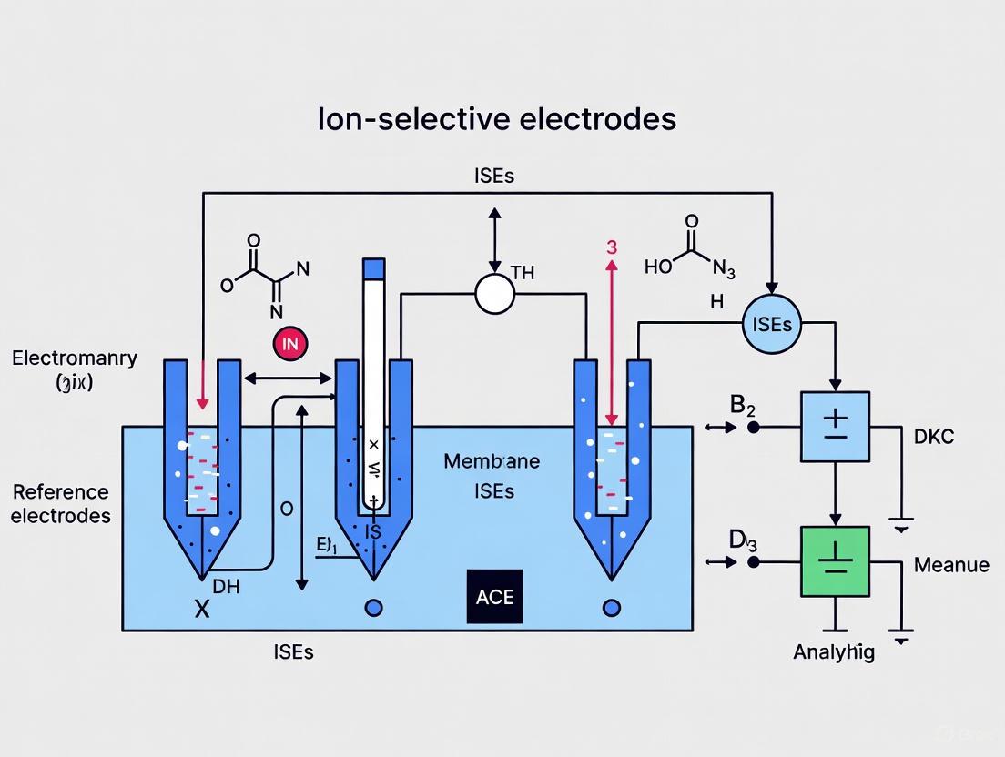
A complete potentiometric measurement system requires two essential components: the ion-selective electrode (indicator electrode) and a reference electrode that maintains a constant potential regardless of the sample composition [1]. The potential difference between these electrodes, measured under conditions of zero current flow, forms the basis of the analytical signal [1] [2].
The following diagram illustrates the fundamental working principle of an ISE and its relationship with the reference electrode within a potentiometric cell:
Figure 1: ISE Potentiometric Measurement Principle
The cell potential (E_cell) is calculated as the difference between the indicator and reference electrode potentials [6]:
[ E{\text{cell}} = E{\text{ise}} - E_{\text{ref}} ]
Where Eise incorporates the potential across the ion-selective membrane (Em) and the internal reference electrode potential [6]. The membrane potential develops due to the selective partitioning of ions between the sample solution and the membrane phase, creating a charge separation at the interface [2].
ISE Architecture and Membrane Types
Fundamental ISE Design Configurations
Ion-selective electrodes are categorized based on their structural design and membrane composition. The primary architectures include:
- Liquid-Contact ISEs: Traditional design with an internal filling solution contacting the ion-selective membrane [4]
- Solid-Contact ISEs (SC-ISEs): Advanced design where the membrane is in direct contact with a solid conductive material, eliminating the internal solution [2] [3]
- Coated Wire Electrodes: Simplified solid-contact design where a wire is directly coated with the ion-selective membrane [4]
Solid-contact ISEs have gained prominence in recent research due to their enhanced mechanical stability, miniaturization potential, and reduced maintenance requirements compared to traditional liquid-contact designs [2] [3]. The solid-contact transducer material (conductive polymers, carbon-based materials, or nanomaterials) serves as an ion-to-electron transducer between the ion-selective membrane and the underlying electrode substrate [2].
Ion-Selective Membrane Typologies
The selectivity of ISEs is primarily determined by the composition of the ion-selective membrane. The four principal membrane types are characterized in the table below:
Table 1: Ion-Selective Membrane Types and Characteristics
| Membrane Type | Composition | Target Ions | Selectivity Mechanism | Applications |
|---|---|---|---|---|
| Glass Membranes | Silicate or chalcogenide glass | H⁺, Na⁺, other monovalent cations | Ion-exchange at glass surface | pH electrodes, sodium ISEs [6] |
| Crystalline Membranes | Poly- or monocrystalline materials | F⁻, Cl⁻, Br⁻, I⁻, CN⁻, S²⁻, Cd²⁺, Pb²⁺ | Crystal lattice permeability | Fluoride ISE (LaF₃ crystal) [1] [6] |
| Polymer (Ion-Exchange Resin) Membranes | PVC or similar polymer with plasticizer and ionophore | Various cations and anions, including drugs | Selective ion complexation | Pharmaceutical compounds, environmental monitoring [1] [3] |
| Gas-Sensing Membranes | Gas-permeable membrane with internal electrolyte | CO₂, NH₃, NOₓ | Gas diffusion and internal pH change | Bacterial cultures, biological samples [1] [7] |
The following diagram illustrates the architectural differences between liquid-contact and solid-contact ISE designs, highlighting their key components:
Figure 2: ISE Architectural Designs Comparison
Performance Parameters and Analytical Characteristics
Key Analytical Performance Metrics
The effectiveness of ion-selective electrodes in research and analytical applications is evaluated through several critical performance parameters:
Selectivity: The ability of an ISE to respond preferentially to the target ion in the presence of interfering ions, quantified by the selectivity coefficient (Kᵢⱼ) [1]. Lower values indicate higher selectivity.
Sensitivity: The change in electrode potential per unit change in analyte concentration, reflected by the slope of the calibration curve [1]. Ideal Nernstian sensitivity is 59.2/z mV per decade at 25°C.
Detection Limit: The lowest concentration that can be reliably detected, typically defined by the IUPAC method as the intersection of the two linear segments of the calibration curve [2]. Modern SC-ISEs can achieve detection limits down to the pM level for certain applications [2].
Response Time: The time required for the electrode to reach a stable potential (typically 95% of final value) after a change in analyte concentration [1]. This parameter depends on membrane thickness, sample stirring, and measurement conditions.
Lifespan: The operational lifetime of the electrode before significant performance degradation. Well-maintained ISEs can remain functional for several months [2] [3].
Quantitative Performance of Representative ISEs
Table 2: Performance Characteristics of Representative Ion-Selective Electrodes
| Target Analyte | Linear Response Range | Slope (mV/decade) | Detection Limit | Response Time | Reference |
|---|---|---|---|---|---|
| Letrozole (PANI sensor) | 1.00 × 10⁻⁸ – 1.00 × 10⁻² M | 20.30 | 1.00 × 10⁻⁸ M | <30 s | [4] |
| Propranolol HCl | 1.0 × 10⁻³ – 3.1 × 10⁻⁶ M | ~59 (theoretical) | 3.1 × 10⁻⁶ M | <10 s | [3] |
| Lidocaine HCl | 1 × 10⁻³ – 2 × 10⁻⁶ M | ~59 (theoretical) | 2 × 10⁻⁶ M | <10 s | [3] |
| Letrozole (GNC sensor) | 1.00 × 10⁻⁶ – 1.00 × 10⁻² M | 20.10 | 1.00 × 10⁻⁶ M | <30 s | [4] |
| Diclofenac | Not specified | Not specified | Not specified | 2-3 s | [2] |
The data demonstrates that modified solid-contact electrodes can achieve exceptionally low detection limits while maintaining rapid response times, making them particularly suitable for pharmaceutical applications requiring high sensitivity.
Experimental Protocols for ISE Implementation
ISE Calibration and Measurement Protocol
Proper calibration is essential for obtaining accurate results with ion-selective electrodes. The following protocol outlines the standard calibration procedure:
Electrode Conditioning: Immerse the ISE in a solution containing the target ion (typically 10⁻³ M) for 15-60 minutes before initial use [3] [5].
Standard Solution Preparation: Prepare a series of standard solutions spanning the expected concentration range (typically 10⁻² to 10⁻⁶ M) using appropriate buffer solutions to maintain constant ionic strength [5].
Measurement Sequence: Immerse the ISE and reference electrode in each standard solution from lowest to highest concentration while stirring consistently at 300 rpm [3].
Potential Recording: Record the stable potential reading for each standard after the response stabilizes (typically 1-3 minutes per solution) [3].
Calibration Curve Construction: Plot potential (mV) versus logarithm of ion activity and perform linear regression to determine the slope, intercept, and correlation coefficient [5].
Sample Measurement: Measure the potential of unknown samples under identical conditions and determine concentration from the calibration curve using the Nernst equation [5].
Protocol for Solid-Contact ISE Preparation (PVC-Based)
The following detailed protocol describes the preparation of a solid-contact ion-selective electrode for pharmaceutical compounds, based on established methodologies [3] [4]:
Membrane Cocktail Preparation:
- Weigh and combine membrane components: 33% PVC, 66% plasticizer (e.g., O-NPOE), and 1% ion exchanger (e.g., KTpClPB) [3]
- Dissolve the "dry" mixture in tetrahydrofuran (THF) to create a 20% (w/w) solution
- Mix thoroughly until a clear, homogeneous solution is obtained
Electrode Body Assembly:
- Cut conductive substrate (carbon cloth) to appropriate dimensions (e.g., 7 × 2 cm²)
- Roll the carbon cloth and mount inside a PVC cylinder with central cylindrical hole
- Ensure secure electrical contact with the connecting wire
Membrane Deposition:
- Apply the membrane cocktail in multiple aliquots (e.g., 100 µL portions every 30 minutes)
- Allow THF to evaporate between applications
- Total membrane cocktail application: approximately 450 µL per electrode
- Air-dry the completed membrane for 48 hours at room temperature [3]
Electrode Conditioning:
- Condition the prepared ISE in a solution containing the target drug (e.g., 1.0 × 10⁻³ M) with appropriate pH adjustment (e.g., 10⁻² M HCl, pH 2.0) for 24 hours [3]
- Store conditioned electrodes in the same solution when not in use
The following workflow diagram illustrates the key stages in the development and application of solid-contact ISEs for pharmaceutical analysis:
Figure 3: Solid-Contact ISE Fabrication Workflow
Research Reagent Solutions for ISE Development
The following table details essential materials and their functions in ISE fabrication, particularly for pharmaceutical applications:
Table 3: Essential Research Reagents for ISE Fabrication
| Material Category | Specific Examples | Function | Application Notes |
|---|---|---|---|
| Polymer Matrices | Polyvinyl chloride (PVC), Ethylcellulose (EC) | Structural backbone of the membrane | Provides mechanical stability; PVC most common [3] [4] |
| Plasticizers | 2-Nitrophenyl octyl ether (NPOE), Di-octyl phthalate (DOP) | Imparts flexibility and governs dielectric constant | Affects ion exchanger solubility and mobility [3] [4] |
| Ion Exchangers | Potassium tetrakis(4-chlorophenyl) borate (KTpClPB), Sodium tetraphenylborate (NaTPB) | Provides initial ionic sites in membrane | Essential for proper electrode response [3] [4] |
| Ionophores | 4-tert-butylcalix-8-arene (TBCAX-8), various crown ethers | Selective complexation of target ions | Determines electrode selectivity [4] |
| Transducer Materials | Polyaniline (PANI), graphene nanocomposite (GNC), multiwall carbon nanotubes (MWCNT) | Ion-to-electron transduction in SC-ISEs | Enhances signal stability and lowers detection limit [2] [4] |
| Solvents | Tetrahydrofuran (THF), cyclohexanone | Dissolves membrane components for casting | THF most commonly used [3] [4] |
Pharmaceutical Applications and Research Implications
The application of ion-selective electrodes in pharmaceutical research has expanded significantly, driven by their unique advantages for drug analysis. Key applications include:
- Drug Release Studies: Continuous monitoring of drug release from dosage forms without sample pretreatment [3]
- Content Uniformity Testing: Rapid determination of active pharmaceutical ingredient distribution in solid dosage forms [2]
- Dissolution Testing: Real-time monitoring of dissolution profiles with high temporal resolution [3]
- Therapeutic Drug Monitoring: Measurement of drug concentrations in biological fluids like plasma and serum [4]
- Quality Control Analysis: Routine assessment of raw materials and finished products [2] [7]
The inherent advantages of ISEs for these applications include their ability to measure ions directly in colored or turbid solutions, minimal sample volume requirements, rapid analysis times (seconds to minutes), and capability for continuous monitoring [3] [4]. Furthermore, the development of miniaturized and wearable ISE-based sensors opens new possibilities for non-invasive therapeutic drug monitoring and point-of-care diagnostics [2].
Recent advancements in solid-contact ISEs incorporating novel materials such as MXene, conductive polymers, and carbon-based nanomaterials have significantly improved detection limits, selectivity, and operational stability [2]. These developments continue to expand the applicability of potentiometric sensors in pharmaceutical research and drug development workflows.
The potentiometric principle, governed by the Nernst equation, provides the fundamental theoretical foundation for ion-selective electrode operation. Through continuous refinement of electrode designs, particularly in solid-contact configurations, and optimization of membrane compositions, ISEs have evolved into sophisticated analytical tools with expanding applications in pharmaceutical research. The ongoing development of novel materials and fabrication techniques promises to further enhance the sensitivity, selectivity, and practicality of these sensors, ensuring their continued importance in both basic research and applied analytical science.
Ion-selective electrodes (ISEs) represent a cornerstone of modern analytical chemistry, enabling the precise quantification of ionic species in complex environments ranging from clinical diagnostics to environmental monitoring [8]. These potentiometric sensors function as electrochemical cells that generate a membrane potential in response to the activity of a specific ion in solution, operating on the fundamental principle of potentiometry where the cell potential is measured at near-zero current [8]. The historical progression of ISE technology spans over a century of innovation, beginning with the pioneering development of glass membrane electrodes and culminating in today's advanced solid-contact designs that offer unprecedented miniaturization, stability, and application versatility [8] [9]. This evolutionary journey reflects continuous interdisciplinary efforts in materials science, electrochemistry, and engineering to overcome fundamental limitations while expanding analytical capabilities. Within the broader context of fundamental research on ISE principles, understanding this historical trajectory provides critical insights into how theoretical advances and material innovations have collectively shaped contemporary sensor design, performance characteristics, and application scope, particularly in demanding fields such as pharmaceutical development and clinical analysis [2].
The Early Foundations: Glass Membrane Electrodes
The genesis of ion-selective electrode technology can be traced to 1906 when Cremer invented the first glass pH electrode, marking the birth of membrane-based potentiometric sensing [8]. This pioneering discovery was rapidly adopted as a routine analytical tool by the 1930s, establishing the glass electrode as the fundamental platform for hydrogen ion activity measurement [8]. These early glass membranes operated on an ion-exchange principle facilitated by specialized silicate or chalcogenide glass formulations that demonstrated selective permeability to specific cations [8] [10].
The core mechanism of glass membrane electrodes involves the development of a phase boundary potential at the glass-solution interface, governed by the selective partitioning of ions between these phases according to the Nernst equation [8] [11]. The glass composition determines ion selectivity; traditional silicate-based glasses exhibit preferential response to single-charged cations such as H⁺, Na⁺, and Ag⁺, while chalcogenide glass formulations extend sensitivity to certain double-charged metal ions including Cd²⁺ and Pb²⁺ [10]. Despite their revolutionary impact, early glass electrodes presented significant limitations including high electrical resistance, susceptibility to alkaline and acidic errors at pH extremes, and limited selectivity for analytes beyond hydrogen ions [10]. Throughout the 1940s and 1950s, numerous attempts were made to develop glass compositions responsive to alternative cations, but these efforts achieved only limited success, highlighting the need for fundamentally different membrane materials and sensing mechanisms [8].
Paradigm Shifts: The Emergence of Alternative Membrane Materials
The 1960s marked a transformative period in ISE development with two groundbreaking innovations that expanded sensing capabilities beyond protons. In 1966, Frant and Ross pioneered the first commercial crystalline membrane ISE utilizing a LaF₃ single-crystal for fluoride ion detection, while Stefanac and Simon simultaneously introduced the revolutionary concept of neutral ionophores with their valinomycin-based potassium-selective electrode [8]. These developments established new paradigms in ion-selective membrane design that quickly superseded earlier attempts to modify glass compositions for non-hydrogen ions.
Crystalline Membrane Electrodes
Crystalline membranes employ polycrystalline or single-crystal materials, typically consisting of insoluble inorganic salts such as LaF₃ for fluoride detection or Ag₂S for silver or sulfide ions [8] [10]. The ion-selectivity mechanism in these systems arises from the crystal lattice structure, which contains vacancies or sites that preferentially interact with specific ions of appropriate size and charge [10] [11]. The fluoride-selective electrode based on LaF₃ crystals remains one of the most successful implementations, offering excellent selectivity with interference primarily limited to hydroxide ions at high pH [8] [10]. These crystalline systems demonstrated superior mechanical robustness and eliminated the internal solution required in glass electrodes, thereby reducing potential junction complications [10].
Polymer-Based Liquid Membrane Electrodes
The introduction of neutral ionophores represented a fundamental advancement in molecular recognition for ISEs. These hydrophobic organic compounds, such as valinomycin for potassium selectivity, are incorporated into plasticized polymer membranes where they selectively complex with target ions and facilitate their transport across the organic phase [8]. The typical membrane composition includes a polymer matrix (commonly polyvinyl chloride or PVC), a plasticizer to impart fluidity, the ionophore for recognition, and lipophilic ionic additives to establish permselectivity and reduce membrane resistance [9]. This design creates a highly tailored molecular environment where the ionophore's specific coordination chemistry dictates selectivity, enabling the development of sensors for numerous cations and anions that were previously undetectable with glass or crystalline membranes [8] [9].
Table 1: Evolution of Ion-Selective Membrane Types and Their Characteristics
| Membrane Type | Key Developments | Representative Analytes | Selectivity Mechanism | Limitations |
|---|---|---|---|---|
| Glass Membranes | Invented by Cremer (1906); routine use by 1930s [8] | H⁺, Na⁺, Ag⁺ [10] | Ion-exchange at glass surface [10] | Alkaline/acidic errors; limited cation range [10] |
| Crystalline Membranes | LaF₃ F⁻ electrode (Frant & Ross, 1966) [8] | F⁻, S²⁻, Cl⁻, Br⁻, I⁻ [8] [10] | Crystal lattice permeability [10] | Limited to ions compatible with crystal structure [10] |
| Liquid/Polymer Membranes | Neutral ionophores (Stefanac & Simon, 1966) [8] | K⁺, Ca²⁺, NH₄⁺, various drugs [8] [12] | Selective complexation by ionophores [8] | Membrane component leaching; limited lifetime [9] |
Theoretical Underpinnings: The Principles of Potentiometric Sensing
The operational foundation of all ISEs rests on the establishment of a stable electrochemical potential at the interface between the ion-selective membrane and the sample solution. This boundary potential (Δφₘₑₘ) follows the Nernst equation, which relates the measured potential to the logarithm of the target ion activity [8] [11]:
Δφₘₑₘ = Δφₘₑₘ⁰ + (RT/zF)ln(aᵢ)
where R is the universal gas constant, T is absolute temperature, z is the ionic charge, F is Faraday's constant, and aᵢ is the activity of the primary ion [8] [11]. Under ideal conditions, this relationship produces a linear response with a Nernstian slope of approximately 59.16/z mV per decade of activity change at 25°C [8].
The complete potentiometric cell includes both the ion-selective electrode and a reference electrode that maintains a constant potential regardless of sample composition [8] [11]. The measured cell potential (Ecell) represents the cumulative potential differences across all interfaces in the system [11]:
Ecell = Eise - Eref + Ej
where Eise is the potential of the ISE, Eref is the reference electrode potential, and Ej represents the liquid junction potential that arises at the reference electrode bridge [11]. To ensure accurate measurements, modern potentiometers feature high input impedance (>10¹³ Ω) and appropriate operational amplifiers to handle the associated minute currents while maintaining near-zero current flow through the cell [8].
The selectivity coefficient (Kᵢⱼᴾᴼᵀ) quantifies an ISE's ability to discriminate between the primary ion and interfering species, representing a critical performance parameter [8]. This coefficient is typically determined using the separate solution method or fixed interference method, with ideal sensors exhibiting very small values (≪1) for all potential interferents [8].
The Solid-Contact Revolution: Overcoming Liquid-Contact Limitations
Traditional liquid-contact ISEs (LC-ISEs) utilizing internal filling solutions presented several operational challenges including evaporation, sensitivity to temperature and pressure variations, osmotic pressure effects causing water flux, and difficulties in miniaturization [9]. The pioneering work by Cattrall and Freiser in 1971 introduced the first "coated wire electrode," eliminating the internal solution and establishing the foundation for solid-contact ISEs (SC-ISEs) [2] [13]. However, these early designs suffered from poor potential stability and reproducibility due to high charge transfer resistance at the conductor-membrane interface [13].
The seminal 1997 publication by the Pretsch Group demonstrated that conventional LC-ISEs were biased by undesirable transmembrane ion fluxes from concentrated internal solutions, degrading analytical selectivity and sensitivity [8]. This critical insight reinvigorated the field, spurring development of SC-ISEs with controlled internal composition or complete elimination of filling solutions, yielding orders of magnitude improvement in both selectivity and detection limits [8].
Contemporary SC-ISEs incorporate a solid-contact (SC) layer between the ion-selective membrane (ISM) and electron-conducting substrate (ECS), serving as an ion-to-electron transducer [9]. This architecture eliminates the internal solution, creating a two-phase system that enhances detection limits and operational robustness [13]. The solid-contact layer typically consists of conductive polymers (e.g., polyaniline, PEDOT), carbon nanomaterials (e.g., MWCNTs, graphene), or other redox-active materials (e.g., ferrocene) that provide either redox capacitance or electric double-layer capacitance to stabilize the potential [9] [13].
Contemporary Applications and Experimental Implementations
The evolution from glass electrodes to advanced SC-ISEs has profoundly expanded practical applications across pharmaceutical analysis, clinical diagnostics, environmental monitoring, and industrial process control [8] [2]. Recent research demonstrates the versatility of modern SC-ISEs in addressing complex analytical challenges, with particular significance in pharmaceutical compound detection where simplicity, cost-effectiveness, rapid analysis, and suitability for on-site monitoring are paramount [2].
Pharmaceutical Analysis Case Studies
Benzydamine Hydrochloride Determination: A 2025 study developed both conventional PVC membrane and coated graphite all solid-state ISEs for detecting benzydamine hydrochloride (BNZ·HCl), a nonsteroidal anti-inflammatory drug [12]. The sensors employed an ion-pair complex formed between BNZ⁺ and tetraphenylborate (TPB⁻) incorporated into plasticized PVC membranes. The conventional PVC electrode demonstrated a Nernstian response of 58.09 mV/decade across a linear range of 10⁻⁵–10⁻² M with a detection limit of 5.81×10⁻⁸ M, while the all solid-state version exhibited comparable performance (57.88 mV/decade, detection limit 7.41×10⁻⁸ M) while eliminating internal solution complications [12]. Both sensors successfully determined BNZ·HCl in pharmaceutical cream and biological fluids without matrix interference and exhibited stability-indicating capability by detecting the drug in the presence of its oxidative degradant [12].
Letrozole Quantification: A 2023 investigation developed green SC-ISEs for the potentiometric determination of the anticancer drug letrozole [4]. The research compared a conventional sensor based on 4-tert-butylcalix-8-arene (TBCAX-8) as ionophore with membranes modified with graphene nanocomposite (GNC) and polyaniline (PANI) nanoparticles. The PANI-modified sensor demonstrated superior performance with the widest linear range (1.00×10⁻⁸–1.00×10⁻³ M), sub-Nernstian slope of 20.30 mV/decade, and successful application for letrozole determination in human plasma with recoveries of 88.00–96.30% [4]. This study highlighted how nanomaterial integration enhances sensor performance while aligning with green analytical chemistry principles.
Venlafaxine Hydrochloride Sensing: A 2024 systematic study compared transduction mechanisms for venlafaxine HCl detection using multiwalled carbon nanotubes (MWCNTs), polyaniline (PANi), and ferrocene as solid-contact materials [13]. The MWCNT-based sensor exhibited optimal electrochemical behavior with a near-Nernstian slope of 56.1 ± 0.8 mV/decade, detection limits of 3.8×10⁻⁶ mol/L, and minimal potential drift (34.6 µV/s) [13]. Comprehensive characterization using electrochemical impedance spectroscopy (EIS), chronopotentiometry (CP), and cyclic voltammetry (CV) revealed that each transducer's unique chemical and physical properties directly influenced sensor performance, with MWCNTs providing superior double-layer capacitance and interfacial stability [13].
Table 2: Performance Comparison of Modern Solid-Contact ISE Applications
| Analyte | Solid-Contact Material | Linear Range (M) | Slope (mV/decade) | Detection Limit (M) | Application Matrix |
|---|---|---|---|---|---|
| Benzydamine HCl [12] | Coated Graphite | 10⁻⁵–10⁻² | 57.88 | 7.41×10⁻⁸ | Pharmaceutical cream, biological fluids |
| Letrozole [4] | Polyaniline (PANI) nanoparticles | 10⁻⁸–10⁻³ | 20.30 | - | Bulk powder, dosage form, human plasma |
| Venlafaxine HCl [13] | Multiwalled Carbon Nanotubes (MWCNTs) | 10⁻²–10⁻⁷ | 56.1 ± 0.8 | 3.8×10⁻⁶ | Pharmaceutical dosage forms, synthetic urine |
Experimental Protocol: Fabrication of Coated Graphite Solid-Contact ISE
The following detailed methodology for constructing a coated graphite all solid-state ISE adapts procedures from recent pharmaceutical applications [12] [13]:
Ion-Pair Complex Preparation: Combine 50 mL of 10⁻² M drug solution (e.g., BNZ·HCl) with 50 mL of 10⁻² M sodium tetraphenylborate solution. Allow the precipitate to equilibrate with supernatant for 6 hours, then collect by filtration, wash thoroughly with bi-distilled water, and air-dry for 24 hours to obtain powdered ion-pair complex [12].
Sensing Membrane Formulation: Precisely weigh 10 mg of ion-pair complex, 45 mg of high-molecular-weight PVC, and 45 mg of plasticizer (e.g., dioctyl phthalate or 2-nitrophenyl octyl ether). Dissolve the mixture in 7 mL tetrahydrofuran (THF) and homogenize thoroughly [12] [13].
Electrode Assembly: Apply the membrane cocktail directly to a graphite electrode substrate using a micropipette, depositing multiple uniform layers with THF evaporation between applications. Alternatively, prepare a master membrane by casting the cocktail in a glass petri dish, allowing THF evaporation overnight, then cutting discs (typically 8-mm diameter) for attachment to electrode bodies using THF as adhesive [12].
Conditioning and Storage: Condition assembled electrodes by immersion in 10⁻² M primary ion solution for 4 hours to establish stable membrane potentials. For storage, keep electrodes dry under refrigeration when not in use to prolong lifetime [12].
Potential Measurements: Perform potentiometric measurements using a high-impedance pH/mV meter with Ag/AgCl reference electrode. Maintain minimal current flow (<1 pA) during measurements. Construct calibration curves by plotting measured potential (mV) versus logarithm of analyte activity [12] [13].
The Scientist's Toolkit: Essential Materials for SC-ISE Research
Table 3: Essential Research Reagents and Materials for Solid-Contact ISE Development
| Material Category | Specific Examples | Function | Application Notes |
|---|---|---|---|
| Polymer Matrices [12] [9] | Polyvinyl chloride (PVC), Polyurethane, Acrylic esters, Silicone rubber | Provides mechanical stability and serves as membrane backbone | PVC most common; alternative polymers offer different hydrophobicity and compatibility |
| Plasticizers [12] [9] [13] | Dioctyl phthalate (DOP), Dibutyl phthalate (DBP), Bis(2-ethylhexyl) sebacate (DOS), 2-Nitrophenyl octyl ether (o-NPOE) | Imparts membrane fluidity and affects dielectric constant | Choice influences ionophore selectivity and membrane longevity |
| Ionophores/Receptors [9] [4] | Valinomycin (K⁺), 4-tert-butylcalix-8-arene (cations), Natural/synthetic ion carriers | Molecular recognition element for selective ion binding | Critical for selectivity; neutral or charged carriers available for various ions |
| Ion-Exchangers [9] | Sodium tetrakis(pentafluorophenyl)borate (NaTFPB), Potassium tetrakis(4-chlorophenyl)borate (KTPCIPB), Sodium tetraphenylborate (NaTPB) | Provides permselectivity and reduces membrane resistance | Essential for establishing Donnan exclusion in neutral carrier membranes |
| Solid-Contact Materials [9] [4] [13] | Polyaniline (PANI), PEDOT, Multiwalled carbon nanotubes (MWCNTs), Graphene nanocomposite, Ferrocene | Ion-to-electron transduction between membrane and conductor | Determines potential stability and capacitance; redox vs. double-layer mechanisms |
| Solvents [12] [4] | Tetrahydrofuran (THF), Cyclohexanone | Dissolves membrane components for homogeneous casting | High purity essential to prevent membrane defects; THF most common |
Future Perspectives and Concluding Remarks
The historical evolution from glass pH electrodes to contemporary solid-contact ISEs represents a remarkable century of innovation in electrochemical sensing technology. Current research frontiers focus on further enhancing SC-ISE performance through advanced nanomaterials, improved transduction mechanisms, and expanded application domains [9]. Key development areas include novel solid-contact materials with higher capacitance and better hydrophobicity to prevent water layer formation, miniaturized designs for wearable and point-of-care applications, and integration with wireless technologies for remote monitoring [2] [9]. The emergence of wearable ISE sensors utilizing Bluetooth or NFC communication protocols for non-invasive health monitoring represents a particularly promising direction [2].
Despite significant advances, challenges remain in achieving long-term stability, ensuring reproducibility across manufacturing scales, and developing environmentally benign membrane components [9]. Ongoing research aims to address these limitations through standardized characterization protocols, improved understanding of interfacial processes, and development of sustainable sensor materials [9] [13]. The integration of SC-ISEs with emerging technologies such as Internet of Things (IoT) platforms and artificial intelligence for data analysis will likely expand their impact across clinical diagnostics, environmental surveillance, and industrial process control [2] [9].
The journey from Cremer's initial glass membrane to today's sophisticated solid-contact designs exemplifies how fundamental research principles coupled with materials innovation can transform analytical capabilities. As ISE technology continues to evolve, its convergence with nanotechnology, materials science, and digital health platforms promises to further advance this century-old technology, ensuring its continued relevance in addressing emerging analytical challenges across scientific disciplines.
Ion-selective electrodes (ISEs) are membrane-based potentiometric sensors that convert the activity of specific ions in a solution into an electrical potential [14] [15]. This transduction forms the cornerstone of a versatile analytical technique widely employed in chemical, biological, environmental, and industrial analyses. The core principle hinges on the use of a selective membrane that creates a potential difference dependent on the logarithm of the ionic activity of the target ion, as described by the Nernst equation [15] [16]. The general setup of an ISE includes the ion-selective membrane, an internal reference electrode, and an external reference electrode that completes the electrochemical cell [14].
The significance of ISE technology lies in its unique advantages. These sensors provide real-time measurements, can detect a wide range of ion concentrations, and require minimal sample preparation [14] [17]. Unlike many analytical methods, ISEs measure ion activity rather than mere concentration, which is often more relevant for understanding chemical behavior and biological activity [14]. The fundamental process can be summarized by the cell potential equation, Ecell = Eise – Eref, where Eise includes the potential of the internal reference electrode and the ion-selective membrane potential, and Eref is the potential of the external reference electrode [15]. The following diagram illustrates the core working principle of a potentiometric ISE.
Ion-Selective Membrane Typology and Function
The ion-selective membrane serves as the heart of the ISE, granting the sensor its specificity. Its primary function is to selectively permit the target ion to interact, generating a boundary potential. This potential arises from an unequal charge distribution across the membrane when the activity of the target ion differs between the sample and internal solutions [16]. Membranes are broadly classified based on their composition and physical properties, each with distinct advantages and selective affinities. The four principal types are glass, crystalline, ion-exchange resin (polymer), and enzyme-based membranes [14] [15].
Glass Membranes are primarily composed of silicate or chalcogenide glass and exhibit excellent selectivity for single-charged cations like H+ (pH electrode), Na+, and Ag+ [14] [15] [17]. Chalcogenide glass variants extend this selectivity to certain double-charged metal ions, such as Pb2+ and Cd2+ [14]. These membranes are noted for their high chemical durability, allowing operation in aggressive media [14]. However, they are susceptible to alkali and acidic errors at pH extremes and can be physically fragile [14] [18].
Crystalline Membranes can be formed from either a single crystal (e.g., LaF3 for fluoride electrodes) or a polycrystalline precipitate (e.g., Ag2S for sulfide or silver electrodes) [15] [17]. Their selectivity is inherently high because only ions capable of entering the crystal lattice can interfere with the electrode response [15]. A key advantage of some crystalline membranes is the absence of an internal solution, which simplifies design and reduces potential junctions [15].
Ion-Exchange Resin (Polymer) Membranes represent the most widespread type of ISE [15]. They consist of a polymer matrix, typically polyvinyl chloride (PVC), plasticizers, and a lipophilic ion-exchange substance or neutral carrier (ionophore) that confers selectivity [15] [19]. This design allows for the creation of selective electrodes for dozens of different ions, both cationic and anionic [15]. While highly versatile, these membranes generally have lower chemical and physical durability compared to glass or crystalline types and a finite lifetime [15].
Enzyme Electrodes are compound sensors that are often categorized under ISEs [14] [15]. They operate via a double-reaction mechanism where an enzyme immobilized in a membrane reacts specifically with a substrate. The product of this reaction (often H+ or OH−) is then detected by a true ISE, such as a pH electrode, housed within the same assembly [15]. A common example is the glucose-selective electrode [15].
Table 1: Comparison of Primary Ion-Selective Membrane Types
| Membrane Type | Composition | Target Ions (Examples) | Key Advantages | Key Limitations |
|---|---|---|---|---|
| Glass | Silicate or Chalcogenide glass | H+, Na+, Ag+, Pb2+, Cd2+ | High chemical durability; Excellent for single-charged cations. | Alkaline/Acidic error; Fragile; Limited ion range. |
| Crystalline | Mono-/Polycrystalline solids (e.g., LaF3, Ag2S) | F-, S2-, CN-, Cl-, Br-, I- | Excellent selectivity; No internal solution needed for some. | Membrane dissolution over time; Limited to compatible ions. |
| Polymer (Ion-Exchange Resin) | Polymer (e.g., PVC), Plasticizer, Ionophore | K+, Ca2+, NH4+, NO3-, Cl- | Highly versatile; Can be made for many ions. | Lower durability; Finite shelf life; Anionic electrodes less stable. |
| Enzyme-Based | Enzyme layer over a standard ISE | Glucose, Urea, etc. | High specificity for neutral molecules. | Complex construction; Response depends on enzyme kinetics. |
Ionophores: The Key to Molecular Recognition
In polymer membrane-based ISEs, the ionophore is the molecular component responsible for imparting high selectivity. Ionophores are lipophilic organic compounds that can selectively and reversibly bind to a target ion, facilitating its extraction from the aqueous sample into the organic membrane phase [19] [18]. The stability constant of the complex formed between the ionophore and the target ion relative to its complexes with potential interfering ions is the primary determinant of the sensor's selectivity [20].
The purity and quality of the ionophore are critical for optimal sensor performance. Impurities such as metal ions or surfactants can leach from the membrane, causing signal drift and anomalous baseline readings [19]. Therefore, application-tested, high-purity ionophores are essential for developing reliable ISEs. The following table details key ionophores used in research and commercial sensors.
Table 2: Key Research Reagent Solutions: Selectophore-Grade Ionophores
| Ionophore Name / Reagent | Target Ion | Function-Tested Performance | Critical Function |
|---|---|---|---|
| Valinomycin (Ammonium Ionophore I) [15] [19] | K+ | Linear Range: 1x10⁻⁶ to 1x10⁻¹ M; Slope: ~60.8 mV/dec [19] | Neutral carrier that forms a selective complex with K+ over Na+. |
| Calcium Ionophore I (ETH 1001) [19] | Ca2+ | Linear Range: 2x10⁻⁷ to 1x10⁻¹ M; Slope: ~28 mV/dec [19] | Neutral carrier with extremely high selectivity for Ca2+ ions. |
| Nonactin (Ammonium Ionophore I) [19] | NH4+ | Linear Range: 1x10⁻⁶ to 1x10⁻¹ M; Slope: ~60.8 mV/dec [19] | Antibiotic ionophore selective for NH4+; also used for urea detection. |
| Tetradodecylammonium Nitrate (TDDAN) [21] | NO3- | Sensitivity: up to -55 mV/pNO₃ [21] | Positively charged ion-exchanger selective for nitrate ions (NO3-). |
| Sodium Ionophore | Na+ | N/A (Typically used in glass membranes) | Components in glass membrane (e.g., Aluminosilicate) confer Na+ selectivity [14]. |
| KTFBP (Ionic Additive) [21] | N/A (Additive) | N/A | Lipophilic anionic additive (e.g., KTFBP) reduces membrane resistance and improves response time. |
Ion-to-Electron Transduction in Solid-Contact ISEs
A critical challenge in ISE design is establishing a stable potential at the interface between the ion-conductive membrane and the electron-conductive measuring instrument. In traditional ISEs, this is achieved with an internal aqueous solution. Solid-Contact ISEs (SC-ISEs) eliminate this internal solution, enabling miniaturization and simpler fabrication [20] [21]. In these designs, an ion-to-electron transducer layer is interposed between the electron-conductive substrate (e.g., a metal electrode) and the ion-selective membrane. This transducer must convert ionic current in the membrane into an electronic current in the substrate, and it must exhibit a stable standard potential and high capacitance to prevent signal drift caused by the formation of thin water layers at the interface [21].
Conducting Polymers are a leading class of transducing materials. Polymers like poly(3,4-ethylenedioxythiophene) (PEDOT) doped with poly(styrenesulfonate) (PSS) or embedded with carbon nanotubes (CNTs) are highly effective [20] [21]. They conduct both ions and electrons and possess high redox capacitance, which stabilizes the potential at the inner interface [20]. When the sample ion activity changes, the resulting shift in the phase-boundary potential at the sample-membrane interface is compensated by a redox reaction within the conducting polymer layer, generating a transient current.
Carbon-Based Materials, such as ordered mesoporous carbon and double-walled carbon nanotubes (DWCNTs), are also used as transducers. Their high surface area provides a large double-layer capacitance, which contributes to potential stability [20] [21]. Recent research focuses on composites, such as PEDOT doped with DWCNTs, which combine the benefits of both materials to achieve improved transduction, lower detection limits, and enhanced long-term stability for sensors, such as those detecting nitrate ions [21].
The experimental workflow for fabricating and testing a solid-contact ISE with a conducting polymer transducer is illustrated below.
Advanced Signal Transduction and Amplification
While traditional ISEs rely on potentiometric (voltage) measurement, recent innovations have focused on transducing the ion-recognition event into other signals to overcome sensitivity limitations imposed by the Nernst equation (theoretical limit of 59.16/z mV per decade of activity change at 25°C) [20].
Constant Potential Coulometry is a prominent example. In this method, the potential between the SC-ISE and a reference electrode is held constant at 0 V [20]. A change in sample ion activity disturbs this equilibrium, causing a transient current to flow as the conducting polymer transducer is oxidized or reduced to compensate for the potential change. The integrated charge over time is proportional to the change in ion activity. This method offers significantly higher sensitivity, enabling the detection of minute activity changes as low as 0.1% for K+ [20]. The signal transduction and amplification principle is detailed below.
Coulometric ISE with Electronic Capacitor: To address baseline drift in constant potential coulometry, a new method introduces an external electronic capacitor in series with the ISE [20]. When the sample is changed, the potential shift is compensated by charging this external capacitor, and the current required to do so is measured. This approach eliminates the baseline drift associated with the slow redox degradation of conducting polymers and shortens the measurement time [20].
Detailed Experimental Protocol: Nitrate SC-ISE with DWCNT/PEDOT Transducer
This protocol details the fabrication of a solid-contact nitrate ion-selective electrode based on a DWCNT/PEDOT transducing layer and a fluoropolysiloxane (FPSX) membrane, as explored in current research [21].
Materials and Equipment
- Working Electrode: Fabricated silicon chip with platinum ultramicroelectrode array (UMEA) [21].
- Chemical Reagents:
- Ion-to-Electron Transducer: Double-walled carbon nanotubes (DWCNTs), 3,4-ethylenedioxythiophene (EDOT) monomer.
- Ion-Selective Membrane: Fluoropolysiloxane (FPSX) polymer, Tetrahydrofuran (THF) solvent.
- Ion-Exchanger: Tetradodecylammonium nitrate (TDDAN).
- Ionic Additive: Potassium tetrakis [3,5-bis(trifluoromethyl)phenyl] borate (KTFPB).
- Standard Solutions: Sodium nitrate (NaNO₃) solutions for calibration (e.g., 10⁻⁵ M to 10⁻¹ M).
- Instrumentation: Potentiostat/Galvanostat, pH/mV meter with high input impedance, Ag/AgCl reference electrode.
Step-by-Step Methodology
- Transducer Layer Deposition: The Pt working electrode is cleaned. A dispersion of DWCNTs is drop-cast onto the electrode surface and allowed to dry. Subsequently, the PEDOT polymer is electrodeposited onto the DWCNT layer from a solution containing EDOT monomer and DWCNTs using cyclic voltammetry or constant potential amperometry [21].
- Ion-Selective Membrane Preparation: The membrane cocktail is prepared by dissolving 200 mg of FPSX polymer in 1.5 mL of THF. To this, 4.2 mg of TDDAN (ion exchanger) and 2.5 mg of KTFPB (ionic additive) are added, resulting in a TDDAN:KTFPB molar ratio of 2:1 [21]. The mixture is thoroughly homogenized.
- Membrane Deposition: A small volume of the prepared membrane cocktail is drop-cast directly onto the DWCNT/PEDOT transducing layer. The device is left undisturbed to allow the THF solvent to evaporate slowly, forming a stable, solid polymeric membrane (approx. 6 µm thick) [21].
- Conditioning: The newly fabricated SC-ISE is conditioned by immersing it in a 0.01 M NaNO₃ solution for several hours (or overnight) to hydrate the membrane and stabilize the electrochemical potential.
- Calibration and Measurement: The conditioned SC-ISE is paired with an external Ag/AgCl reference electrode. The potential (EMF) is measured while the electrodes are immersed in a series of standard NaNO₃ solutions of known concentration (e.g., from 10⁻⁵ M to 10⁻¹ M), with gentle stirring. The potential is recorded once stable.
- Data Processing: The measured EMF (mV) is plotted against the logarithm of the NO₃⁻ activity (log a_NO₃⁻). The data is fitted using a linear regression to establish the calibration curve, from which the slope (sensitivity, in mV/decade) and detection limit can be determined.
Performance Metrics and Troubleshooting
- Expected Performance: A well-optimized sensor may exhibit a sensitivity of up to -55 mV per decade of NO₃⁻ activity, with a linear range from approximately 10⁻⁵ M to 10⁻¹ M [21].
- Common Issues:
- High Temporal Drift: Can be caused by the formation of a thin water layer between the membrane and the transducer. Using hydrophobic transducers (like DWCNT/PEDOT) and membranes (like FPSX) mitigates this [21].
- Slow Response Time: Can result from a membrane that is too thick or poor adhesion between layers. Optimizing the membrane thickness and deposition process is key.
- Reduced Sensitivity: Often linked to the leaching of membrane components or degradation of the ionophore/exchanger. Ensuring high-purity reagents and a well-formulated membrane is critical [19].
Ion-selective electrodes (ISEs) are transducer devices that convert the activity of specific ions in solution into an electrical potential, functioning as membrane-based potentiometric sensors [15]. While glass membrane electrodes, particularly pH electrodes, are widely recognized, recent decades have witnessed substantial advancement in solid-state, polymer, and crystalline membrane electrodes that offer remarkable improvements in detection limits, selectivity, and application versatility [22]. These developments have fundamentally transformed potentiometric analysis, pushing detection limits from the micromolar range down to the picomolar level—an improvement factor of up to one million—while enhancing interference discrimination by factors of up to one billion [22]. This technical guide examines the fundamental principles, material innovations, and experimental methodologies underlying these advanced membrane electrodes, providing researchers and drug development professionals with comprehensive insights into their capabilities and implementation.
Fundamental Principles of Membrane Electrodes
The Potentiometric Sensing Mechanism
The operational principle of all ion-selective electrodes centers on the development of a membrane potential that correlates with the activity of target ions in solution. When an ion-selective membrane separates two solutions containing different activities of the analyte ion, a boundary potential develops across the membrane according to a Nernst-like relationship [16]. The overall electrochemical cell potential is measured between reference electrodes immersed in the sample and internal solutions, described by:
Ecell = Eref(int) - Eref(samp) + Emem
Where Emem represents the membrane potential, which for a target ion A with charge z follows the relationship:
Emem = Easym - (RT/zF)ln[(aA)int/(aA)samp]
This simplifies to the practical working equation:
Ecell = K + (0.05916/z)log(aA)samp at 25°C
where K is a constant incorporating all other potentials [16]. The membrane thus generates a measurable electrical potential that depends logarithmically on the activity of the target ion in the sample solution.
Membrane Ion Selectivity and Permeability
The fundamental requirement for any ion-selective membrane is its ability to preferentially permit the passage of target ions while excluding interferents. This selective permeability arises from specific molecular interactions between the membrane components and the target ion, whether through ion-exchange processes, carrier complexation, or structural compatibility with crystalline lattices [15]. The membrane's selective nature ensures that the boundary potential responds primarily to changes in the activity of the target ion, with minimal interference from other ions present in the sample matrix.
Figure 1: Working principle of an ion-selective electrode showing the key components and ion transport mechanism that generates the measurable potential difference.
Classification and Properties of Advanced Membrane Electrodes
Crystalline Membrane Electrodes
Crystalline membranes represent a sophisticated class of ISEs fabricated from mono- or polycrystalline materials that provide exceptional ionic selectivity through their defined crystal structures [23] [15]. These membranes are typically formed from low-solubility inorganic salts, with heavy metal sulfides and silver salts being particularly common [24]. The crystalline lattice structure permits only ions that can integrate into the crystal matrix to interfere with electrode response, making these membranes inherently highly selective [15].
A paradigmatic example is the fluoride selective electrode based on LaF₃ crystals, which exhibits remarkable selectivity for fluoride ions due to the perfect matching of fluoride ions with the lanthanum fluoride crystal lattice [23] [15]. The membrane operates by allowing fluoride ions to migrate through the crystal lattice defects and vacancies, generating a potential dependent on the fluoride ion activity in solution. The primary advantage of crystalline membranes is their lack of internal solution, which reduces potential junctions and enhances measurement stability [15]. Additionally, these membranes demonstrate excellent chemical durability and can function in aggressive media where other membrane types might degrade.
Solid-State and Polymer Membrane Electrodes
Solid-state and polymer membranes encompass diverse materials systems that enable ion selectivity through various mechanisms, from glass analogues to advanced polymer composites.
Glass Membranes for Monovalent Cations While traditional glass membranes are well-established for pH measurements, advanced formulations using chalcogenide glass extend selectivity to double-charged metal ions like Pb²⁺ and Cd²⁺ [23] [15]. These membranes operate through an ion-exchange mechanism at the glass surface, where specific cations in the solution interact with binding sites in the glass matrix. The membrane potential develops due to the differential mobility of cations within the glass structure. However, users must account for alkali error (at high pH with low H⁺ concentration) and acidic error (at low pH with high H⁺ concentration), which can introduce non-linear responses outside optimal pH ranges [23].
Solvent Polymeric Membranes Polymer membranes represent the most widespread type of ion-selective electrodes, typically utilizing polyvinyl chloride (PVC) matrices plasticized with specific compounds to create flexible, ion-sensitive films [22] [15]. These membranes incorporate ionophores—molecular recognition agents that selectively complex with target ions—and ion exchangers to establish permselectivity. The ionophores can be electrically charged or neutral compounds designed with specific binding pockets for target ions. The extensive versatility of polymer membranes allows preparation of selective electrodes for dozens of different ions through appropriate selection of ionophores and membrane compositions [15].
Composite Solid-State Electrolytes Recent advances have integrated inorganic fillers into polymer matrices to create composite electrolytes with enhanced properties. For example, PEO–LiTFSI–LATP (PELT) composite electrolytes incorporate nanosized Li₁.₃Al₀.₃Ti₁.₇(PO₄)₃ fillers into a polyethylene oxide matrix, effectively reducing crystallinity while providing additional Li⁺ transport channels [25]. These composites demonstrate improved mechanical robustness, wider electrochemical stability windows (up to 4.9 V), and enhanced Li⁺ transference numbers compared to pure polymer electrolytes [25].
Table 1: Comparison of Advanced Ion-Selective Membrane Types and Their Characteristics
| Membrane Type | Composition | Primary Ions Detected | Selectivity Mechanism | Key Advantages | Limitations |
|---|---|---|---|---|---|
| Crystalline | Mono-/polycrystallites (e.g., LaF₃, Ag₂S) | F⁻, CN⁻, S²⁻, Cl⁻, Br⁻, I⁻ | Crystal lattice compatibility | Excellent selectivity, no internal solution, chemical durability | Limited to ions matching crystal structure, mechanical brittleness |
| Glass | Silicate or chalcogenide glass | H⁺, Na⁺, Ag⁺, Pb²⁺, Cd²⁺ | Surface ion-exchange | High chemical durability, works in aggressive media | Limited to single-charged and some double-charged cations, pH errors |
| Polymer | PVC with plasticizers and ionophores | K⁺, Na⁺, Ca²⁺, NO₃⁻, Cl⁻ | Ionophore complexation | Versatile for many ions, flexible, customizable | Lower physical durability, limited lifespan |
| Composite Solid-State | Polymer matrices with inorganic fillers (e.g., PEO-LATP) | Li⁺ | Hybrid transport pathways | Enhanced conductivity, mechanical strength, wide voltage window | Complex fabrication, interfacial resistance challenges |
Recent Advancements in Membrane Performance
The last decade has witnessed revolutionary improvements in ISE technology, particularly regarding lower limits of detection (LOD) and selectivity. Traditional ISEs were limited to concentrations around 10⁻⁶ M, but contemporary designs now achieve detection limits in the range of 10⁻⁸ to 10⁻¹¹ M for numerous ions [22]. This million-fold improvement stems from understanding and controlling ion fluxes through the membrane. By optimizing the composition of the inner solution and reducing ion diffusion in the membrane, researchers have successfully minimized the ion fluxes that previously established a limiting concentration near the membrane surface, thereby enabling trace-level measurements [22].
Furthermore, selectivity coefficients have improved dramatically, now often reaching values smaller than 10⁻¹⁰, with some systems demonstrating selectivity better than 10⁻¹⁵ [22]. These extraordinary improvements have opened new application domains for ISEs, particularly in environmental monitoring of trace metals and bioanalysis using metal nanoparticle labels, where they can now compete with sophisticated analytical techniques like ICP-MS for specific applications [22].
Experimental Protocols and Methodologies
Fabrication of Polymer Membrane ISEs
Materials and Reagents:
- High-molecular-weight Polyvinyl Chloride (PVC) - matrix material
- Plasticizers (e.g., phthalates) - impart flexibility and influence dielectric constant
- Ionophore (specific to target ion) - provides ion selectivity
- Salt solutions for internal filling - establish stable reference potential
- Tetrahydrofuran or cyclohexanone - solvent for membrane components
- Silver/silver chloride wire - internal reference electrode
Procedure:
- Dissolve 100-200 mg PVC in 2-3 mL tetrahydrofuran with continuous stirring
- Add plasticizer (typically 2:1 plasticizer to PVC ratio) and appropriate ionophore (0.5-5 mg depending on selectivity requirements)
- Stir the mixture thoroughly until a homogeneous solution is obtained
- Pour the solution into a glass ring placed on a glass plate and cover loosely to allow slow solvent evaporation over 24-48 hours
- Once solidified, punch membrane disks of appropriate diameter (typically 6-8 mm)
- Mount the membrane in an electrode body and fill with internal solution containing fixed activity of target ion
- Condition the electrode in a solution containing the target ion for 24-48 hours before use [15]
Preparation of Composite Solid-State Electrolytes
Materials and Reagents:
- Polyethylene oxide (PEO) - polymer matrix
- Lithium bis(trifluoromethane)sulfonimide (LiTFSI) - lithium salt
- Li₁.₃Al₀.₃Ti₁.₇(PO₄)₃ (LATP) - ceramic filler
- Anhydrous acetonitrile - solvent
- Aluminum oxide-coated polyethylene separator - mechanical scaffold
Procedure:
- Dissolve 1.0 g high-molecular-weight PEO (Mw = 600,000) in anhydrous acetonitrile
- Add LiTFSI at EO:Li ratio of 3:1 (mass ratio of 1:3 against polymer)
- Stir continuously at 25°C for 4 hours, then at 60°C for 2 hours
- Incorporate 0.15 g nanosized LATP ceramic filler into the PEO-LiTFSI solution
- Continue stirring for 6 hours to achieve homogeneous dispersion
- Cast the slurry into a polytetrafluoroethylene mold
- Allow solvent evaporation at ambient temperature for 6 hours
- Transfer to vacuum oven and dry at 50°C for 24 hours to remove residual solvent
- Punch electrolyte disks (19 mm diameter) in an argon-filled glovebox [25]
Integrated Electrode-Electrolyte Architecture Fabrication
To address interfacial resistance challenges in solid-state batteries, an integrated electrode-electrolyte architecture can be fabricated:
- Prepare conventional LiFePO₄ cathode by mixing active material, conductive carbon, and PVDF binder (8:1:1 ratio) in appropriate solvent
- Cast slurry onto aluminum foil and dry at 80°C for 24 hours in vacuum oven
- Uniformly coat PELT precursor solution onto the surface of dried LFP cathode
- Allow precursor infiltration at room temperature for 6 hours
- Dry in vacuum oven at 50°C for 12 hours to form compact composite interface
- Punch integrated electrode disks (16 mm diameter) in argon-filled glovebox [25]
Figure 2: Experimental workflow for fabricating polymer membrane ion-selective electrodes, highlighting key steps from component preparation to final conditioning.
The Scientist's Toolkit: Essential Research Reagents and Materials
Table 2: Key Research Reagents and Materials for Advanced Membrane Electrode Development
| Material/Reagent | Function | Application Examples | Technical Considerations |
|---|---|---|---|
| Polyvinyl Chloride (PVC) | Polymer matrix for membrane | Cation and anion selective electrodes | High molecular weight grades provide better mechanical stability |
| Ionophores (e.g., Valinomycin) | Molecular recognition element | K⁺-selective electrodes | Selectivity depends on molecular structure and complexation constants |
| Plasticizers (e.g., Phthalates) | Impart flexibility and adjust dielectric constant | Polymer membrane electrodes | Influence dielectric constant and ionophore solubility |
| LiTFSI | Lithium salt for ion conduction | Solid polymer electrolytes | High solubility and dissociation constant in polymer matrices |
| LATP Filler | Ceramic ion conductor | Composite solid electrolytes | Nanosized particles provide greater surface area for enhanced conduction |
| Polyethylene Oxide (PEO) | Polymer matrix for solid electrolytes | Lithium metal batteries | High molecular weight PEO provides better mechanical properties |
| Tetrahydrofuran | Solvent for membrane casting | Polymer membrane preparation | Anhydrous conditions prevent phase separation |
| Silver/Silver Chloride | Reference electrode material | Internal reference systems | Requires electrochemical conditioning for stable potential |
Analytical Performance and Applications
Quantitative Performance Metrics
The analytical performance of advanced membrane electrodes is characterized by several key parameters. Sensitivity is reflected in the electrode slope, which ideally approaches the Nernstian value (59.16/z mV per decade of activity at 25°C). The lower limit of detection (LOD), traditionally defined by the IUPAC method as the activity where the calibration curve deviates from linearity, has been dramatically improved in modern ISEs, now reaching the 10⁻⁹ to 10⁻¹² M range for many ions [22]. Selectivity coefficients (Kₚₒₜ) quantify the electrode's preference for the primary ion over interfering ions, with contemporary membranes achieving values below 10⁻¹⁰ in optimal cases [22]. Response time ranges from seconds to minutes depending on membrane thickness, composition, and the magnitude of activity change.
Applications in Research and Industry
The improved performance characteristics of modern membrane electrodes have enabled their application across diverse fields:
Environmental Monitoring ISEs with enhanced lower detection limits have been successfully applied to trace metal monitoring in environmental samples. For example, lead-selective electrodes can now measure Pb²⁺ activities down to 10⁻¹¹ M, enabling direct speciation analysis in water samples [22]. The ability to distinguish free ion activities from total concentrations makes ISEs particularly valuable for environmental speciation studies, as demonstrated by the pH-dependent response of Pb²⁺-ISEs in the presence of carbonate, which aligns perfectly with ICP-MS measurements of total lead content [22].
Bioanalysis and Medical Applications Miniaturized ISEs serve as powerful tools for bioanalysis, with sub-femtomole detection limits reported for various cations when sample volumes are reduced [22]. Enzyme electrodes coupling enzymatic reactions with potentiometric detection enable measurement of substrates like glucose, urea, and neurotransmitters [15]. In clinical settings, ISEs routinely perform billions of measurements annually for ions like Na⁺, K⁺, Ca²⁺, and Cl⁻ in blood, serum, and plasma samples [22]. Recent advances in solid-contact electrodes have further improved stability for in vivo measurements.
Energy Storage Systems Solid-state and composite electrolytes play critical roles in advanced battery technologies. The puzzle-like molecular assembly strategy using triallyl phosphate and 2,2,3,3,4,4,4-heptafluorobutyl methacrylate segments spliced into a vinyl ethylene carbonate matrix produces solid-state polymer electrolytes with high ionic conductivity (0.432 mS cm⁻¹) and Li⁺ transference numbers (0.70) at 25°C [26]. These materials enable high-voltage operation (up to 5.15 V) and support stable cycling in Li||LiNi₀.₆Co₀.₂Mn₀.₂O₂ cells for over 300 cycles, demonstrating the electrochemical robustness of modern membrane materials [26].
Future Perspectives and Research Directions
The ongoing evolution of membrane electrode technology continues to expand analytical capabilities. Several promising research directions are emerging, including the development of pulsed amperometric methods that extend beyond traditional potentiometry, novel calibration procedures that reduce demands on signal stability and reproducibility, and multifunctional membranes that combine sensing with other properties like self-powering or self-healing [22]. The integration of computational materials design with high-throughput experimentation promises to accelerate the discovery of novel ionophores and membrane compositions tailored for specific analytical challenges. As fundamental understanding of interfacial processes and mass transport in membrane systems deepens, further improvements in detection limits, selectivity, and operational stability are anticipated, solidifying the role of advanced membrane electrodes as indispensable tools in analytical chemistry, biomedical research, and energy storage technologies.
Ion-selective electrodes (ISEs) have undergone a revolutionary transformation with the development of solid-contact architectures, fundamentally addressing the limitations inherent in traditional liquid-contact designs. Solid-contact ion-selective electrodes (SC-ISEs) represent a significant technological advancement by eliminating the internal filling solution, thereby enabling unprecedented miniaturization, enhanced stability, and expanded application potential in fields ranging from pharmaceutical development to wearable environmental monitors [9] [27]. This transition from liquid-contact ISEs (LC-ISEs) to solid-contact systems has not only resolved fundamental operational challenges but has also opened new frontiers in sensor technology, particularly for applications requiring portability, continuous monitoring, and integration with miniaturized analytical devices.
The evolution began in 1971 with Cattrall and Freiser's pioneering "coated wire electrodes," which first demonstrated the possibility of eliminating the internal solution [27]. However, these early designs suffered from potential drift due to insufficiently defined transduction mechanisms at the membrane-conductor interface. The breakthrough came in 1992 when Lewenstam and Ivaska introduced an intermediate polypyrrole layer functioning as an "ion-to-electron transducer," establishing the modern SC-ISE architecture that has since become the state-of-the-art standard [27]. Subsequent research has focused on refining this core concept through novel materials and improved interfacial designs, leading to the current generation of high-performance SC-ISEs with exceptional potential stability, reproducibility, and detection capabilities [9] [28].
Fundamental Limitations of Liquid-Contact ISEs
Traditional liquid-contact ISEs feature an internal filling solution that serves as a bridge between the ion-selective membrane (ISM) and an internal reference electrode. While this design has proven effective for laboratory applications, it presents several inherent limitations that restrict its practical implementation beyond controlled environments [9].
Table 1: Key Limitations of Liquid-Contact ISEs
| Limitation Category | Specific Technical Challenges | Impact on Performance and Application |
|---|---|---|
| Physical Instability | Evaporation, permeation, and pressure/temperature-induced volume changes of internal solution [9] | Signal drift, requirement for careful maintenance, limited operational environments |
| Miniaturization Barriers | Difficulty reducing internal solution volume below milliliter level [9] | Bulky designs, incompatible with wearable/microfabricated devices |
| Ionic Flux Issues | Steady-state ionic flux between inner filling and test solutions [9] | Limited detection range, shortened electrode lifetime |
| Mechanical Complexity | Osmotic pressure differences causing water transfer and membrane stratification [9] | Compromised membrane integrity, potential delamination |
These limitations collectively restricted LC-ISEs from achieving their full potential in field-deployable, miniaturized, and continuous monitoring applications. The internal solution component fundamentally constrained the physical design, necessitating a paradigm shift toward all-solid-state architectures [9] [27].
SC-ISE Architecture and Working Principles
Core Structural Components
The modern SC-ISE features a sophisticated three-layer architecture that enables its superior performance characteristics:
Ion-Selective Membrane (ISM): The recognition element typically composed of a polymer matrix (usually PVC), plasticizer, ionophore (selective molecular recognition agent), and ion exchanger [9]. This membrane selectively interacts with target ions in the sample solution, generating an ion-specific potential.
Solid-Contact (SC) Layer: The ion-to-electron transducer positioned between the ISM and conductive substrate. This critical component replaces the internal solution of traditional ISEs and exists in two primary varieties based on transduction mechanism: redox-capacitive materials (conducting polymers, ferrocene derivatives) and electric double-layer (EDL) capacitive materials (carbon nanomaterials, nanostructured metals) [9] [13].
Electron-Conducting Substrate (ECS): The underlying electrode material (typically glassy carbon, gold, or screen-printed electrodes) that provides electrical connection to the measurement instrumentation [9] [29].
Ion-to-Electron Transduction Mechanisms
The fundamental operation of SC-ISEs relies on two distinct transduction mechanisms for converting ionic currents in the ISM to electronic currents in the ECS:
Redox Capacitance Mechanism: Utilizes materials with reversible redox properties, such as conducting polymers (PEDOT, PANi, polypyrrole) or molecular redox couples (ferrocene) [13] [27]. When target ions enter the SC layer, they trigger compensatory redox reactions that generate electron flow while maintaining charge neutrality. For example, in PEDOT-based transducers, the overall reaction can be represented as:
This mechanism establishes a thermodynamically defined potential that follows Nernstian behavior [27].
Electric Double-Layer (EDL) Capacitance Mechanism: Employed by high-surface-area materials such as carbon nanotubes (CNTs), graphene, and porous carbon structures [13] [29]. These materials function as electrochemical capacitors where ions accumulate at the SC/ISM interface, creating separated ionic and electronic charge layers that behave as a capacitor. The resulting potential stability is proportional to the capacitance of the SC layer, with higher capacitance yielding improved stability against current-induced polarization [9] [29].
Critical Materials and Experimental Methodologies
The Researcher's Toolkit: Essential Materials for SC-ISE Development
Table 2: Key Research Reagents and Materials for SC-ISE Fabrication
| Material Category | Specific Examples | Function and Importance |
|---|---|---|
| Polymer Matrices | Polyvinyl chloride (PVC), polyurethane, polystyrene, acrylic esters [9] | Provides structural backbone for ISM, determines mechanical properties and compatibility |
| Plasticizers | Bis(2-ethylhexyl) sebacate (DOS), 2-nitrophenyl octyl ether (NPOE), dibutyl phthalate (DBP) [9] [3] | Enhances membrane fluidity, controls dielectric constant, influences ionophore selectivity |
| Ionophores | Calix[n]arenes, crown ethers, cyclodextrins, natural/synthetic ion carriers [9] [29] | Provides selective molecular recognition of target ions through specific binding |
| Ion Exchangers | Sodium tetrakis(pentafluorophenyl)borate (NaTFPB), potassium tetrakis[3,5-bis(trifluoromethyl)phenyl]borate (KTFPB) [9] [3] | Introduces counter-ions for charge balance, facilitates ion exchange, establishes Donnan exclusion |
| Redox Transducers | PEDOT, PANi, polypyrrole, ferrocene derivatives [13] [27] | Enables redox capacitance mechanism with reversible ion-to-electron transduction |
| EDL Capacitive Materials | Multi-walled carbon nanotubes (MWCNTs), single-walled CNTs, graphene, 3D-ordered porous carbon [13] [29] | Provides high surface area for double-layer capacitance, enhances potential stability |
| Conductive Substrates | Glassy carbon electrodes, screen-printed electrodes (SPEs), gold films [9] [29] | Serves as electron-conducting foundation, enables electrical connection to instrumentation |
Standard Fabrication Protocol for SC-ISEs
Based on methodologies successfully implemented in recent studies [13] [3] [29], the following protocol represents current best practices for SC-ISE fabrication:
Step 1: Substrate Preparation
- Polish glassy carbon electrodes (GCE, 3mm diameter) successively with 1.0, 0.3, and 0.05 μm alumina slurries
- Rinse thoroughly with deionized water and ethanol between polishing steps
- Alternatively, use commercially available screen-printed electrodes (SPEs) for disposable configurations
Step 2: Solid-Contact Layer Deposition
- For CNT-based contacts: Prepare 1 mg/mL dispersion of MWCNTs in ethanol and deposit 10-20 μL onto substrate
- For conducting polymers: Electropolymerize monomer solution (e.g., 0.1 M EDOT in acetonitrile) via cyclic voltammetry (typically 10 cycles between -0.5 and +1.2 V)
- Allow deposited layers to dry thoroughly under ambient conditions or controlled temperature
Step 3: Ion-Selective Membrane Formulation
- Prepare membrane cocktail containing:
- 1.0 wt% ionophore (e.g., calix[4]arene for Ag⁺ sensing)
- 0.5 wt% ion exchanger (e.g., NaTFPB)
- 32.5 wt% PVC polymer matrix
- 66.0 wt% plasticizer (e.g., NPOE)
- Dissolve components in 2-3 mL tetrahydrofuran (THF) and mix thoroughly
Step 4: Membrane Deposition and Conditioning
- Deposit 50-100 μL of membrane cocktail onto solid-contact layer
- Allow THF solvent to evaporate slowly for 24-48 hours at room temperature
- Condition completed electrode in 0.01 M solution of target ion for 12-24 hours before use
Performance Validation Methodologies
Comprehensive characterization of SC-ISEs requires multiple electrochemical techniques to evaluate different performance aspects [13]:
Potentiometric Measurements:
- Calibrate in primary ion solutions across concentration range (typically 10⁻⁷ to 10⁻¹ M)
- Determine slope, linear range, and detection limit from EMF vs. log concentration plot
- Calculate selectivity coefficients using Separate Solution Method or Fixed Interference Method
Chronopotentiometry:
- Apply constant current (±1 nA) for 60 seconds
- Measure potential drift (ΔE/Δt) as stability indicator
- Calculate capacitance from C = i/(dE/dt), where higher capacitance indicates better stability
Electrochemical Impedance Spectroscopy (EIS):
- Scan frequency range from 0.1 Hz to 100 kHz at open-circuit potential
- Fit data to equivalent circuit to determine bulk resistance (Rᵦ), double-layer capacitance (C𝒹𝓁), and geometric capacitance (Cℊ)
Water Layer Test:
- Immerse electrode in primary ion solution (e.g., 0.01 M)
- Transfer to interfering ion solution (e.g., 0.1 M) of different primary ion concentration
- Monitor potential response; slow drift indicates minimal water layer formation
Performance Comparison and Optimization Strategies
Quantitative Performance Metrics of SC-ISE Transducer Materials
Table 3: Comparative Performance of Common Solid-Contact Materials
| Transducer Material | Mechanism | Potential Drift (μV/s) | Specific Capacitance (F/g) | Slope (mV/decade) | Detection Limit (M) |
|---|---|---|---|---|---|
| MWCNTs [13] [29] | EDL Capacitance | 34.6 | 12.8 | 56.1 ± 0.8 | 3.8 × 10⁻⁶ |
| PEDOT [27] | Redox Capacitance | 28.9 | 15.3 | 59.2 ± 1.1 | 2.1 × 10⁻⁶ |
| Polyaniline (PANi) [13] | Redox Capacitance | 41.3 | 9.7 | 57.8 ± 1.3 | 5.6 × 10⁻⁶ |
| Ferrocene [13] | Redox Capacitance | 67.2 | 6.2 | 55.3 ± 1.7 | 8.9 × 10⁻⁶ |
| 3D-Ordered Porous Carbon [28] | EDL Capacitance | 22.5 | 21.4 | 59.1 ± 0.6 | 1.2 × 10⁻⁶ |
Key Optimization Strategies for Enhanced SC-ISE Performance
Recent research has identified several critical strategies for optimizing SC-ISE performance:
Interfacial Hydrophobicity Control: Incorporating hydrophobic nanomaterials (e.g., MWCNTs) prevents formation of detrimental water layers between ISM and SC interfaces, a major source of potential drift [29]. This approach maintains interface stability even during prolonged immersion.
Capacitance Enhancement: Utilizing three-dimensionally structured materials with high specific surface area significantly increases double-layer capacitance, providing superior charge storage capacity and resistance to current-induced polarization [28] [30].
Redox System Stabilization: Employing conducting polymers with appropriate dopant ions that match the ISM ion-exchanger creates thermodynamically well-defined interfaces with minimal phase boundary potentials [27].
Miniaturization Compatibility: Optimizing material viscosity and deposition techniques enables seamless integration with microfabrication processes, particularly for wearable sensors and screen-printed configurations [13] [29].
Current Research Challenges and Future Perspectives
Despite significant advances, several challenges remain in the widespread implementation of SC-ISEs. Recent investigations have revealed that kinetic constraints at solid-solid and solid-liquid interfaces can significantly impact performance, with ion-selective membranes potentially inhibiting the full utilization of transducer material capacitance [30]. This underscores the need for co-optimization of membrane and transducer materials rather than independent development.
The reproducibility of standard potentials between different electrode batches remains challenging, with potential drifts on the order of 10 μV/h even after extensive conditioning representing a barrier to calibration-free operation [28]. Additionally, the long-term stability of transducer materials, particularly conducting polymers under continuous polarization, requires further investigation for applications requiring extended deployment.
Future research directions focus on developing novel composite materials that combine redox-active and capacitive properties, implementing advanced interfacial engineering to control charge transfer processes, and establishing standardized protocols for evaluating and reporting SC-ISE performance [9] [30]. The integration of machine learning approaches for analyzing complex interfacial phenomena, as recently demonstrated, represents a promising avenue for accelerating material development and optimization [30].
As these challenges are addressed, SC-ISEs are poised to enable transformative applications in wearable health monitors, implantable medical devices, environmental sensor networks, and high-throughput pharmaceutical screening systems, fulfilling their potential as robust, miniaturized analytical tools for the 21st century.
Transforming Pharmaceutical and Clinical Analysis: Practical Applications of ISEs
Direct potentiometry is a powerful analytical technique that measures the electrical potential of an electrochemical cell under static conditions to determine the concentration of ionic species in solution. This method is particularly valuable in pharmaceutical analysis for its ability to provide rapid, real-time measurements of Active Pharmaceutical Ingredients (APIs) without extensive sample preparation. The foundation of modern potentiometry was established with the formulation of the Nernst equation in 1889, which mathematically relates an electrochemical cell's potential to the concentration of electroactive species in the cell [31].
The significance of potentiometry expanded considerably with the development of ion-selective electrodes (ISEs). These are membrane-based potentiometric devices designed to measure specific ion activities in solution through the measurement of electrical potential [32]. Unlike many analytical sensors, ISEs measure ion activity rather than concentration, providing unique insights into the behavior of pharmaceutical compounds in solution. The technique has gained prominence in pharmaceutical applications due to several advantages: ease of operation, affordability, wide concentration measurement range, real-time measurement capability, and the ability to measure both negatively and positively charged ions [32].
Within the framework of fundamental research on ion-selective electrodes, understanding the core principles of direct potentiometry is essential for advancing pharmaceutical analysis techniques. This guide explores the theoretical foundations, practical methodologies, and specific applications of direct potentiometry for API quantification in pharmaceutical development and quality control.
Theoretical Foundations of Direct Potentiometry
The Nernst Equation and Its Significance
The operational principle of all ion-selective electrodes is governed by the Nernst equation, which describes the relationship between the measured electrical potential and the activity of the target ion in solution. The Nernst equation indicates that the voltage across the ion-selective membrane depends on the logarithm of the specific ionic activity [32]. The fundamental form of the equation for a cell potential (Ecell) is:
Ecell = K + (RT/zF)ln(a)
Where:
- K is a constant that includes the standard potential of the electrode and the potential of the reference electrode
- R is the universal gas constant
- T is the temperature in Kelvin
- z is the charge number of the ion
- F is Faraday's constant
- a is the ionic activity of the target ion
For practical analytical applications, this equation is often simplified to:
E = K + S·logC
Where:
- E is the millivolt reading
- S is the electrode slope (theoretical Nernstian slope is 59.16/z mV/decade at 25°C)
- C is the concentration of the ion [32]
In potentiometric measurements, the difference between activity and concentration is significant. While concentration represents the total amount of an ion in solution, activity reflects the effective concentration that accounts for interionic interactions. In pharmaceutical applications, where solutions often contain multiple ionic species, this distinction becomes particularly important for accurate quantification [31].
Potentiometric Electrochemical Cells
A typical potentiometric electrochemical cell consists of two half-cells, each containing an electrode immersed in a solution of ions whose activities determine the electrode's potential [31]. The complete cell includes:
- Indicator/Working Electrode: The ion-selective electrode whose potential responds to the analyte's activity
- Reference Electrode: An electrode with a known, fixed potential that remains constant throughout the measurement
- Salt Bridge: Contains an inert electrolyte (e.g., KCl) that connects the two half-cells while preventing mixing of solutions
- Ion-Selective Membrane: The heart of the ISE that provides selectivity for the target ion
The cell potential is measured under conditions of zero or negligible current flow to ensure the composition of the electrochemical cell remains unchanged during measurement [31].
Ion-Selective Electrodes: Types and Components
Membrane Types and Their Selectivity Mechanisms
The selectivity of ISEs is determined by the composition and properties of the ion-selective membrane. Four primary types of membranes are used in pharmaceutical applications:
Table 1: Types of Ion-Selective Membranes and Their Characteristics
| Membrane Type | Composition | Selectivity Profile | Advantages | Limitations |
|---|---|---|---|---|
| Glass Membranes | Chalcogenide or silicate glass | Single-charged cations (H⁺, Na⁺, Ag⁺) [32] | High durability, resistant to aggressive media [32] | Alkali error (pH >12), acidic error (pH <1) [32] |
| Crystalline Membranes | Poly- or monocrystalline substances (e.g., LaF₃ for fluoride) [32] | Ions that can enter crystal structure (anions and cations) [32] | Good selectivity, no internal solution required [32] | Limited to specific crystal-compatible ions |
| Ion-Exchange Resin Membranes | Organic polymer membranes with ion-exchange substances [32] | Wide range of single-atom and multi-atom ions [32] | Versatile selectivity, most common type [32] | Lower physical/chemical durability for anionic electrodes [32] |
| Enzyme Electrodes | Enzyme-containing membrane covering a true ISE [32] | Substrates of specific enzymes (e.g., glucose) [32] | Enables measurement of non-ionic analytes | Double-reaction mechanism, more complex [32] |
Electrode Components and Setup
A complete ISE measurement system consists of several key components:
- Ion-Selective Electrode: Comprises an internal reference electrode (typically silver wire coated with solid silver chloride) embedded in a filling solution containing the ions to be measured [32]
- Reference Electrode: Features a similar structure to the ISE but with the selective membrane replaced by fritted glass (a porous substance) that allows formation of a liquid junction with the external solution [32]
- Ion-Selective Membrane: The critical component that provides selectivity by allowing only specific ions to pass through, making the membrane selectively permeable [32]
- Voltmeter: A high-impedance potentiometer that measures the potential difference between the ISE and reference electrode without drawing significant current [31]
The potential of the complete electrochemical cell is determined by the equation:
Ecell = Eise - Eref
Where Eise represents the potential of the ion-selective membrane and internal reference electrode, and Eref is the potential of the external reference electrode [32].
Methodological Approaches for API Quantification
Analytical Procedure for Direct Potentiometry
The quantification of APIs using direct potentiometry follows a systematic experimental workflow:
Calibration and Method Validation
Calibration is critical for accurate API quantification. A series of standard solutions with known concentrations of the target ion is prepared, covering the expected concentration range of samples. The potential of each standard is measured, and a calibration curve is constructed by plotting potential (E) versus logarithm of concentration (log C). The curve should display a linear relationship with a slope close to the theoretical Nernstian value [32].
For fluoride determination as an example, a calibration curve is established using the equation E = K + S·logC, where E is the millivolt reading and C is the concentration in mg/L. Specific measurements might show -35.6 mV at 200 mg/L (log C = 2.301), 16.8 mV at 25 mg/L (log C = 1.396), and 89.3 mV at 1.563 mg/L [32].
Method validation for pharmaceutical applications should include:
- Accuracy: Assessment through recovery studies (typically 98-102%)
- Precision: Evaluation of repeatability and intermediate precision (RSD <2%)
- Linearity: Correlation coefficient (r) >0.999 across the working range
- Selectivity: Evaluation against potentially interfering ions
- Limit of Quantification (LOQ): Sufficiently low to detect APIs at specification levels
Research Reagent Solutions and Materials
Table 2: Essential Research Reagents and Materials for Potentiometric API Analysis
| Item | Function/Application | Technical Specifications |
|---|---|---|
| Ion-Selective Electrodes | Target-specific API quantification | Selectivity coefficients <0.01 for interfering ions; Nernstian slope (50-60 mV/decade) [7] |
| Reference Electrodes | Provide stable reference potential | Double junction design; stable potential (±0.2 mV); compatible filling solutions [7] |
| Ionic Strength Adjuster | Maintain constant ionic background | High-purity salts (e.g., KCl, NaClO₄); typically 0.1-1.0 M concentration; analyte-compatible |
| Standard Solutions | Calibration curve preparation | Certified reference materials; minimum 5 points across concentration range; prepared in matrix-matched solvents |
| pH Buffers | Control solution pH | Appropriate buffer capacity; non-interfering ions; pH range suitable for API stability |
| Organic Solvents | Dissolve hydrophobic APIs | HPLC-grade solvents (methanol, acetonitrile); low water content; membrane-compatible [33] |
Pharmaceutical Applications and Case Studies
API and Excipient Analysis
Direct potentiometry finds extensive application in pharmaceutical analysis for both APIs and excipients. Current USP-NF monographs recommend potentiometric titration (a related technique) for the assay of approximately 630 active pharmaceutical ingredients and 110 excipients in both aqueous and non-aqueous media [33].
Specific pharmaceutical applications include:
- Cation Analysis: Sodium, potassium, calcium, and lithium monitoring in various pharmaceutical formulations [7]
- Anion Analysis: Chloride, fluoride, nitrate, and bromide quantification in tablets, vitamins, and toothpaste [7]
- Surfactant Characterization: Determination of anionic, cationic, and nonionic surfactants used as dispersing and solubilizing agents [33]
- Edible Oil and Fat Analysis: Determination of acid value, ester value, hydroxyl value, iodine value, peroxide value, and saponification value in pharmaceutical formulations [33]
Measurement Ranges and Performance Characteristics
Table 3: Typical Measurement Ranges for Pharmaceutical Ions Using ISEs
| Target Ion | Application Context | Measurement Range | Electrode Technology |
|---|---|---|---|
| Lithium (Li⁺) | Psychiatric medications | 0.2 - 10,000 ppm [7] | PVC Membrane [7] |
| Sodium (Na⁺) | Injectable solutions, WFI purity | 0.01 - 100,000 ppm [7] | Sensing Glass [7] |
| Potassium (K⁺) | Electrolyte preparations | 0.04 - 39,000 ppm [7] | PVC Membrane [7] |
| Calcium (Ca²⁺) | Calcium supplements | 0.5 - 40,100 ppm [7] | PVC Membrane [7] |
| Chloride (Cl⁻) | Saline solutions, raw materials | 1.8 - 35,000 ppm [7] | Solid State Pellet [7] |
| Fluoride (F⁻) | Dental products, vitamins | 0.02 - 19,000 ppm [7] | Solid State Crystal [7] |
Case Studies in Pharmaceutical Analysis
Case Study 1: Sulfanilamide Purity Determination
The purity of sulfanilamide, used in treating vaginal yeast infections, can be determined in aqueous solution by automatic, potentiometric titration using sodium nitrite as the titrant. Potassium bromide is added to the solution as bromide ions act as catalysts for the diazotization titration. Using a Pt Titrode electrode, purity of the sample is determined in as little as three to five minutes, including electrode maintenance time [33].
Case Study 2: Ketoconazole Analysis
Ketoconazole, an antifungal drug, presents analytical challenges due to its low solubility point (less than 1 mg/mL). The concentration can be determined by non-aqueous acid-base titration using perchloric acid as titrant and a Solvotrode easyClean electrode. The analysis requires only three to five minutes, or up to 10 minutes including electrode conditioning time [33].
Case Study 3: Lidocaine in Ointments
Lidocaine, used as an anesthetic and anti-arrhythmic, can be assayed via potentiometric titration with sodium tetraphenylborate using a nonionic surfactant electrode. Methanol and heat are used to dissolve or destroy emulsion formulations, then glacial acetic acid is added to the prepared sample solution prior to titration. Automated potentiometric titration improves accuracy and repeatability of results while reducing human error in this complex matrix [33].
Advanced Applications and Future Perspectives
The continuing development of ion-selective membranes extends potentiometry to an increasingly diverse array of analytes relevant to pharmaceutical analysis [31]. Recent advances in membrane technology include nanochannel-based systems with precisely engineered channels that have shown exceptional potential for selective ion extraction due to their ability to control ion transport at the molecular level [34].
Critical parameters influencing selectivity in advanced membrane systems include surface charge distribution, nanochannel dimensions, morphology, and wettability. These factors interact with external driving forces to enable selective ion transport, providing crucial insights for optimizing membrane selectivity and performance [34].
Future directions in pharmaceutical potentiometry include:
- Development of miniaturized sensors for in-process monitoring
- Multi-array sensors for simultaneous determination of multiple APIs
- High-throughput systems for pharmaceutical quality control
- Advanced materials with enhanced selectivity and durability
- Integration with process analytical technology (PAT) initiatives
Direct potentiometry remains a vital technique in pharmaceutical analysis due to its unique combination of selectivity, sensitivity, and practicality. As membrane technology continues to advance, the application of ion-selective electrodes in pharmaceutical development and quality assurance will expand, providing robust solutions for the challenging analytical needs of modern drug development.
In the development of new pharmaceutical products, understanding and controlling drug release from delivery systems is paramount to ensuring therapeutic efficacy and safety. Traditional methods, particularly UV spectrophotometry, have long been the standard for quantifying drug release in vitro. However, these conventional approaches present significant limitations, including the necessity for sample withdrawal, inability to provide real-time data in biologically relevant environments, and challenges in differentiating between released drug and drug still encapsulated within carrier systems. These limitations have prompted the exploration of more advanced analytical techniques that can provide real-time, size-resolved monitoring under physiological conditions.
Within this context, ion-selective electrodes (ISEs) have emerged as a powerful alternative technology for drug release profiling. Building upon decades of fundamental research into ISE principles, modern applications demonstrate exceptional capability for monitoring pharmaceutical compounds in complex media. The core advantage of ISE technology lies in its ability to provide continuous, non-destructive measurements of ion activity in solution, making it ideally suited for tracking drug release kinetics without disrupting the physiological environment. This technical guide explores the fundamental principles of ISEs, their application in drug release monitoring, and provides detailed experimental protocols for researchers seeking to implement this powerful technology in their pharmaceutical development workflows.
Fundamental Principles of Ion-Selective Electrodes
Ion-selective electrodes are potentiometric sensors that measure the electrical potential of a solution relative to a specific ionic activity. The operation of ISEs is grounded in the Nernst equation, which describes the relationship between the measured electrochemical potential and the activity of the target ion in solution [35]. According to this fundamental principle, the voltage across an ISE membrane depends logarithmically on the ionic activity of the target analyte, enabling highly sensitive detection across a wide concentration range.
Core Components and Working Mechanism
The basic architecture of an ISE consists of several key components that work in concert to generate a selective potentiometric response:
- Ion-Selective Membrane: The heart of the ISE, this membrane contains specific ionophores or ion-exchange sites that selectively interact with the target ion. The membrane's composition determines the electrode's selectivity and sensitivity [36].
- Internal Reference Electrode: Typically a silver/silver chloride wire immersed in a solution containing a fixed concentration of the target ion, this component provides a stable reference potential [35].
- External Reference Electrode: Completes the electrochemical cell and maintains a constant reference potential against which the ISE potential is measured [35].
- Voltmeter: Measures the potential difference between the internal and external reference electrodes with high impedance to ensure negligible current flow [36].
The working mechanism involves the selective partitioning of ionic species between the sample solution and the membrane phase, creating a potential difference that follows the Nernstian relationship: E = E⁰ + (RT/zF)ln(a), where E is the measured potential, E⁰ is the standard potential, R is the gas constant, T is temperature, z is the ionic charge, F is Faraday's constant, and a is the ionic activity [35].
Evolution of ISE Technology and Relevance to Pharmaceutical Analysis
The development of ISE technology has progressed through several significant stages, with each advancement expanding its applicability to pharmaceutical analysis:
- Early ISEs (1900s-1960s): Characterized by the invention and refinement of the glass pH electrode, which became a standard laboratory tool by the 1930s [36].
- Modern ISEs (1960s-1980s): Marked by the development of various membrane types beyond glass, including crystalline membranes (e.g., LaF₃ for fluoride detection) and liquid membranes incorporating neutral ionophores like valinomycin for potassium detection [36] [22].
- Contemporary ISEs (1990s-Present): Featuring solid-contact electrodes, improved lower limits of detection (extending to 10⁻⁸–10⁻¹¹ M for some ions), and dramatically enhanced selectivity coefficients (<10⁻¹⁰ for some interfering ions) [2] [22].
The critical advancement that revolutionized ISE performance was the recognition that controlling ion fluxes through the membrane significantly impacts detection limits. By optimizing the composition of the inner solution and reducing undesirable ion diffusion, researchers achieved improvements in lower detection limits by factors of up to one million [22]. This breakthrough, combined with the inherent advantages of ISEs—including simplicity, affordability, rapid analysis, and the ability to measure ion activity rather than concentration—makes them exceptionally suitable for pharmaceutical applications where real-time monitoring under physiological conditions is essential [2].
Figure 1: Working Mechanism of an Ion-Selective Electrode (ISE). The diagram illustrates how target ions selectively partition into the membrane, generating a potential difference measured against a reference electrode.
ISE Versus UV Spectrophotometry for Drug Release Monitoring
The selection of an appropriate analytical technique for drug release profiling depends on multiple factors, including the specific drug delivery system, the required detection limits, and the complexity of the release medium. The following comparison highlights the distinct advantages of ISE technology over traditional UV spectrophotometry for real-time drug release applications.
Table 1: Comparative Analysis of ISE and UV Spectrophotometry for Drug Release Monitoring
| Parameter | Ion-Selective Electrodes (ISEs) | UV Spectrophotometry |
|---|---|---|
| Measurement Principle | Potentiometric measurement of ion activity | Absorption of ultraviolet-visible light |
| Sample Requirements | Minimal preparation; tolerates turbid samples | Requires clear solutions; susceptible to interference from particulates |
| Measurement Environment | Direct measurement in complex media including saline | Often requires sample dilution or extraction |
| Temporal Resolution | Real-time, continuous monitoring | Typically discrete time-point measurements |
| Selectivity | High for specific ions; can be tailored with ionophores | Limited to chromophores; susceptible to spectral overlap |
| Detection Limit | Can reach 10⁻⁸–10⁻¹¹ M for optimized electrodes [22] | Typically 10⁻⁶–10⁻⁷ M |
| Analysis Time | Seconds to minutes for stable readings | Minutes per sample including preparation |
| Multi-analyte Capability | Requires multiple specialized electrodes | Possible with advanced deconvolution |
| Physiological Relevance | Measures bioactive ion concentration | Measures total drug concentration (free + bound) |
| Automation Potential | High for continuous monitoring | Limited to automated sampling systems |
Advantages of ISE Technology in Pharmaceutical Contexts
The transition from UV spectrophotometry to ISE-based monitoring offers several distinct advantages in pharmaceutical development:
- Direct Activity Measurement: ISEs measure ion activity, which often correlates more closely with pharmacological activity than the concentration measurements provided by UV spectrophotometry [35]. This is particularly valuable when developing formulations where the free ion concentration determines therapeutic efficacy.
- Real-time Monitoring Capability: ISEs enable continuous, non-invasive monitoring of drug release without the need for sample withdrawal, enabling truly real-time release profiling [2]. This contrasts with UV methods that typically require discrete sampling and often involve disrupting the release environment.
- Tolerance to Complex Media: Modern solid-contact ISEs demonstrate excellent performance in biologically relevant media, including saline solutions, buffers, and complex matrices like blood serum and urine [2]. This eliminates the need for extensive sample preparation that often complicates UV analysis.
- Miniaturization and Portability: ISE technology readily lends itself to miniaturization, enabling the development of implantable or wearable sensors for in vivo monitoring applications [2]. Such miniaturization is considerably more challenging for UV spectrophotometric systems.
Implementation Strategies: ISE for Drug Release Profiling
Experimental Design Considerations
Successful implementation of ISE technology for drug release monitoring requires careful experimental design. Several critical factors must be addressed to ensure reliable and physiologically relevant data:
- Electrode Selection: Choose an ISE with appropriate selectivity for the target ion. For drug compounds that are not inherently ionic, consider derivatization strategies or the use of enzyme-based ISEs that convert the target analyte to a detectable ion [36].
- Calibration Approach: Develop a comprehensive calibration protocol that accounts for the specific release medium. Standard addition methods are particularly valuable when analyzing complex matrices where the background ionic strength may vary [35].
- Temperature Control: Maintain constant temperature throughout the experiment, as ISE response is temperature-dependent according to the Nernst equation [35].
- Reference Electrode Configuration: Ensure proper placement of the reference electrode to maintain a stable liquid junction potential, particularly when working with non-aqueous or mixed solvents [36].
Integrated Monitoring Systems
For comprehensive drug release characterization, ISEs can be integrated with complementary analytical techniques in multi-detector platforms. For example, asymmetric flow field-flow fractionation (AF4) coupled with UV-Vis detection and multi-angle light scattering (MALS) has been successfully used to monitor drug loading and release from nanoparticle systems while providing size-resolved information [37]. In such integrated systems, ISEs provide specific ion activity data while complementary techniques address other parameters of interest.
Experimental Protocols for ISE-Based Drug Release Monitoring
Protocol 1: Standard Calibration and Drug Release Profiling
This protocol outlines the fundamental procedures for establishing a reliable ISE-based drug release monitoring system, using a hydrochloride drug salt as a model compound.
Table 2: Research Reagent Solutions for ISE-Based Drug Release Monitoring
| Reagent/Material | Function | Specifications |
|---|---|---|
| Ion-Selective Electrode | Target ion detection | Selectivity coefficient <10⁻⁴ for primary ion |
| Reference Electrode | Stable potential reference | Double junction for complex media |
| Ionic Strength Adjuster | Constant background ionic strength | Typically 0.1-1.0 M inert salt (e.g., NaNO₃) |
| Standard Solutions | Calibration curve generation | 10⁻² to 10⁻⁶ M in release medium |
| Release Medium | Physiologically relevant environment | Buffer at pH 7.4 ± 0.1 with 0.9% NaCl |
| Temperature Control System | Maintain constant temperature | 37 ± 0.2°C for physiological studies |
Procedure:
- Electrode Conditioning: Immerse the ISE in a solution containing 10⁻³ M of the target ion for at least 1 hour before initial use. For storage, keep in a dilute solution of the primary ion.
- Calibration Curve:
- Prepare standard solutions across the concentration range 10⁻² to 10⁻⁶ M in the release medium.
- Add ionic strength adjuster to both standards and samples to maintain constant ionic background.
- Measure the potential of each standard solution, beginning with the most dilute and proceeding to the most concentrated.
- Plot potential (mV) versus logarithm of concentration. The slope should approximate the theoretical Nernstian value (59.16/z mV per decade at 25°C).
- Drug Release Experiment:
- Place the drug delivery system in the release medium maintained at 37°C with continuous agitation.
- Immerse the ISE and reference electrode in the release vessel, ensuring proper positioning relative to the agitation pattern.
- Record the potential at predetermined intervals (e.g., every 10 seconds initially, then less frequently as release stabilizes).
- Convert measured potentials to concentration values using the established calibration curve.
- Data Analysis:
- Plot cumulative drug release versus time to generate release profiles.
- Apply appropriate mathematical models (e.g., Higuchi, Korsmeyer-Peppas) to characterize release mechanisms.
Protocol 2: Solid-Contact ISE for Continuous Monitoring
Solid-contact ISEs (SC-ISEs) offer advantages for continuous monitoring applications by eliminating the internal filling solution, enhancing mechanical stability, and facilitating miniaturization.
Procedure:
- Electrode Preparation:
- Select a solid conductor substrate (glassy carbon, gold, or conducting polymer).
- Apply a layer of ion-to-electron transducer material (conducting polymer or carbon nanomaterial).
- Coat with ion-selective membrane containing PVC, plasticizer, ionophore, and ionic additive.
- Condition the prepared SC-ISE in a solution of the target ion overnight.
- Performance Validation:
- Determine linear range, detection limit, and selectivity coefficients against potentially interfering ions.
- Evaluate response time and potential stability over extended periods (24-48 hours).
- Continuous Release Monitoring:
- Implement the SC-ISE in a flow-through cell or directly in the release vessel.
- Use a data acquisition system for continuous potential recording.
- Apply chemometric tools for signal processing if monitoring multiple ions simultaneously.
Figure 2: Experimental Workflow for ISE-Based Drug Release Profiling. The diagram outlines the key steps in implementing ISE technology for monitoring drug release from delivery systems.
Advanced Applications and Future Perspectives
Emerging Applications in Pharmaceutical Development
The application of ISE technology in pharmaceutical analysis continues to expand, with several emerging areas showing particular promise:
- Nanoparticle Drug Delivery Systems: ISEs enable real-time monitoring of drug release from nanocarriers without the need for separation techniques that might disturb the release equilibrium. The high sensitivity of modern ISEs makes them ideal for tracking the relatively small amounts of drug released from nanoparticle systems [37].
- Stimuli-Responsive Delivery Systems: For delivery systems designed to respond to specific physiological stimuli (e.g., pH, enzyme activity, or biomarker concentration), ISEs provide a direct means of monitoring triggered release events in real-time [38].
- Continuous Manufacturing: The pharmaceutical industry's shift toward continuous manufacturing requires real-time monitoring techniques for quality control. ISEs integrated into manufacturing equipment can provide immediate feedback on drug release characteristics, enabling real-time release testing (RTRT) [39].
- Wearable and Implantable Sensors: Miniaturized ISEs can be incorporated into wearable devices or implantable sensors for monitoring drug release in clinical settings, potentially enabling personalized dosing regimens based on real-time pharmacokinetic data [2].
Integration with Complementary Analytical Techniques
While ISEs provide exceptional capability for monitoring specific ions, their utility is enhanced when integrated with complementary analytical techniques:
- Multi-detector Platforms: Combining ISEs with techniques such as AF4-UV-MALS provides comprehensive characterization of complex drug delivery systems, simultaneously monitoring drug release while tracking changes in nanoparticle size and distribution [37].
- Spectroscopic Integration: Coupling ISE measurements with fluorescence or vibrational spectroscopy enables correlation of drug release with structural changes in the delivery system or cellular responses [40].
- Microfluidic Systems: Incorporating ISEs into microfluidic "lab-on-a-chip" devices creates powerful platforms for high-throughput screening of formulation performance under physiologically relevant flow conditions [41].
Ion-selective electrode technology represents a powerful alternative to traditional UV spectrophotometry for drug release profiling, offering significant advantages in temporal resolution, physiological relevance, and application flexibility. The fundamental principles underlying ISE operation—rooted in the Nernst equation and selective ion partitioning—provide a robust foundation for quantitative analysis of drug release kinetics. Modern ISE designs, particularly solid-contact configurations with optimized membranes, achieve detection limits and selectivity coefficients that enable sensitive and specific monitoring of pharmaceutical compounds in complex biological media.
The experimental protocols outlined in this technical guide provide researchers with practical frameworks for implementing ISE technology in their drug development workflows. As the pharmaceutical industry continues to advance toward more sophisticated delivery systems and manufacturing approaches, the real-time monitoring capabilities of ISEs will play an increasingly vital role in formulation optimization, quality control, and ultimately, the development of more effective and reliable therapeutic products.
Ion-selective electrodes represent a class of potentiometric sensors that enable the specific measurement of ion activity in solution through a selective membrane [36]. The operational principle of ISEs is governed by the Nernst equation, which describes the relationship between the electrical potential across an ion-selective membrane and the logarithmic activity of the target ion [42]. This fundamental physicochemical relationship forms the theoretical foundation for modern wearable sweat sensing technologies, allowing researchers to transform biological ion concentrations into quantifiable electrical signals. The earliest ISE, the glass pH electrode, was invented in 1906, with significant advancements occurring in the 1960s with the development of the fluoride ISE with a lanthanum fluoride membrane and the potassium ISE using valinomycin as a neutral ionophore [36]. These pioneering developments established the membrane-based sensing architecture that continues to underpin contemporary wearable electrolyte monitoring systems.
Wearable sweat sensors represent a revolutionary application of ISE technology, enabling non-invasive, real-time monitoring of physiological status through the detection of electrolytes in perspiration [43]. These biosensors leverage the principles of ISEs while addressing unique challenges associated with wearable applications, including miniaturization, signal stability during movement, and biocompatibility. The integration of ISEs into wearable platforms has created unprecedented opportunities for personalized health monitoring by providing continuous, non-invasive tracking of key electrolytes such as sodium, potassium, chloride, and calcium [44]. This technical guide explores the fundamental ISE principles, material innovations, and experimental methodologies driving the development of wearable sweat sensors for electrolyte monitoring within the broader context of personalized health.
Fundamental Principles of Ion-Selective Electrodes
Core Components and Working Mechanism
Ion-selective electrodes function based on a potentiometric measurement principle, where the electrical potential across a selective membrane is measured under conditions of near-zero current [36]. The fundamental components of a conventional ISE include an ion-selective membrane, internal reference electrode, internal filling solution, and external reference electrode [42]. The heart of the ISE is the permselective membrane, which facilitates species recognition through selective interaction with target ions while excluding interfering ions [36]. When the ISE is immersed in a sample solution, a boundary potential develops at the membrane-solution interface due to selective ion exchange or extraction processes. This potential, which follows the Nernstian relationship, is measured against a stable reference potential to determine the target ion activity [42] [36].
The measurable cell potential (Ecell) in an ISE setup is defined by the equation: Ecell = Eise - Eref, where Eise represents the potential of the ion-selective membrane and internal reference electrode, while Eref denotes the potential of the external reference electrode [42]. The key membrane potential (Em) is controlled by the analyte's activity on both sides of the selective membrane. For a target ion with charge z, the membrane potential follows the Nernst equation: Em = E0 + (RT/zF)ln(a), where E0 is a constant reference potential, R is the universal gas constant, T is temperature in Kelvin, F is Faraday's constant, and a is the ionic activity of the target ion [42]. This logarithmic relationship enables the ISE to respond across a wide concentration range, typically several orders of magnitude, making it particularly suitable for monitoring dynamic physiological concentrations of electrolytes in sweat.
ISE Membrane Types and Selectivity Mechanisms
The selectivity and performance of ISEs are primarily determined by the composition and properties of the ion-selective membrane. Four principal membrane types have been developed for different sensing applications:
Glass Membranes: Primarily used for single-charged cations like H+, Na+, and Ag+, glass membranes consist of ion-exchange glass (silicate or chalcogenide) that demonstrates high durability in aggressive media [42]. These membranes operate through an ion-exchange mechanism at the glass surface, where target cations replace mobile ions in the glass matrix, creating the boundary potential.
Crystalline Membranes: Fabricated from poly- or monocrystalline materials like lanthanum fluoride (for fluoride detection) or silver sulfide, these membranes offer excellent selectivity as only ions capable of entering the crystal lattice structure can interfere with the electrode response [42]. The conductivity in these membranes occurs through lattice defects or vacancies that permit ion migration.
Ion-Exchange Resin Membranes: Utilizing organic polymer membranes impregnated with ion-exchange substances, these represent the most common ISE type and can be engineered for a wide range of single-atom and multi-atom ions [42]. These membranes typically incorporate ionophores—molecular recognition agents that selectively complex with target ions—embedded in a plasticized polymer matrix, most commonly polyvinyl chloride (PVC).
Enzyme Electrodes: While not true ISEs, enzyme electrodes incorporate a biochemical recognition layer (enzyme) that reacts with a specific substrate, producing ions detectable by an underlying ISE [42]. This enables the detection of non-ionic analytes like glucose or urea through enzymatic conversion to measurable ions.
The selectivity coefficient is a critical parameter for evaluating ISE performance, quantifying the electrode's preference for the primary ion over interfering ions. This selectivity is achieved through molecular design of ionophores or ion-exchange sites that exhibit preferential molecular recognition for the target ion based on size, charge density, and coordination geometry.
Wearable Sweat Sensor Architecture and Materials
Sensor Design and Integration Platforms
Wearable sweat sensors based on ISE technology require careful integration of multiple components into a compact, flexible platform suitable for continuous skin contact. The fundamental architecture typically consists of a sweat collection system, ISE array for multi-ion detection, reference electrode, signal processing electronics, and data transmission module [43] [44]. Recent advances have focused on miniaturizing these components while maintaining analytical performance and ensuring user comfort during prolonged wear. Sweat collection approaches range from direct skin contact using microfluidic channels to non-contact designs that leverage capillary action or natural sweat flow [44]. The sensor platform must maintain stable mechanical and electrical properties during movement, flexing, and varying environmental conditions encountered in real-world use.
The integration of ISEs into wearable devices has been facilitated by the development of solid-contact transducer systems that eliminate the need for liquid internal filling solutions [36]. These solid-contact ISEs typically employ conductive polymer layers or nanostructured carbon materials (such as carbon nanotubes) as ion-to-electron transducers between the ion-selective membrane and the underlying electrode [44]. This architectural innovation significantly enhances the mechanical robustness of wearable sensors while simplifying miniaturization and mass manufacturing processes. Additionally, the elimination of liquid components prevents leakage and extends operational lifetime, both critical considerations for consumer healthcare devices.
Advanced Materials for Enhanced Performance
Material innovation has been instrumental in advancing wearable sweat sensor capabilities, particularly in addressing challenges related to signal stability, skin compatibility, and continuous monitoring:
Carbon Nanotube (CNT) Substrates: CNTs provide high mechanical flexibility, electrical conductivity, and large surface area, making them ideal substrates for wearable ISEs [44]. Their fibrous structure facilitates the formation of highly conductive, porous networks that maintain functionality during repeated deformation.
Bio-Inspired Microtextured Membranes: Recent research has developed rose petal-inspired ion-selective membranes with microsurface textures that enhance wettability while maintaining self-cleaning properties [44]. These membranes replicate the unique wetting behavior of rose petals, which are hydrophilic at low water volumes (facilitating sweat retention for measurement) but become hydrophobic at higher volumes (enabling self-cleaning).
Polymer Composites: Plasticized PVC remains the most common matrix for ion-selective membranes, but recent formulations have incorporated various additives and alternative polymers to enhance flexibility, biocompatibility, and adhesion to transducer layers [36] [44]. These composites are typically optimized for specific manufacturing processes like screen printing or inkjet deposition to enable high-volume production.
The table below summarizes key material components and their functions in wearable sweat sensors:
Table 1: Essential Materials for Wearable Sweat Sensor Fabrication
| Material Component | Function | Common Examples | Key Properties |
|---|---|---|---|
| Ion-Selective Membrane | Target recognition | Plasticized PVC with ionophores | Selectivity, stability, ion exchange capacity |
| Ionophore | Molecular recognition | Valinomycin (K+), nonactin (NH4+) | Binding constant, selectivity coefficient |
| Transducer Layer | Signal transduction | CNTs, conductive polymers, graphene | Conductivity, capacitance, stability |
| Substrate | Structural support | Polyimide, PET, silicone | Flexibility, biocompatibility, conformability |
| Reference Electrode | Stable potential reference | Ag/AgCl with polymer electrolyte | Potential stability, low drift, robustness |
Innovative Approaches: Bio-Inspired Sensor Designs
Recent breakthrough research has demonstrated the potential of biologically inspired designs to overcome fundamental limitations in wearable sweat monitoring. A pioneering study published in August 2025 developed novel ion-selective membranes with microtextures inspired by rose petals that significantly enhance sensor performance [44]. This bio-inspired approach addresses the inherent hydrophobicity of conventional ISMs, which typically repels water and sweat, leading to poor signal stability and responsiveness. The research team from Waseda University created two distinct microtextured ISMs layered onto CNT-forest substrates: Sensor A replicated the fine micro-wrinkles of inner rose petals, while Sensor B mimicked the polygonal islands with spike-like protrusions characteristic of outer petals [44].
Both bio-inspired sensors demonstrated markedly improved water retention in static conditions compared to conventional smooth membranes, with Sensor A exhibiting the highest retention capacity, making it particularly suitable for sweat monitoring during physical movement [44]. Notably, both sensors maintained the self-cleaning behavior observed in actual rose petals, where surfaces transition from hydrophilic to hydrophobic when water volume exceeds a specific threshold, effectively repelling excess fluid and contaminants. This dual functionality addresses two significant challenges in wearable sweat sensing: maintaining consistent sample contact for stable measurements while preventing fouling from accumulated sweat components. Additionally, the microtextured membranes demonstrated enhanced electrochemical activity compared to conventional designs, potentially improving signal-to-noise ratios in real-world applications.
The research team successfully integrated these bio-inspired sensors into 3D-printed wearable devices that incorporated microchannels to transport sweat to the sensing elements while maintaining a 2-millimeter gap to avoid direct skin contact [44]. This non-contact design significantly reduces skin irritation and improves user comfort compared to conventional sensors requiring tight skin adhesion. During running tests, the devices accurately monitored dynamic sodium concentration changes, providing real-time assessment of electrolyte loss. The self-cleaning capability further enabled a sweat-recirculation mechanism, where sensors retained fluid within channels during low-sweat production periods and triggered cleaning once sweat levels increased beyond a specific limit [44]. This innovative approach represents a significant advancement toward comfortable, durable, and accurate wearable sweat monitoring systems suitable for long-term health tracking.
Experimental Protocols for Wearable Sweat Sensor Evaluation
Sensor Fabrication and Characterization Methodology
The development and validation of wearable sweat sensors requires systematic experimental protocols to ensure analytical reliability and physiological relevance. A comprehensive fabrication and characterization methodology includes the following key steps:
Substrate Preparation and Electrode Patterning: Begin with cleaning and surface treatment of flexible substrate materials (e.g., polyimide, PET). Deposit adhesion promotion layers if required, then pattern electrode structures using photolithography, screen printing, or inkjet printing. For CNT-forest substrates, grow or deposit aligned carbon nanotubes using chemical vapor deposition or filtration/transfer processes [44].
Ion-Selective Membrane Formulation: Prepare ion-selective membrane cocktails by dissolving high-molecular-weight PVC, plasticizer (e.g., DOS, o-NPOE), ionophore, and lipophilic additive in tetrahydrofuran (typical composition: 1-2% ionophore, 30-33% PVC, 65-68% plasticizer, 0.5-1% additive) [36]. For bio-inspired microtextured membranes, use replication techniques from rose petal molds as described in Section 4.
Membrane Deposition and Curing: Apply membrane cocktail to electrode surfaces via drop-casting, spin-coating, or printing methods. Allow solvent evaporation under controlled conditions (typically 24 hours at room temperature). For microtextured membranes, ensure proper replication of surface structures during curing [44].
Electrochemical Characterization: Characterize sensor performance using potentiometric measurements in standard solutions. Determine linear range, detection limit, sensitivity (Nernstian slope), and selectivity coefficients using the separate solution method or fixed interference method. Evaluate response time and potential stability over extended periods (≥24 hours).
Mechanical Testing: Subject sensors to bending cycles (typically 1000+ cycles at relevant radii) and stretching deformation where applicable. Monitor potential stability during and after mechanical stress to assess robustness for wearable applications.
The following workflow diagram illustrates the key stages in sensor development and validation:
Analytical Performance Assessment Protocol
Rigorous analytical validation is essential to establish the reliability of wearable sweat sensors for health monitoring applications. A comprehensive performance assessment should include:
Calibration Procedure: Prepare standard solutions covering the physiological range for each target analyte (e.g., Na+: 10-100 mM, K+: 1-20 mM, Cl-: 10-100 mM). Measure potential values in order of increasing concentration, with continuous stirring. Rinse sensors thoroughly between measurements. Construct calibration curves by plotting potential (mV) versus logarithm of ion activity. Calculate slope, intercept, linear correlation coefficient, and theoretical detection limit (typically taken as the concentration where the calibration curve deviates from linearity by a specific potential value) [42].
Selectivity Determination: Evaluate sensor selectivity using the separate solution method (SSM) or fixed interference method (FIM). For SSM, measure potential responses in separate solutions of primary ion and interfering ions at identical activities. Calculate selectivity coefficients (logK^pot_A,B) using the appropriate equation for the method employed. For wearable sweat applications, particularly relevant interferents include Na+ for K+ sensors, Ca2+ for Mg2+ sensors, and lactate for chloride sensors [42] [36].
Stability and Drift Assessment: Monitor potential output in standardized solutions over extended periods (≥24 hours) under controlled temperature conditions. Calculate drift rates (mV/hour) and evaluate reproducibility between multiple sensors from the same fabrication batch. For wearable applications specifically, assess the impact of temperature fluctuations typical of skin surface variations (28-36°C).
In Vitro Sweat Analysis: Validate sensor performance in artificial sweat solutions with compositions matching human perspiration. Test across physiological ranges under dynamic concentration changes to simulate real sweat patterns during exercise. Compare sensor results with reference analytical methods (e.g., ion chromatography, atomic absorption spectroscopy) to establish accuracy and reliability.
The table below summarizes key analytical performance metrics for typical wearable sweat sensors:
Table 2: Analytical Performance Requirements for Wearable Sweat Sensors
| Performance Parameter | Target Specification | Test Method | Physiological Relevance |
|---|---|---|---|
| Detection Limit | <0.1 mM for all electrolytes | Calibration curve extrapolation | Ensures detection at low sweat rates |
| Linear Range | Covers 5-100% of physiological range | Potentiometric measurement in standards | Matches expected sweat concentrations |
| Response Time | <30 seconds for 95% response | Step-change in concentration | Enables real-time monitoring |
| Drift Rate | <0.5 mV/hour | Extended measurement in standard | Ensures reliability during prolonged wear |
| Selectivity Coefficient | logK^pot < -2.0 for key interferents | SSM or FIM | Prevents false readings from interfering ions |
| Batch Reproducibility | <5% variation in slope | Multiple sensors from same batch | Ensures consistent performance across devices |
Research Reagent Solutions and Essential Materials
The development and implementation of wearable sweat sensors requires specific research-grade materials and reagents carefully selected for their electrochemical and biocompatibility properties. The following table comprehensively details essential research reagents and their specific functions in sensor fabrication and operation:
Table 3: Essential Research Reagents and Materials for Wearable Sweat Sensor Development
| Category | Specific Reagents/Materials | Function/Purpose | Technical Specifications |
|---|---|---|---|
| Polymer Matrix | Polyvinyl chloride (PVC), Polyurethane, Silicone rubber | Membrane matrix providing structural integrity | High molecular weight (>40,000), low impurity content |
| Plasticizers | bis(2-ethylhexyl) sebacate (DOS), o-nitrophenyl octyl ether (o-NPOE) | Impart flexibility and regulate membrane permittivity | High purity, low water solubility, appropriate lipophilicity |
| Ionophores | Valinomycin (K+), BME-44 (Ca2+), Sodium ionophore X (Na+) | Molecular recognition elements for target ions | High selectivity, appropriate complex stability constants |
| Lipophilic Additives | Potassium tetrakis(4-chlorophenyl)borate, Tridodecylmethylammonium chloride | Control membrane permselectivity and reduce membrane resistance | Compatible with membrane matrix, minimal leaching |
| Transducer Materials | Poly(3,4-ethylenedioxythiophene) (PEDOT), Carbon nanotubes (CNTs) | Convert ionic to electronic signals in solid-contact ISEs | High conductivity, redox stability, high capacitance |
| Reference Electrode | Polyvinyl chloride covalently modified with methyl methacrylate and 2-hydroxyethyl methacrylate | Provide stable reference potential in solid-contact systems | Low permeability to water and ions, stable potential |
| Sensor Substrates | Polyimide, Polyethylene terephthalate (PET), Polydimethylsiloxane (PDMS) | Flexible support for sensor components | Biocompatibility, appropriate Young's modulus, low moisture absorption |
The selection and quality of these research reagents directly impact sensor performance parameters including detection limit, selectivity, response time, and operational stability. Particularly critical are the ionophores which determine the fundamental molecular recognition capability, and the plasticizer systems which influence both the dielectric properties of the membrane and the mobility of ionophore-ion complexes [36]. For wearable applications specifically, additional consideration must be given to biocompatibility of all materials contacting skin or sweat, with rigorous testing required to ensure no leaching of membrane components occurs during prolonged wear. Recent advances in material science have enabled the development of increasingly specialized reagents optimized for the unique requirements of wearable sweat monitoring, including enhanced adhesion to flexible substrates and resistance to degradation by sweat components.
Data Analysis and Interpretation in Sweat Sensing
The transformation of raw potentiometric signals into physiologically meaningful information requires careful data analysis and interpretation strategies. The fundamental relationship between measured potential and ion activity follows the Nernst equation: E = E0 + (RT/zF)ln(a), where E is the measured potential, E0 is a constant, R is the universal gas constant, T is temperature, z is ion charge, F is Faraday's constant, and a is ion activity [42]. For practical applications, activity is often approximated with concentration, though this requires maintaining relatively constant ionic strength across measurements. In sweat analysis, where total ionic strength can vary significantly, appropriate calibration strategies are essential for accurate concentration determination.
Data presentation follows specific conventions for scientific clarity. Tables should be numbered sequentially, include clear concise titles, and present data in logical order with appropriate units specified [45] [46]. For frequency distributions of quantitative variables like electrolyte concentrations, data should be organized into class intervals of equal size, with customary recommendations suggesting between 6-16 classes for optimal presentation [45]. The following diagram illustrates the complete data workflow from acquisition to physiological interpretation:
For continuous monitoring applications, time-series analysis of electrolyte concentrations provides insights into dynamic physiological processes. Sodium trends in sweat correlate with hydration status and electrolyte balance, while potassium fluctuations may indicate muscle fatigue and metabolic activity [44]. Chloride monitoring offers information about electrolyte loss and cystic fibrosis screening potential. Proper data interpretation requires consideration of contextual factors including sweat rate, collection location on body, time after exercise initiation, and individual baseline concentrations. Advanced data processing approaches including signal filtering, baseline correction, and drift compensation algorithms further enhance the quality and reliability of extracted physiological information.
Wearable sweat sensors represent a transformative application of ion-selective electrode technology that brings laboratory-grade electrochemical sensing to non-invasive personalized health monitoring. The successful implementation of these devices relies on the fundamental principles of ISEs while incorporating innovative materials and design strategies to address the unique challenges of wearable operation. Recent breakthroughs in bio-inspired membrane designs, such as the rose petal-inspired microtextured sensors, demonstrate the potential for significant performance enhancements through nature-inspired engineering [44]. These advances, coupled with ongoing developments in flexible electronics, microfluidics, and data analytics, are paving the way for a new generation of comfortable, reliable, and information-rich wearable monitoring systems.
The future development of wearable sweat sensors will likely focus on several key areas: further improvement of sensor stability and selectivity through novel membrane materials and architectures; expansion of detectable analytes to include metabolites, proteins, and inflammatory markers; enhanced integration with complementary sensing modalities like pH and temperature measurement; and development of sophisticated data interpretation algorithms that translate complex multi-analyte temporal patterns into actionable health insights. As these technologies mature and validation studies establish their clinical utility, wearable sweat sensors based on ISE principles are poised to become powerful tools for personalized health assessment, athletic performance optimization, and management of various physiological disorders, ultimately fulfilling the promise of true continuous, non-invasive physiological monitoring.
In the rapidly advancing field of biopharmaceutical manufacturing, achieving precise and reliable control over bioreactor environments is paramount for optimizing product yields, ensuring quality compliance, and facilitating scalable production. Ion-Selective Electrodes (ISEs) represent a critical class of potentiometric sensors that enable real-time, selective monitoring of specific ionic species directly within complex fermentation broth matrices [47] [2]. Their operational principles are rooted in fundamental electrochemical thermodynamics, where the potential difference across an ion-selective membrane is measured under near-zero current conditions, providing a quantitative relationship with the activity of the target ion in solution [36] [48].
The integration of ISEs into bioprocess control systems aligns with the industry's move towards flexible, intensified, and data-driven manufacturing processes [49]. This technical guide examines the core principles of ISE technology, its specific applications in monitoring critical ions during fermentation, and the practical methodologies for implementing these sensors to enhance control in biotechnological processes, particularly within the context of pharmaceutical development and production.
Fundamental Principles of Ion-Selective Electrodes
Operational Mechanism and the Nernst Equation
Ion-Selective Electrodes are potentiometric sensors that measure the activity of specific ions in a solution. The core component is a permselective membrane that creates a potential difference by selectively interacting with the target ion [36]. This potential, which develops at the interface between the membrane and the sample solution, is measured against a reference electrode to form a complete electrochemical cell [48].
The fundamental relationship describing the electrode response is the Nernst equation:
Where E is the measured potential, E⁰ is a constant standard potential, R is the universal gas constant, T is the temperature in Kelvin, z is the ionic charge, F is Faraday's constant, and a is the activity of the ion [47] [36]. For practical analytical purposes, the equation is often adapted to use concentration instead of activity when the ionic strength of the solution is kept constant by using a suitable buffer [36] [48].
Key Components and Membrane Types
The performance and selectivity of an ISE are primarily determined by its membrane composition. The four primary types of ion-selective membranes are:
- Glass Membranes: Typically used for measuring single-charged cations like H⁺ (pH), Na⁺, and Ag⁺. They are durable and suitable for use in aggressive media, but are subject to alkali and acidic error ranges at pH extremes [47].
- Crystalline Membranes: Composed of poly- or monocrystalline materials (e.g., LaF₃ for fluoride sensing). They offer excellent selectivity, as only ions that can enter the crystal lattice interfere with the electrode response [47] [36].
- Ion-Exchange Resin Membranes: These utilize organic polymer membranes containing an ion-exchange substance, making them the most common ISE type. They can be prepared for a wide range of single-atom and multi-atom ions [47].
- Enzyme Electrodes: Not true ISEs, but utilize a coupled reaction where an enzyme reacts with a specific substance, and the product (often an ion) is detected by a true ISE (e.g., a pH electrode) [47].
The following diagram illustrates the basic structure and functioning of a typical liquid-contact ISE system.
Figure 1: Schematic Structure of an Ion-Selective Electrode (ISE) Measuring System. The system comprises an ISE and a reference electrode immersed in the sample solution. The ion-selective membrane is the core sensing component, generating a potential specific to the target ion activity. The internal solution and reference electrode provide a stable reference potential, and the high-impedance voltmeter measures the potential difference with minimal current draw [47] [48].
Critical Ions in Fermentation and Their Monitoring with ISEs
Fermentation is a complex biological process where microorganisms convert raw materials into valuable products. Maintaining optimal process conditions is crucial for consistent output, and tracking specific ions provides vital insights into the metabolic state and progress of the culture [50] [51].
Key Ions and Their Significance
The table below summarizes critical ions monitored in bioprocesses, their significance, and the corresponding ISE membrane technology.
Table 1: Key Ions Monitored in Fermentation and Biotech Processes Using ISEs
| Target Ion | Significance in Bioprocesses | Typical ISE Membrane Type | Application Example |
|---|---|---|---|
| Ammonium (NH₄⁺) | Key nitrogen source; indicator of metabolic status [50]. | PVC Membrane [7] | Monitoring nitrogen consumption in microbial fermentations [7]. |
| Potassium (K⁺) | Essential electrolyte for cell growth and enzyme function [50]. | PVC Membrane [7] | Optimizing growth media for bacterial and yeast cultures [7]. |
| Sodium (Na⁺) | Osmolarity regulation; critical for cell viability [50]. | Sensing Glass [7] | Ensuring purity of Water for Injection (WFI) and controlling media osmolarity [7]. |
| Calcium (Ca²⁺) | Signaling molecule; cofactor for enzymes [50]. | PVC Membrane [7] | Screening process fluids and monitoring cellular functions [7]. |
| Nitrate (NO₃⁻) | Alternative nitrogen source in some microbial cultures. | PVC Membrane [7] | Tracking nutrient utilization in environmental and agricultural bioprocesses [47]. |
| Chloride (Cl⁻) | Osmolarity control; anion balance [50]. | Solid State Pellet [47] [7] | Monitoring salt levels in urine, blood, and in food/beverage production [47]. |
| Carbon Dioxide (CO₂) | Metabolic product in bacterial cultures; impacts pH [50] [7]. | Gas Sensing [7] | Monitoring metabolic activity and controlling pH in bacterial fermentations [7]. |
The Role of ISEs in Bioreactor Control Systems
The control architecture for a modern bioreactor operates at multiple levels. At the basic device/activator level, classical PID controllers adjust individual parameters like pump speeds or heater power based on sensor feedback [49]. ISEs provide the critical data for these control loops. For instance, a pH sensor (a type of ISE) provides feedback for the automatic addition of acid or base, while a dissolved oxygen probe controls aeration and agitation rates [50] [51].
More advanced Distributed Control Systems (DCS) integrate data from all sensors, including ISEs, to execute sophisticated control and optimization strategies across the entire plant [49]. The real-time data from ISEs allows for:
- Nutrient Feeding Strategies: Monitoring ammonium or potassium levels enables feedback-controlled feeding, preventing nutrient depletion or toxic accumulation [50] [7].
- Metabolic State Analysis: Tracking ion consumption and by-product generation (e.g., CO₂) provides insights into the metabolic health and phase of the culture [50] [51].
- Process Consistency and Quality Control: Ensuring consistent ion concentrations across batches is vital for product quality, especially in the production of biopharmaceuticals [7] [2].
Practical Implementation and Experimental Protocols
Quantitative Measurement Ranges for Key Ions
The practical application of ISEs requires knowledge of their operational ranges to ensure accurate quantification within the expected concentrations of the fermentation broth. The following table compiles the measurement ranges for ISEs targeting ions relevant to bioprocesses.
Table 2: Operational Ranges of Select Ion-Selective Electrodes in Bioprocessing [7]
| Ion-Selective Electrode | Measurement Range @ 25°C | Technology Type |
|---|---|---|
| Ammonia (NH₃) | 0.01 – 17,000 ppm as NH₃ | Gas Sensing |
| Ammonium (NH₄⁺) | 0.1 – 14,000 ppm as N | PVC Membrane |
| Calcium (Ca²⁺) | 0.5 – 40,100 ppm | PVC Membrane |
| Carbon Dioxide (CO₂) | 4.4 – 440 ppm | Gas Sensing |
| Potassium (K⁺) | 0.04 – 39,000 ppm | PVC Membrane |
| Sodium (Na⁺) | 0.01 – 100,000 ppm | Sensing Glass |
| Nitrate (NO₃⁻) | 0.1 – 14,000 ppm as N | PVC Membrane |
A Generalized Protocol for Monitoring Ammonium in a Fermentation Broth
The following workflow outlines a standard methodology for quantifying ammonium ion concentration in a bioreactor using an ammonium ISE, adaptable for other ions.
Figure 2: Workflow for Ion Concentration Measurement Using an ISE. The process begins with a critical calibration step using standard solutions, followed by prepared sample measurement, and concludes with data analysis against the calibration curve [47] [52].
Step 1: Calibration Curve Generation Calibrate the ISE with standard solutions of known ammonium concentrations (e.g., 0.1, 1, 10, 100 ppm). To account for the logarithmic response and maintain a constant ionic strength, all standards and samples must be diluted in a consistent background of Ionic Strength Adjustment Buffer (ISAB). Measure the potential (mV) for each standard and plot the potential versus the logarithm of the concentration to establish a calibration curve [47] [48].
Step 2: Sample Preparation Aseptically withdraw a sample from the bioreactor. Centrifuge or filter the sample to remove cells and obtain a clear supernatant. Mix an aliquot of the supernatant with an equal volume of ISAB to ensure a consistent ionic background [51].
Step 3: Potential Measurement Immerse the calibrated ammonium ISE and reference electrode in the prepared sample. Allow the potential reading to stabilize (typically under one minute unless concentrations are very low). Record the stable millivolt (mV) value [47] [48].
Step 4: Concentration Determination Use the calibration curve to convert the measured mV reading from the sample into the corresponding ammonium ion concentration.
The Scientist's Toolkit: Essential Reagents and Materials
Table 3: Key Research Reagent Solutions for ISE-based Monitoring
| Item | Function | Example from Protocol |
|---|---|---|
| Ionic Strength Adjustment Buffer (ISAB) | Masks the effect of varying background ionic strength, fixes pH, and eliminates interference from complexing agents. Allows concentration to be used in the Nernst equation instead of activity [36] [48]. | A buffer specific to the ion being measured (e.g., for fluoride, TISAB II is used) [52]. |
| Standard Solutions | Used to generate the calibration curve for quantifying the target ion in unknown samples. | Ammonium chloride solutions of known concentration (e.g., 1, 10, 100 ppm) for calibrating an NH₄⁺ ISE. |
| Reference Electrode | Provides a stable, constant half-cell potential against which the potential of the ISE is measured, completing the electrochemical cell [47] [48]. | A sealed Ag/AgCl reference electrode with a liquid junction. |
| Oxygen Combustion Vessel | (For solid samples) Used in sample preparation for total element analysis (e.g., Total Fluorine) by combusting the sample in an oxygen atmosphere [52]. | Parr oxygen combustion vessel with ignition wire and gelatin capsules. |
Recent Advances and Future Perspectives
The field of ISEs is continuously evolving, with recent research focused on overcoming limitations related to long-term stability, miniaturization, and sensitivity for a wider range of analytes.
Solid-Contact ISEs (SC-ISEs): A major trend involves eliminating the internal liquid filling solution of traditional ISEs. SC-ISEs use a solid conductive material (e.g., conductive polymers, carbon nanomaterials, metal oxides) as an ion-to-electron transducer. This simplifies manufacturing, enables miniaturization, improves mechanical stability, and reduces the drift associated with liquid-filled electrodes [36] [2]. These advancements are crucial for developing robust, disposable sensors for single-use bioreactors, which are increasingly common in the biopharmaceutical industry [49] [2].
Enhanced Selectivity and Sensitivity: The incorporation of novel materials like MXenes, advanced polymers, and composite-based transducers is pushing the detection limits of SC-ISEs down to the pico-molar (pM) range. This enhances their utility in detecting trace-level metabolites or impurities in complex biological matrices without requiring extensive sample pre-treatment [2].
Integration with Advanced Control Systems: The robustness of new ISE designs supports their integration into sophisticated process control strategies, including model-based predictive control and real-time optimization algorithms. This contributes to the evolution of Industry 4.0 in biomanufacturing, facilitating flexible and intensified production processes [49].
Ion-Selective Electrodes provide an indispensable tool for the real-time monitoring and control of critical ionic species within bioreactor and fermentation processes. Their operational principles, grounded in the Nernst equation and membrane selectivity, allow for direct insights into the metabolic state of microbial and cell cultures. The practical implementation of ISEs, from careful calibration to sample preparation, is essential for generating accurate and actionable data. With ongoing advancements in solid-contact technology and the integration of novel materials, ISEs are poised to become even more robust, sensitive, and integral to the advanced control systems that will define the next generation of biopharmaceutical manufacturing. Their role in ensuring process consistency, optimizing yields, and guaranteeing product quality underscores their fundamental value in biotechnology research and production.
High-Throughput Content Uniformity and Dissolution Testing in Drug Development
The pursuit of efficient and reliable analytical methods is paramount in drug development. High-throughput content uniformity and dissolution testing are critical for ensuring that every dosage unit contains the intended amount of Active Pharmaceutical Ingredient (API) and releases it consistently. Ion-Selective Electrodes (ISEs) represent a powerful analytical technology grounded in fundamental potentiometric principles, offering a pathway to automate and accelerate these essential quality control tests. ISEs are membrane-based sensors that measure the ionic activity of a specific analyte in a solution, providing real-time data based on the electrical potential generated across a selective membrane [53]. Their inherent advantages—including wide concentration measurement ranges, real-time output, and the ability to measure both positively and negatively charged ions—make them exceptionally suitable for integrated, high-throughput pharmaceutical analysis [53].
The core principle of ISE operation is governed by the Nernst equation, which states that the voltage across the membrane depends logarithmically on the ionic activity of the target ion [53]. This relationship allows for the precise quantification of specific ions directly in dissolution media or sample solutions, without the need for complex sample preparation that can bottleneck traditional techniques like High-Performance Liquid Chromatography (HPLC). This document details how the fundamental principles of ISEs can be leveraged to design robust, high-throughput workflows for content uniformity and dissolution testing, framing the discussion within the broader context of membrane potential and ion transport theory [11].
Fundamental Principles of Ion-Selective Electrodes
Operational Theory and Membrane Selectivity
Ion-Selective Electrodes function by generating an electrical potential that is specific to the activity of a particular ion in solution. The complete measurement setup involves an ISE and a reference electrode, both immersed in the analyte solution and connected to a voltmeter [53]. The key component is the ion-selective membrane, which is designed to be selectively permeable to the target ion. When the membrane interacts with the sample solution, a potential difference, known as the phase-boundary potential, develops at the interface. This potential is a direct function of the ion's activity in the sample compared to its activity inside the electrode [11].
The total cell potential ((E{cell})) is a composite signal described by the equation (E{cell} = E{ise} - E{ref}), where (E{ise}) encompasses the potential of the ion-selective membrane and the internal reference electrode, and (E{ref}) is the potential of the external reference electrode [53]. The potential across the selective membrane ((E_m)) is controlled by the analyte's activity on both of its sides. The selectivity of the membrane is the cornerstone of ISE technology. It is determined by the membrane's composition, which is engineered to facilitate a specific interaction—such as ion exchange or carrier complexation—with the primary ion, while minimizing interference from other ions present in the solution [11].
Key Types of Ion-Selective Membranes
The selectivity and performance of an ISE are dictated by the material of its membrane. Different membrane types are selected based on the target ion and the application requirements.
Table 1: Types of Ion-Selective Electrode Membranes
| Membrane Type | Composition | Primary Ions Measured | Key Characteristics |
|---|---|---|---|
| Glass Membranes | Silicate or chalcogenide glass [53] | Single-charged cations (H⁺, Na⁺, Ag⁺) [53] | High durability in aggressive media; subject to alkali and acidic error at pH extremes [53] |
| Crystalline Membranes | Poly- or monocrystalline substances (e.g., LaF₃ for fluoride) [53] | Ions that can enter the crystal lattice (e.g., F⁻, Cl⁻) [53] | Excellent selectivity; no internal solution; selectivity can apply to anion/cation of membrane substance [53] |
| Ion-Exchange Resin Membranes | Organic polymer membranes with ion-exchange substances [53] | Wide range of single- and multi-atom ions [53] | Most common ISE type; anionic electrodes have lower physical/chemical durability [53] |
| Enzyme Electrodes | Enzyme-containing membrane covering a true ISE [53] | Substances that react with the enzyme (e.g., glucose) [53] | Not a true ISE; uses enzyme reaction, with products detected by a underlying ISE like pH [53] |
High-Throughput Applications in Drug Development
Content Uniformity Testing
Content uniformity testing verifies that the amount of API in individual dosage units falls within a specified range. ISEs can be integrated into automated systems to provide rapid, non-destructive analysis. For ionic drugs or APIs that can be converted into an ionic form, an ISE can directly measure the ion concentration in a dissolved dosage unit. A high-throughput system can utilize a multi-well plate format, with an array of ISEs and automated liquid handling to sequentially or simultaneously measure multiple samples.
Recent advancements in automated compounding systems, such as 3D printing-based technologies, highlight the need for in-process content uniformity checks. One study utilized High-Performance Liquid Chromatography (HPLC) to validate the content uniformity of personalized hydrocortisone dosage forms (gel tablets, troches, and orodispersible films) produced via an automated system [54]. The results confirmed that all formulations met pharmacopeial criteria for mass and content uniformity, demonstrating the potential for automated production coupled with rigorous analytical testing [54]. ISEs could serve as a complementary or alternative technique for such in-process controls, especially for ions like sodium or chloride in the formulation.
Dissolution Testing
Dissolution testing measures the rate and extent at which an API is released from its dosage form into a dissolution medium. ISEs enable real-time, in-situ monitoring of ion release without the need for manual sampling and off-line analysis. This is a significant advantage for high-throughput applications, as it eliminates the need to stop the test for aliquot removal and processing.
In a typical ISE-based dissolution setup, the electrode is placed directly into the dissolution vessel. As the ionic API dissolves, the ISE continuously monitors the change in ion activity. The potentiometric signal is logged, providing a complete and continuous release profile. This methodology was exemplified in a study on 3D-printed hydrocortisone forms, where traditional dissolution testing showed that orodispersible films and troches achieved over 75% drug release within 5 minutes, while gel tablets had a slower profile, reaching 86% by 60 minutes [54]. For ionic drugs, ISEs can provide similar kinetic data with greater efficiency and less manual intervention.
Experimental Protocols and Methodologies
Calibration of Ion-Selective Electrodes
Calibration is a critical step to ensure the accuracy of ISE measurements. A series of standard solutions with known concentrations of the target ion must be prepared, covering the expected concentration range of the samples.
- Preparation of Standard Solutions: Create at least three standard solutions spanning the concentration range of interest. For example, for a fluoride ISE, standards might be 1.0 mg/L, 10.0 mg/L, and 100.0 mg/L.
- Measurement of Potential: Immerse the ISE and reference electrode in each standard solution, starting with the most dilute. Allow the potential reading (in millivolts, mV) to stabilize, then record the value.
- Plotting the Calibration Curve: Plot the recorded potential (E) against the logarithm of the ion concentration (log C). The plot should yield a linear relationship, consistent with the Nernst equation: (E = K + S \log C), where (S) is the slope and (K) is a constant [53].
- Validation: Periodically check the calibration by measuring a standard solution as an unknown to verify the accuracy of the curve.
Detailed Protocol for Content Uniformity Using ISEs
Objective: To determine the content of an ionic API in individual dosage units using an Ion-Selective Electrode.
Materials:
- Ion-Selective Electrode for the target ion
- Reference electrode
- Precision voltmeter/pH meter with ISE capability
- Magnetic stirrer and stir bars
- Volumetric flasks, beakers, and pipettes
- Ionic Strength Adjustment Buffer (ISAB), if required
Procedure:
- Standard Preparation: Prepare a stock solution of the API reference standard. Dilute this stock to prepare a series of at least 3 standard solutions covering the expected concentration range (e.g., 50%, 100%, 150% of label claim).
- Sample Preparation: For each dosage unit, transfer it to an appropriate container and dissolve in a known volume of solvent (e.g., dissolution medium or water). Dilute to volume if necessary. Note: The solvent and ionic strength should match the calibration standards as closely as possible; use ISAB if needed.
- Calibration: Measure the potential of each standard solution as described in Section 4.1 and construct the calibration curve.
- Sample Measurement: Immerse the electrodes in the prepared sample solution. Record the potential after stabilization.
- Calculation: Use the calibration curve equation to calculate the concentration of the target ion in the sample solution. Based on the dilution factor, calculate the content of the API in the dosage unit.
- Acceptance Criteria: Evaluate the results according to pharmacopeial guidelines (e.g., USP <905>).
Detailed Protocol for Dissolution Testing Using ISEs
Objective: To continuously monitor the in-vitro release profile of an ionic API from a solid dosage form using an Ion-Selective Electrode.
Materials:
- Standard dissolution apparatus (e.g., USP Apparatus 1 (Baskets) or 2 (Paddles))
- Ion-Selective Electrode and reference electrode designed for in-situ use
- Data acquisition system connected to the voltmeter
- Dissolution medium (e.g., buffer at specified pH)
Procedure:
- Apparatus Setup: Fill the dissolution vessel with the specified volume of medium and equilibrate to 37°C ± 0.5°C.
- Electrode Calibration: Calibrate the ISE in a solution with ionic strength and pH similar to the dissolution medium prior to the test.
- Electrode Placement: Securely position the ISE and reference electrode in the vessel, ensuring they do not interfere with the paddle or basket rotation.
- Baseline Reading: Record the baseline potential of the medium before introducing the dosage form.
- Initiation of Test: Start the apparatus and simultaneously drop the dosage form into the vessel. Begin data acquisition.
- Data Collection: Continuously record the potential at predetermined short intervals (e.g., every 10-30 seconds) for the duration of the test.
- Data Conversion: After the test, convert the recorded potential values to concentration values using the calibration curve.
- Profile Generation: Plot the cumulative percentage of drug released versus time to generate the dissolution profile.
The Scientist's Toolkit: Essential Research Reagents and Materials
The following table details key materials and reagents essential for implementing ISE-based methods in a pharmaceutical development setting.
Table 2: Essential Research Reagents and Materials for ISE-Based Testing
| Item | Function/Application |
|---|---|
| Ion-Selective Electrode | The core sensor; selected based on the target ion (e.g., chloride, sodium, nitrate) [53]. |
| Reference Electrode | Provides a stable, constant potential against which the ISE's potential is measured (e.g., double-junction Ag/AgCl) [53]. |
| Ionic Strength Adjustment Buffer (ISAB) | Added to standards and samples to maintain a constant ionic background, minimizing the effect of varying sample composition on the measured potential [53]. |
| API Reference Standard | A highly purified material used to prepare calibration standards for accurate quantification. |
| Dissolution Media Buffers | To maintain a physiologically relevant pH during dissolution testing (e.g., phosphate buffers at pH 6.8). |
| Voltmeter/pH Meter with ISE Input | A high-impedance meter capable of accurately measuring the millivolt potential generated by the ISE. |
| Automated Liquid Handling System | Enables high-throughput sample preparation and standard dilution for content uniformity testing. |
| Data Acquisition Software | For logging and processing continuous potentiometric data during dissolution testing. |
Data Analysis, Calibration, and Overcoming Challenges
Data Interpretation and Calibration Curves
The foundation of quantitative analysis with ISEs is the calibration curve. As per the Nernst equation, a plot of potential (E) versus the logarithm of concentration (log C) should be linear. The slope of this line indicates the sensitivity of the electrode. A slope close to the theoretical Nernstian value (e.g., ~59.16 mV per decade for a monovalent ion at 25°C) confirms proper electrode function. An example calibration data set for a fluoride ISE is shown below [53]:
Table 3: Example Calibration Data for a Fluoride Ion-Selective Electrode
| Concentration (mg/L) | Log C | Measured Potential (mV) |
|---|---|---|
| 1.563 | 0.194 | 89.3 |
| 25.0 | 1.398 | 16.8 |
| 200.0 | 2.301 | -35.6 |
Common Challenges and Mitigation Strategies
While ISEs offer significant advantages, users must be aware of potential challenges to ensure data integrity.
- Interfering Ions: Other ions in the sample can cross-react with the membrane, leading to inaccurate readings.
- Mitigation: Use an ISAB to mask interferents and choose an ISE with high selectivity coefficients for the primary ion over common interferents [53].
- Membrane Fouling: Proteins or other macromolecules in complex samples can adsorb to the membrane surface, degrading performance.
- Mitigation: Use appropriate cleaning protocols and consider membrane guards or specialized membrane materials for challenging matrices.
- Drift and Stability: The electrode potential may drift over time, affecting long-term measurements like dissolution.
- Mitigation: Allow sufficient equilibration time, maintain stable temperature, and perform frequent calibration checks. For dissolution, a stable baseline reading before test initiation is crucial.
- Sample Matrix Effects: Variations in pH, ionic strength, or viscosity between standards and samples can affect the potential.
- Mitigation: Matrix-match calibration standards with samples as closely as possible, and use ISAB consistently [53].
Achieving Precision: A Guide to ISE Calibration, Maintenance, and Error Mitigation
Ion-Selective Electrodes (ISEs) represent a cornerstone of modern potentiometric analysis, providing researchers with a direct means to measure ion activity in complex matrices. The fundamental principle governing ISE response is the Nernst equation, which establishes a logarithmic relationship between the measured electrochemical potential and the activity of the target ion [55]. For monovalent ions, this relationship predicts a slope of approximately 59.16 mV per decade at 25°C, while divalent ions exhibit a slope of approximately 29.58 mV per decade [56]. However, the theoretical ideal is often compromised by practical realities, making rigorous calibration not merely a preparatory step but a critical research activity that validates electrode performance and ensures data integrity across diverse applications from pharmaceutical development to environmental monitoring [3].
This guide establishes a comprehensive framework for ISE calibration, bridging fundamental potentiometric principles with advanced experimental protocols. By mastering standard preparation, interpolation strategies, and diagnostic slope evaluation, researchers can transform raw millivolt readings into reliable, publication-quality concentration data, thereby advancing foundational knowledge in ISE technology and its applications in drug development and beyond.
Theoretical Foundations of ISE Calibration
The Nernst Equation and Slope Evaluation
The entire edifice of ISE calibration is built upon the Nernst equation, which for a cation C⁺ with charge z is expressed as: E = E⁰ + (RT/zF)ln(a_C⁺) where E is the measured potential, E⁰ is the standard potential, R is the universal gas constant, T is the absolute temperature, F is the Faraday constant, and a_C⁺ is the ion activity [55].
In practical terms, this translates to a linear relationship between the electrode potential and the logarithm of the ion activity. The slope of this line, particularly its conformance to the theoretical Nernstian value, serves as the primary diagnostic for electrode health and measurement validity [56]. Significant deviation from the expected slope indicates potential issues with the electrode membrane, reference junction, or experimental conditions.
Activity versus Concentration: A Critical Distinction
A fundamental concept often overlooked in ISE research is that these electrodes respond to ion activity, not concentration [55] [57]. Activity (a) relates to concentration (C) through the activity coefficient (γ): a = γC. In dilute solutions, γ approaches 1, making activity and concentration nearly identical. However, as ionic strength increases, electrostatic interactions between ions reduce their effective activity, causing γ to fall below 1 [57].
This distinction has profound implications for calibration accuracy. The following table illustrates how ionic strength affects the activity coefficient and consequently introduces errors when measuring concentration:
Table 1: Activity Coefficients and Measurement Error at Different Ionic Strengths
| Ion Type | Ionic Strength (mol/L) | Activity Coefficient | % Error in Concentration |
|---|---|---|---|
| Monovalent (e.g., K⁺, Cl⁻) | 0.5 | 0.688 | 31% |
| 0.1 | 0.771 | 23% | |
| 0.01 | 0.901 | 10% | |
| 0.001 | 0.965 | 4% | |
| Divalent (e.g., Pb²⁺, Cd²⁺) | 0.5 | 0.341 | 66% |
| 0.1 | 0.413 | 59% | |
| 0.01 | 0.674 | 33% | |
| 0.001 | 0.869 | 13% |
Failure to account for the difference between activity and concentration, especially in samples with high or variable ionic strength, represents a major source of inaccuracy in ISE measurements [57].
Experimental Protocols for Standard Preparation and Calibration
Preparation of Calibration Standards
The accuracy of any ISE measurement is directly contingent upon the quality of the calibration standards. The following protocol ensures the preparation of reliable standards:
- Bracketing Concentrations: Select at least two, and preferably three or more, standard solutions that bracket the expected sample concentration. The highest and lowest standards should differ by at least one order of magnitude (e.g., 1 mg/L and 10 mg/L) to adequately define the calibration curve [56] [58].
- Serial Dilution: Employ serial dilution as the most accurate method for preparing standards from a concentrated stock solution. This technique minimizes cumulative volumetric errors compared to parallel dilution [56].
- Use of Ionic Strength Adjuster (ISA): Add an equal volume of ISA to all standards and samples. The ISA "masks" the varying ionic background of different solutions, ensuring that both standards and samples have a consistent, high ionic strength. This practice effectively equalizes activity coefficients, making the measured potential dependent primarily on the concentration of the target ion rather than the sample matrix, and also reduces interference from other ions [56].
- Solution Conditions: Prepare standards fresh on the day of use with analytically clean glassware to prevent contamination. Ensure all standards and samples have the same temperature, ideally calibrating at 25°C, as temperature significantly affects ion activity and electrode response [56] [58].
The Calibration Workflow: A Step-by-Step Guide
Adherence to a systematic calibration procedure is vital for obtaining repeatable results. The following workflow, depicted in the diagram below, outlines the critical steps:
Diagram 1: ISE Calibration and Measurement Workflow
The logical sequence is critical. Begin by conditioning the electrode by soaking it in a mid-range standard for approximately two hours before first use to hydrate the membrane and establish a stable equilibrium [56] [59]. Before starting, ensure the refill hole is open to allow hydrostatic equilibrium, and fill the reference chamber with fresh electrolyte [56].
During measurement, always calibrate in order of increasing concentration to minimize carry-over contamination [56] [58]. Rinse the electrode with deionized water between standards and gently blot dry with a lint-free cloth to avoid dilution errors [56]. Finally, evaluate the calibration slope against acceptance criteria before proceeding with sample analysis.
Data Interpretation: Interpolation and Slope Diagnostics
The Critical Role of Interpolation
The "bracket method," where samples are interpolated between standards that are close in concentration, is strongly recommended over extrapolation [59]. Extrapolation beyond the range of defined standards is not acceptable for accurate work, as it assumes a linear Nernstian response where non-linearity may exist due to changes in ionic strength or other matrix effects [59]. Using a two-standard bracket method approximately compensates for minor non-linearity in the analytical curve [60].
Evaluating Electrode Slope and Troubleshooting
The calibration slope is the most important parameter for diagnosing electrode performance. The following table provides acceptance criteria for new calibrations and guidance for troubleshooting non-conforming values.
Table 2: Slope Evaluation and Troubleshooting Guide
| Ion Type | Ideal Slope Range (mV/decade) | Common Issues & Corrective Actions |
|---|---|---|
| Monovalent (e.g., NH₄⁺, K⁺, NO₃⁻, Cl⁻) | 52 - 62 [56] | Low Slope (<52 mV): Often indicates membrane aging, contamination, or incomplete conditioning. Soak electrode in a mid-range standard for longer period [56] [59]. High Slope (>62 mV): Less common; can indicate electrical short or specific interference. Check for cracks in the membrane. |
| Divalent (e.g., Pb²⁺, Ca²⁺, Cd²⁺) | 26 - 31 [56] | Low Slope (<26 mV): Suggests membrane degradation or fouling. Clean or replace the sensor module according to manufacturer instructions [56]. |
| All Types | - | Drifting Potential: Can be caused by clogged reference junction, low fill solution, or temperature fluctuations. Ensure refill hole is open, top up electrolyte, and allow more time for thermal equilibrium [57] [59]. |
A slope outside the acceptable range indicates that the electrode is not responding correctly, and data collected with such a calibration should be treated with extreme caution.
Advanced Considerations for Research Applications
Managing Ionic Interference and Selectivity
A fundamental limitation of ISEs is that they are not perfectly ion-specific. All ISEs are sensitive to some extent to other ions, which is quantified by the Selectivity Coefficient (KPQ) [57]. If the primary ion is *P* and an interfering ion is *Q*, a KPQ of 0.1 means the electrode is ten times more sensitive to P than to Q. A common example is the ammonium ISE, which has a selectivity coefficient of approximately 0.1 for potassium. In a sample with equal concentrations of NH₄⁺ and K⁺, the potassium will contribute about 10% to the measured signal for ammonium [57].
Correcting for interference requires measuring the concentration of the interferent and applying a correction based on an experimentally determined selectivity coefficient. However, this coefficient is not a constant and can vary with concentration and ionic strength, making accurate correction complex [57]. The use of ISA and, where possible, removing interferents (e.g., precipitating chloride with silver salts when measuring nitrate) are more practical approaches [57] [56].
The Scientist's Toolkit: Essential Research Reagents and Materials
Table 3: Key Research Reagent Solutions for ISE Experiments
| Item | Function & Importance |
|---|---|
| Ionic Strength Adjuster (ISA) | Critical for negating the matrix effect. It fixes the ionic background, making the activity coefficient constant and allowing concentration to be measured directly [56]. |
| Primary Standard Solutions | High-purity solutions of the target ion used to create the calibration curve. Accuracy begins with these standards [56] [58]. |
| Polymer Membrane ISEs (e.g., for NH₄⁺, NO₃⁻) | The most common type, using an ionophore in a PVC membrane for selectivity. Offer a wide range of target ions [55] [3]. |
| Glass Membrane ISEs (e.g., for H⁺, Na⁺) | Used primarily for single-charged cations. Known for high durability in aggressive media but subject to alkali and acidic errors at pH extremes [55]. |
| Crystalline Membrane ISEs (e.g., for F⁻) | Made from single crystals (e.g., LaF₃ for fluoride). Offer excellent selectivity and do not contain an internal solution [55]. |
| Reference Electrode Fill Solution | Maintains a stable liquid junction potential. Must be kept topped up above the sample level to prevent back-flow and clogging [56]. |
Mastering the calibration of Ion-Selective Electrodes is a multifaceted discipline that integrates theoretical knowledge with meticulous practical execution. This guide has detailed the journey from foundational principles—understanding the Nernstian response and the critical distinction between activity and concentration—to the advanced implementation of robust experimental protocols. For the researcher, rigorous attention to standard preparation, adherence to a logical calibration workflow, and diligent diagnostic evaluation of the electrode slope are non-negotiable prerequisites for generating valid and reliable data. By embracing these practices, scientists can leverage the full potential of ISE technology as a powerful, real-time analytical tool in fundamental research and sophisticated application fields such as pharmaceutical development and environmental analysis.
Ion-selective electrodes (ISEs) are potentimetric sensors that measure the activity of specific ions in solution, finding indispensable applications in environmental monitoring, clinical diagnostics, and drug development [61]. Their operational principle is rooted in the Nernst equation, which describes the relationship between the measured electrical potential and the logarithm of the target ion's activity [62] [63]. Despite their theoretical simplicity and operational advantages, the practical deployment of ISEs is often challenged by three fundamental pitfalls: proper conditioning, membrane contamination, and response time variability. These factors critically influence the sensor's detection limit, sensitivity, selectivity, and long-term stability [64] [65]. This technical guide examines the underlying principles of these challenges within the context of ISE fundamental research and provides detailed protocols for their mitigation, enabling researchers to achieve reliable and reproducible potentiometric measurements.
Pitfall 1: Electrode Conditioning
The Science of Conditioning
Conditioning is the foundational process that prepares the ion-selective membrane (ISM) for measurement by establishing stable equilibrium conditions at all phase boundaries. A newly fabricated or dried ISE membrane contains no ions in its bulk. Conditioning facilitates the exchange of ions between the membrane and the conditioning solution, allowing the ionophore and ion-exchanger to become optimally functional [65]. This process hydrates the membrane surface, minimizes the formation of an undesired water layer between the membrane and the solid contact in solid-contact ISEs (SC-ISEs), and ensures a stable standard electrode potential (E₀) [65] [66]. Inadequate conditioning manifests as signal drift, extended response times, and reduced sensitivity.
Optimized Conditioning Protocol
The following protocol is designed for solid-contact ISEs, which are increasingly common in research and commercial applications due to their ease of miniaturization and integration [65] [67].
- Step 1: Hydration. Soak the newly fabricated or dried ISE in an electrolyte solution (e.g., 0.1 M KCl or a solution of the target ion) for a minimum of 12 hours (overnight is optimal). This allows the polymer matrix to swell and enables ion-exchange sites to become active.
- Step 2: Potential Stabilization. Connect the ISE to a high-impedance potentiometer and monitor the potential versus a reference electrode in a constant, low-activity solution of the target ion (e.g., 1.0 × 10⁻³ M). The electrode can be considered conditioned when the potential drift is less than 0.1 mV per minute for at least 15 minutes [65].
- Step 3: Calibration. Perform a full calibration (e.g., from 1.0 × 10⁻⁷ M to 1.0 × 10⁻¹ M) immediately after conditioning to establish the electrode's sensitivity (slope) and E₀. The measured slope for a monovalent ion should be close to the theoretical Nernstian value of 59.2 mV/decade at 25°C [63].
The diagram below illustrates the conditioning workflow and its critical role in achieving a stable and functional ISE.
Pitfall 2: Membrane Contamination & Selectivity
Membrane contamination poses a severe threat to ISE performance by altering the membrane's composition and its interfacial properties with the sample solution. The primary sources of contamination are:
- Biofouling: The accumulation of microorganisms, algae, or proteins on the membrane surface, particularly problematic in long-term environmental or biological monitoring [64] [63]. This can block ion-access to sensing sites and create a localized microenvironment.
- Lipophilic Interferents: Sample components (e.g., surfactants, lipids from biological fluids) that can solubilize in the organic membrane phase, displacing the ionophore or ion-exchanger and degrading selectivity [64] [62].
- Component Leaching: The loss of critical membrane components (ionophore, ion-exchanger, plasticizer) into the sample solution, especially in low-ionic-strength matrices, leading to signal drift and ultimately sensor failure [64] [63].
The interference from non-target ions is quantitatively described by the Nikolsky-Eisenman equation: E = E₀ + (RT/zF) ln[aᵢ + Kᵢⱼᵖᵒᵗ (aⱼ)^(zᵢ/zⱼ)] where aᵢ and aⱼ are the activities of the primary and interfering ions, and Kᵢⱼᵖᵒᵗ is the selectivity coefficient. A small Kᵢⱼᵖᵒᵗ value indicates high selectivity for the primary ion over the interferent [62] [63].
Experimental Assessment of Contamination and Selectivity
Researchers must rigorously test for contamination and selectivity issues. The following table summarizes key parameters and methods for this assessment.
Table 1: Key Parameters for Assessing ISE Contamination and Selectivity
| Parameter | Description | Experimental Method | Target Value/Outcome |
|---|---|---|---|
| Selectivity Coefficient (Kᵢⱼᵖᵒᵗ) | Quantifies response to interfering ion j relative to primary ion i. | Separate Solution Method (SSM) or Fixed Interference Method (FIM) [68]. | Log Kᵢⱼᵖᵒᵗ << 0 (e.g., -2 to -9) indicates high selectivity. |
| Detection Limit | The lowest detectable activity of the primary ion. | Calibration curve; intersection of the two linear segments [68] [69]. | Should meet application requirements (e.g., nanomolar for trace analysis). |
| Response Time | Time to reach a stable potential (e.g., 95% of total change) after a concentration change. | Measuring potential after switching between standard solutions [69]. | Typically seconds to a few minutes; increases signal drift if too slow [68]. |
| Lifetime | Operational period before performance degrades significantly. | Periodic calibration over days/weeks; monitoring slope and detection limit. | Weeks to months, depending on membrane composition and use [63]. |
Strategies for Mitigating Contamination
- Membrane Formulation: Use highly hydrophobic and lipophilic membrane components (e.g., perfluorocarbons, polyacrylates) to reduce biofouling propensity and prevent leaching [64] [65].
- Sample Pre-treatment: For complex matrices like food, biological fluids, or wastewater, employ filtration, centrifugation, or digestion to remove particulates and macromolecules that could foul the membrane [62].
- Protective Coatings: Apply a protective biocompatible layer (e.g., Nafion, polyurethane) over the ISM to block interferents while allowing the target ion to diffuse through [64].
Pitfall 3: Response Time Variability
Factors Governing Response Kinetics
Response time is a critical figure of merit for ISEs, especially in high-throughput or real-time monitoring applications. It is defined as the time required for the electrode potential to reach a stable value after a change in the sample ion activity. Slow or variable response times can lead to significant measurement errors. The kinetics are governed by:
- Ion Transport: The diffusion of ions through the stagnant layer at the membrane-solution interface (Nernstian diffusion layer) and within the bulk of the membrane itself [68].
- Membrane Composition and Viscosity: The type and amount of plasticizer affect membrane fluidity, which directly influences how quickly ions can move within the membrane [65] [62].
- Membrane Thickness: Thinner membranes generally yield faster response times due to shorter diffusion paths [67].
- Complexation Kinetics: The speed at which the ionophore binds and releases the target ion within the membrane. The simultaneous formation of multiple complexes (e.g., 1:1 and 1:2 ionophore:ion complexes) can lead to non-Nernstian responses and longer stabilization times [68].
Protocol for Characterizing Response Time
- Setup: Place the ISE and a stable reference electrode in a stirred solution of a low concentration of the target ion (e.g., 1.0 × 10⁻⁴ M).
- Measurement: Rapidly add a small volume of a concentrated standard to achieve a final concentration one decade higher (e.g., 1.0 × 10⁻³ M). Simultaneously, start recording the potential at a high frequency (e.g., 1 data point per second).
- Analysis: Plot potential versus time. The response time (t₉₅) is typically calculated as the time taken to reach 95% of the total potential change between the two steady-state readings. Response times in the Nernstian region are usually under 20 seconds, but can extend to several minutes in regions where small activity changes cause major shifts in membrane composition [68].
The Scientist's Toolkit: Essential Research Reagents
The performance of an ISE is directly determined by the quality and properties of its components. The following table details key reagents used in the construction of polymeric membrane ISEs.
Table 2: Essential Reagents for Ion-Selective Membrane Fabrication
| Reagent | Function | Common Examples | Technical Notes |
|---|---|---|---|
| Polymer Matrix | Provides mechanical stability and is the backbone of the membrane. | Polyvinyl chloride (PVC), Polyurethane, Silicone Rubber [65] [62]. | PVC is most common; acrylic esters and polyurethane offer better biocompatibility [64]. |
| Plasticizer | Imparts fluidity to the membrane, dissolving components and facilitating ion transport. | bis(2-ethylhexyl) sebacate (DOS), 2-Nitrophenyl octyl ether (NOPE) [65] [62]. | Polarity and dielectric constant of the plasticizer can optimize selectivity based on the ionophore [65]. |
| Ionophore | The key sensory element; selectively binds to the target ion. | Valinomycin (for K⁺), Crown ethers, Calixarenes [62] [66]. | Must be highly lipophilic to prevent leaching. Defines electrode selectivity [62]. |
| Ion Exchanger | Introduces ionic sites into the membrane to ensure permselectivity and reduce interference. | Sodium tetrakis(pentafluorophenyl)borate (NaTFPB), Potassium tetrakis[3,5-bis(trifluoromethyl)phenyl]borate (KTFPB) [65] [68]. | Critical for achieving low detection limits. The ratio of ion exchanger to ionophore is key for optimizing selectivity [65] [68]. |
| Solid-Contact Material | Transduces ion flux in the membrane to electron flow in the conductor. | Poly(3-octylthiophene) (POT), Multi-walled Carbon Nanotubes (MWCNTs), Nanocomposites [65] [66]. | Must be hydrophobic to prevent water layer formation. Nanocomposites can enhance stability across temperatures [66]. |
Mastering the intricacies of conditioning, contamination, and response time is not merely a procedural necessity but a fundamental requirement for advancing research with ion-selective electrodes. A deep understanding of the underlying principles—such as phase boundary potentials, complexation thermodynamics, and ion transport kinetics—enables scientists to move beyond simply operating these sensors to truly designing and optimizing them for specific, challenging applications. By adhering to the detailed protocols and strategies outlined in this guide, researchers can significantly enhance the reliability, longevity, and data quality of their potentiometric measurements, thereby strengthening the foundation of analytical results in drug development, environmental science, and clinical diagnostics. Future advancements will continue to focus on novel materials, such as engineered nanomaterials and highly stable ionophores, to further push the boundaries of selectivity and robustness against these perennial challenges [34] [67].
Within the field of ion-selective electrode (ISE) fundamental principles research, the stability of the measurement system is paramount. The "slope," a critical parameter derived from the Nernst equation, directly reflects an ISE's sensitivity and accuracy. This technical guide examines temperature as a foundational variable controlling the stability of this slope and, by extension, the overall performance and reliability of solid-contact ion-selective electrodes (SC-ISEs). While the term "slope stability" in a broader engineering context refers to the structural integrity of geological formations [70] [71], within the electrochemical domain of ISEs, it pertains to the consistency of the electrode's potentiometric response. Temperature fluctuations induce physicochemical changes in the electrode's components—including the ion-selective membrane (ISM), the solid-contact (SC) transducer layer, and the aqueous sample solution—which can destabilize the potential across the membrane-solution interface [72]. This whitepaper synthesizes current research to provide an in-depth analysis of these mechanisms, supported by experimental data and protocols, to guide researchers and drug development professionals in designing robust, temperature-resilient potentiometric sensors.
Theoretical Foundations: Temperature in the Potentiometric System
The core potentiometric response of an ISE is described by the Nernst equation: [ E = E^0 + \frac{RT}{zF} \ln a ] where (E) is the measured potential, (E^0) is the standard potential, (R) is the gas constant, (T) is the absolute temperature, (z) is the ion's charge, (F) is the Faraday constant, and (a) is the activity of the target ion [72]. The slope of the electrode's calibration curve (potential vs. logarithm of activity) is thus inherently temperature-dependent, theoretically equal to (RT/zF).
- Ideal vs. Real Behavior: While the theoretical slope increases linearly with temperature, real-world systems deviate due to temperature-induced changes in the ISM's selectivity and resistance, the solubility of membrane components, the hydration of the solid-contact layer, and the equilibrium at the membrane-solution interface [72].
- Impact on Analytical Parameters: Temperature variations affect not only sensitivity but also the lower limit of detection (LLOD), the linear working range, potential stability (drift), and selectivity coefficients [72] [73]. For instance, the stability of the standard potential (E^0) is often a dominant source of error in practical measurements.
The following diagram illustrates the logical chain through which temperature impacts the key performance metrics of a solid-contact ISE.
Figure 1: The pathway of temperature impact on SC-ISE performance.
Experimental Investigations and Data
Impact of Solid-Contact Material on Temperature Resistance
A comparative study systematically evaluated the temperature resistance of potassium SC-ISEs using a valinomycin-based model membrane and different solid-contact materials. The electrodes were tested at 10°C, 23°C, and 36°C to assess key analytical parameters [72].
Table 1: Performance of SC-ISEs with Different Solid-Contact Materials at Varying Temperatures [72]
| Solid-Contact Material | Temperature (°C) | Slope (mV/decade) | Linear Range (M) | Detection Limit (M) | Potential Stability (µV/s) |
|---|---|---|---|---|---|
| Perinone Polymer (PPer) | 10 | 56.18 (Theo.) | 1x10⁻¹ – 1x10⁻⁶ | 3.2x10⁻⁷ | 0.11 |
| 23 | 59.16 (Theo.) | 1x10⁻¹ – 1x10⁻⁶ | 2.9x10⁻⁷ | 0.05 | |
| 36 | 61.37 (Theo.) | 1x10⁻¹ – 1x10⁻⁶ | 5.1x10⁻⁷ | 0.06 | |
| Nanocomposite (NC)(MWCNTs & CuO NPs) | 10 | 56.18 (Theo.) | 1x10⁻¹ – 1x10⁻⁶ | 4.2x10⁻⁷ | 0.12 |
| 23 | 59.16 (Theo.) | 1x10⁻¹ – 1x10⁻⁶ | 3.5x10⁻⁷ | 0.08 | |
| 36 | 61.37 (Theo.) | 1x10⁻¹ – 1x10⁻⁶ | 5.6x10⁻⁷ | 0.09 | |
| Conductive Polymer (POT) | 10 | 53.21 | 1x10⁻¹ – 1x10⁻⁵ | 6.4x10⁻⁷ | 0.23 |
| 23 | 57.89 | 1x10⁻¹ – 1x10⁻⁵ | 5.8x10⁻⁷ | 0.11 | |
| 36 | 60.12 | 1x10⁻¹ – 1x10⁻⁵ | 8.2x10⁻⁷ | 0.14 | |
| Unmodified (GCE/ISM) | 10 | 49.85 | 1x10⁻¹ – 1x10⁻⁴ | 2.1x10⁻⁶ | 1.85 |
| 23 | 51.77 | 1x10⁻¹ – 1x10⁻⁴ | 1.9x10⁻⁶ | 0.94 | |
| 36 | 52.93 | 1x10⁻¹ – 1x10⁻⁴ | 3.4x10⁻⁶ | 1.27 |
Key Findings: The data demonstrates that electrodes with a perinone polymer (PPer) or a nanocomposite (NC) intermediate layer exhibited the most significant temperature resistance. Their slopes were closest to the theoretical Nernstian values across the entire temperature range, and they maintained the widest linear range and lowest detection limits. Furthermore, their potential stability, quantified by a low potential drift in µV/s, was superior to other materials, including the unmodified electrode [72].
Temperature-Dependent Stability of Ag/AgCl Electrodes
Ag/AgCl electrodes, widely used as reference electrodes and chloride sensors, also exhibit strong temperature dependence, particularly when deployed in harsh environments like cement-based materials [73].
Table 2: Factors Influencing Ag/AgCl ISE Stability and Durability [73]
| Factor | Impact on Electrode | Effect on Measurement & Stability |
|---|---|---|
| Temperature | Affects AgCl solubility, reaction kinetics, and standard potential of the Ag/AgCl system. | Directly influences measured potential via Nernst equation; impacts long-term stability of the AgCl layer. |
| Interfering Ions (e.g., Br⁻, I⁻, S²⁻, OH⁻) | Forms less soluble salts (AgBr, AgI) or reacts with AgCl, altering the surface composition. | Causes significant potential drift and reduces selectivity for chloride ions, leading to measurement errors. |
| Alkalinity (High pH) | Accelerates dissolution and exfoliation of the AgCl film in strongly alkaline environments. | Leads to premature and irreversible degradation of the sensor, causing durability failure. |
| AgCl Film Characterization (Thickness, Porosity, Adhesion) | Determines ionic conductivity and mechanical robustness. Looser films from high current density fabrication have weaker adhesion. | A compact, uniformly adhered film is crucial for stable potential response and longer service life. |
Detailed Experimental Protocols
Protocol: Evaluating SC-ISE Temperature Resistance
This protocol is adapted from a study on potassium SC-ISEs [72].
- Objective: To characterize the effect of temperature on the analytical parameters of solid-contact ion-selective electrodes.
- Materials:
- Fabricated SC-ISEs (e.g., with PPer, NC, POT, MWCNTs, CuONPs solid contacts).
- Thermostated electrochemical cell with a magnetic stirrer.
- High-impedance pH/mV meter (e.g., Jenway 3510).
- Ag/AgCl reference electrode (e.g., Orion 900200).
- Standard solutions of primary ion (e.g., KNO₃) from 1 M to 1x10⁻⁷ M, prepared with bi-distilled water.
- Method:
- Conditioning: Condition each SC-ISE in a 1x10⁻² M solution of the primary ion for a defined period (e.g., 4 hours) at room temperature.
- Temperature Equilibration: Place the measurement cell containing a background electrolyte (or the most dilute standard) in a thermostated bath. Set the temperature to the first test point (e.g., 10°C). Allow the SC-ISE, reference electrode, and solution to equilibrate for at least 30 minutes.
- Calibration: Using a separate aliquot for each measurement, sequentially add standards from the most dilute (1x10⁻⁷ M) to the most concentrated (1x10⁻¹ M), under continuous stirring. Record the stable potential reading at each concentration.
- Replication: Repeat step 3 at other relevant temperatures (e.g., 23°C and 36°C).
- Data Analysis:
- Slope & Linear Range: Plot potential (E) vs. logarithm of activity (log a) for each temperature. Perform linear regression on the linear portion to determine the slope and correlation coefficient.
- Detection Limit: Calculate from the intersection of the two extrapolated linear segments of the calibration curve.
- Potential Stability: At a fixed temperature and constant ion activity, record the potential over a prolonged period (e.g., 1 hour). The slope of a linear fit of potential vs. time (in µV/s) indicates the potential drift.
Protocol: Fabrication of a MWCNT-Modified Solid-Contact ISE
This protocol outlines the construction of a stable, solid-contact electrode, as demonstrated for silver ion sensing [29].
- Objective: To fabricate a screen-printed solid-contact ISE modified with multi-walled carbon nanotubes (MWCNTs) to enhance stability and prevent water layer formation.
- Materials:
- Screen-printed electrode (SPE) substrates.
- Multi-walled carbon nanotubes (MWCNTs).
- Selective ionophore (e.g., Calix[4]arene for Ag⁺ ions).
- Polyvinyl chloride (PVC), plasticizer (e.g., NPOE), and ionic additive (e.g., NaTFPB).
- Tetrahydrofuran (THF) as solvent.
- Method:
- Ion-Selective Membrane (ISM) Cocktail: Weigh the following components into a glass vial: 1.0 wt% ionophore, 0.5 wt% ionic additive, 32.5 wt% PVC, and 65.0 wt% plasticizer. Dissolve in a suitable volume of THF (e.g., 3 mL) and vortex until fully mixed.
- MWCNT Dispersion: Disperse a defined amount of MWCNT powder (e.g., 10 mg) in an organic solvent (e.g., 1 mL xylene) via sonication for 5-10 minutes.
- Electrode Modification:
- Step 1 (Transducer Layer): Drop-cast a small volume (e.g., 5 µL) of the MWCNT dispersion onto the working electrode area of the SPE. Allow the solvent to evaporate completely, forming a conductive, hydrophobic base layer.
- Step 2 (Sensing Membrane): Drop-cast a defined volume (e.g., 50 µL) of the ISM cocktail directly onto the MWCNT-modified working electrode.
- Step 3 (Curing): Cover the electrode and allow it to cure overnight at room temperature for the THF to evaporate slowly, forming a uniform, adherent polymeric membrane.
- Conditioning: Before first use, condition the finished SC-ISE in a solution of the target ion (e.g., 1x10⁻² M) for several hours to establish a stable equilibrium potential.
The workflow for this fabrication and subsequent temperature testing is summarized below.
Figure 2: Workflow for fabricating and testing a MWCNT-modified SC-ISE.
The Scientist's Toolkit: Key Research Reagents & Materials
Table 3: Essential Materials for Developing Temperature-Resilient SC-ISEs
| Item | Function / Rationale | Example Use Case |
|---|---|---|
| Conductive Polymers (e.g., PEDOT:PSS, Poly(3-octylthiophene), Polyaniline) | Serves as an ion-to-electron transducer. Their high capacitance and hydrophobicity can enhance potential stability and resist water layer formation [72] [4] [74]. | Used as a solid-contact layer in potassium and sodium ISEs for wearable sweat sensing [74]. |
| Carbon Nanomaterials (e.g., Multi-Walled Carbon Nanotubes - MWCNTs, Graphene Nanocomposite - GNC) | Provide a high surface area, excellent electrical conductivity, and hydrophobicity. They act as efficient transducers and help prevent the formation of a detrimental water layer between the membrane and substrate [72] [29]. | MWCNTs were used as a mediating layer in silver ion-selective electrodes to improve signal stability and prevent water layer formation [29]. |
| Nanocomposites (e.g., MWCNTs + CuO Nanoparticles) | Combines the benefits of individual materials (e.g., conductivity and catalytic properties) to create a synergistic effect, often resulting in superior thermal and mechanical stability [72]. | A MWCNT/CuO nanocomposite was used as a solid contact to create potassium ISEs with outstanding resistance to temperature changes [72]. |
| Hydrophobic Ionophores (e.g., Valinomycin, Calix[n]arenes) | The molecular recognition element that selectively binds the target ion. Hydrophobic ionophores minimize leaching and maintain membrane integrity under varying temperature conditions [72] [29]. | Valinomycin is the standard ionophore for potassium-selective membranes [72]. Calix[4]arene was selected for its high affinity and selectivity for silver ions [29]. |
| Polymeric Matrices (e.g., PVC, Polyurethane) | Forms the bulk of the ion-selective membrane, hosting the ionophore, plasticizer, and additives. The polymer's mechanical and chemical stability is crucial for consistent performance [72] [12] [4]. | PVC is the most common matrix for polymeric ISE membranes, used across countless applications for drug analysis and environmental monitoring [12] [4]. |
Temperature is a non-negotiable critical variable in the design, calibration, and application of ion-selective electrodes. Its impact on the physicochemical properties of every component within an SC-ISE directly governs the stability of the potentiometric slope and the electrode's fundamental analytical parameters. Experimental evidence clearly shows that the choice of solid-contact material—with hydrophobic, conductive materials like perinone polymers, MWCNTs, and specialized nanocomposites providing superior performance—is a decisive factor in mitigating temperature-induced instabilities. As ISEs continue to evolve for demanding applications in pharmaceutical analysis, wearable biosensors, and environmental monitoring, a fundamental understanding and systematic control of temperature effects will be instrumental in developing reliable, high-precision, and robust sensing platforms. Future research should focus on the discovery and characterization of novel nanocomposite materials designed explicitly for thermal stability across a wide operational range.
Ion-selective electrodes (ISEs) represent a powerful class of analytical tools for determining ion activity in solutions across chemical, biological, and environmental disciplines. While the core technology of membrane-based potentiometric sensing is well-established, the accuracy and reproducibility of measurements are profoundly influenced by sample preparation. This whitepaper examines the critical, yet often overlooked, role of Ionic Strength Adjustors (ISAs) in optimizing ISE performance. By creating a uniform ionic background, ISAs mitigate the matrix effects that compromise data integrity. Framed within fundamental potentiometric principles, this guide provides researchers with detailed methodologies and best practices for incorporating ISAs into experimental protocols, ensuring reliable and analytically sound results.
Ion-selective electrodes (ISEs) are membrane-based potentiometric devices capable of accurately measuring the activity of specific ions in a solution [75] [76]. Their operation is grounded in the Nernst equation, which describes the relationship between the measured electrical potential and the logarithm of the ionic activity of the target analyte [76]. This relationship allows ISEs to provide real-time measurements over a wide concentration range, making them preferable to more complex and expensive techniques like atomic absorption spectroscopy (AAS) or inductively coupled plasma mass spectrometry (ICP-MS) for many applications [77] [76].
A typical ISE setup consists of an ion-selective membrane, which is the core of the sensor's selectivity, along with internal and external reference electrodes [76] [78]. The potential is measured at equilibrium under zero-current conditions, and the signal is a function of the potential difference between the ion-selective membrane and the external reference electrode [78]. The membrane itself can be composed of various materials, including glass, crystalline substances, or ion-exchange resins embedded in polymers like polyurethane or polyvinyl chloride (PVC), each offering selectivity for different ions [77] [76].
The Critical Need for Ionic Strength Adjustors (ISAs)
A fundamental challenge in ISE measurements is that the electrode responds to the activity of an ion, not its concentration. Ionic activity is influenced by the total ionic strength of the sample solution. Variations in ionic strength between samples and standards can lead to significant matrix effects, causing inaccurate measurements [79] [80].
Ionic Strength Adjustors (ISAs) are specialized reagents added to all samples and standards to overcome this challenge. Their primary function is to create a uniform background ionic strength [79] [80]. This practice provides several key benefits:
- Reproducible Measurements: By minimizing the variability in ionic strength, ISAs ensure that the activity coefficient of the target ion remains constant, leading to highly reproducible potential readings [79] [80].
- Accurate Calibration: A uniform ionic matrix allows the calibration curve, generated using standards, to be directly applicable to unknown samples.
- Control of Chemical Interferences: Many ISAs contain additional components that address specific chemical interferences. For instance, Ammonia pH-adjusting ISA prevents the complexation of ammonia and inhibits the formation of metal hydroxides, ensuring the measurement of total ammonia in the sample [79] [80]. Total Ionic Strength Adjustment Buffer (TISAB), used for fluoride measurements, also decomposes complexes to free the target ion and maintains a constant pH [80].
Experimental Protocols for ISA Utilization
The following protocols outline the methodologies for integrating ISAs into ISE measurements, from basic use to advanced optimization, as demonstrated in recent research.
General Protocol for ISA Use with ISEs
This is a standard procedure applicable to a wide range of ion measurements.
Materials:
- Ion-selective electrode and corresponding reference electrode
- Potentiometer (e.g., Thermo Orion Scientific Star A211)
- Appropriate Ionic Strength Adjustor (ISA)
- Deionized water
- Standard solutions of the target ion
- Unknown samples
- Volumetric flasks and pipettes
Methodology:
- Preparation of Standards: Prepare a series of standard solutions covering the expected concentration range of the analyte (e.g., 10⁻¹⁰ M to 10⁻¹ M) [77].
- ISA Addition: Add a constant, predetermined volume of ISA to a fixed volume of each standard solution and each unknown sample. The typical requirement is 1-2 mL of ISA per 100 mL of sample, but the manufacturer's instructions should be followed [79] [80].
- Calibration: Immerse the ISE and reference electrode in the ISA-adjusted standard solutions, from the lowest to the highest concentration. Measure and record the stable potential (in mV) for each standard.
- Sample Measurement: Immerse the electrodes in the ISA-adjusted unknown sample and record the stable potential.
- Data Analysis: Plot the calibration curve of potential (mV) vs. the logarithm of ion activity (or concentration). Use the equation of this line to determine the concentration of the unknown sample based on its measured potential.
Case Study: Optimization of a Polyurethane-Based Al³⁺ ISE
A 2025 study on an Al³⁺ ISE based on a castor oil polyurethane membrane modified with 1,10-phenanthroline provides a detailed example of ISE development and characterization [77].
Materials Specific to the Study:
- Polyurethane membrane (synthesized from castor oil and toluene diisocyanate)
- Active substance: 1,10-phenanthroline
- Internal solution: 0.1 M KCl and 0.1 M Al(NO₃)³ [77]
Performance Characterization Methodology:
- Nernstian Slope, Linear Range, and LOD: The potential of standard Al³⁺ solutions (10⁻¹⁰–10⁻¹ M) was measured. A plot of potential vs. log[Al³⁺] was used to determine the sensitivity (mV/decade), linear range, and limit of detection (LOD). The LOD was calculated using the formula YLOD = average blank + 3SD, with the result derived from the calibration curve equation [77].
- Response Time: The time required to achieve a stable potential response when measuring the series of standard solutions was recorded [77].
- pH Effect: The potential of a 10⁻³ M Al(NO₃)³ solution in phosphate buffers with pH values ranging from 4 to 9 was measured. A plot of pH vs. potential identified the optimal working pH range [77].
- Lifetime Study: The calibration procedure was repeated on different days (e.g., days 10, 15, 20, 25, and 30) to monitor the stability of the electrode's sensitivity and linear range over time [77].
Key Findings: The optimized Al³⁺ ISE demonstrated an average sensitivity of 19.94 ± 0.26 mV/decade, a wide linear range of 10⁻¹⁰–10⁻⁴ M, and a very low detection limit of 5.17 × 10⁻¹² M. The electrode had a response time of 180 seconds and was stable in the pH range of 6–8 for up to 33 days [77].
Workflow for ISE Analysis with ISA
The following diagram visualizes the logical workflow for a typical ISE analysis incorporating ISA.
Essential Reagents and Materials for ISE Research
Successful ISE experimentation requires a suite of specialized reagents and materials. The table below catalogs key items, drawing from commercial sources and recent research.
Table 1: Key Research Reagent Solutions for Ion-Selective Electrode Work
| Reagent/Material | Function | Example Application | Source/Reference |
|---|---|---|---|
| Ionic Strength Adjustor (ISA) Buffer | Creates a uniform ionic background for reproducible potential measurements. | Sodium ISE measurements. | Thermo Scientific Orion [79] [80] |
| Total Ionic Strength Adjustment Buffer (TISAB) | Adjusts ionic strength, masks interfering ions, and fixes pH. | Fluoride ISE measurements. | Thermo Scientific Orion [80] |
| pH-Adjusting ISA | Adjusts sample pH to optimal range and prevents complexation of target species. | Ammonia electrode measurements. | Thermo Scientific Orion [79] [80] |
| Sulfide Anti-Oxidant Buffer | Prevents oxidation of the target ion, preserving its activity for measurement. | Sulfide ISE measurements. | Thermo Scientific Orion [80] |
| Reconditioning Solution | Restores the electrode's performance by cleaning the membrane surface. | Maintenance of Sodium and other ISEs. | Thermo Scientific Orion [80] |
| Storage Solution | Prevents membrane dehydration and maintains electrode readiness during storage. | Long-term storage of ISEs. | Thermo Scientific Orion [79] [80] |
| Polymer Matrix (e.g., Polyurethane) | Serves as a flexible, robust host for ionophores, eliminating the need for external plasticizers. | Matrix for Al³⁺ and Pb²⁺ selective membranes [77] [81]. | Safitri et al., 2025 [77] |
| Ionophore (e.g., 1,10-Phenanthroline) | The active substance in the membrane that selectively binds to the target ion. | Selective recognition of Al³⁺ ions [77]. | Safitri et al., 2025 [77] |
Quantitative Data from Recent ISE Research
Recent studies highlight the performance achievable with optimized ISEs and proper sample preparation. The following table summarizes key analytical figures of merit from two studies utilizing polyurethane membranes.
Table 2: Performance Metrics of Recent Polyurethane-Based ISEs
| Target Ion | Membrane Composition | Sensitivity (mV/decade) | Linear Range (M) | Limit of Detection (LOD) (M) | Response Time | Lifetime |
|---|---|---|---|---|---|---|
| Al³⁺ | Castor oil, TDI, 1,10-phenanthroline [77] | 19.94 ± 0.26 | 10⁻¹⁰ – 10⁻⁴ | 5.17 × 10⁻¹² | 180 s | 33 days [77] |
| Pb²⁺ | Castor oil, TDI, D2EHPA [81] | 26.24 ± 0.12 / 27.11 ± 0.11* | 10⁻⁷ – 10⁻¹ | 1.44 × 10⁻⁷ | 10 s | 6 days [81] |
*Sensitivity improved with the addition of TISAB [81].
The path to robust and reliable data from ion-selective electrodes is inextricably linked to rigorous sample preparation. As detailed in this guide, Ionic Strength Adjustors are not mere optional additives but are fundamental components of the potentiometric method. By ensuring a consistent ionic matrix, controlling pH, and mitigating interferences, ISAs allow researchers to unlock the full potential of ISEs, achieving high sensitivity, wide linear ranges, and low detection limits as demonstrated in contemporary research. Adopting the protocols and best practices outlined herein will empower scientists and drug development professionals to generate high-quality analytical data, reinforcing the value of ISEs as indispensable tools in modern chemical analysis.
The reliability of experimental data in research and drug development is fundamentally tied to the proper maintenance of analytical instrumentation. For ion-selective electrodes (ISEs), appropriate storage is not merely a recommendation but a critical practice to preserve the integrity and functionality of the sensing membrane. The performance of ISEs—whether used for environmental monitoring, clinical analysis, or pharmaceutical research—is highly dependent on the conditioning and storage protocols employed between measurements [82]. Proper storage prevents membrane dehydration, maintains a stable equilibrium of the measuring ion, and mitigates against sensor drift, thereby ensuring consistent response characteristics, preserving the linear range defined by the Nernst equation, and extending the usable lifetime of the electrode [82] [83]. This guide details evidence-based protocols for both short-term and long-term storage of ISEs, framed within the core principles of potentiometric sensor operation.
ISE Storage Fundamentals: Principles and Membrane-Specific Protocols
The primary objective of ISE storage is to maintain the hydrated and conditioned state of the ion-selective membrane. Different membrane materials have distinct physicochemical properties and therefore require tailored storage strategies to prevent dehydration, chemical degradation, or crystallization of active components [82].
Table 1: Summary of Storage Recommendations for Different ISE Membrane Types
| Membrane Material | Short-Term Storage | Long-Term Storage | Critical Considerations |
|---|---|---|---|
| Crystal Membrane | In a standard solution of the target ion (c = 0.1 mol/L) [82] | Dry, with a protective cap installed [82] | The membrane can be regenerated by polishing if performance degrades [82] |
| Polymer Membrane | Dry [82] | Dry [82] | Cannot be used with organic solvents, which may attack the membrane [82] |
| Polymer Membrane (Combined ISE) | In a standard solution of the target ion (c = 0.01 – 0.1 mol/L) [82] | Dry, but with some residual moisture preserved [82] | Avoid letting the membrane dry out completely [82] |
| Glass Membrane | In a standard solution of the target ion (c = 0.1 mol/L) [82] | In deionized water [82] | The classic glass pH electrode falls into this category [82] |
Core Principles of Storage
- Preventing Dehydration: The polymer or glass membrane of an ISE must remain hydrated to facilitate ion exchange and maintain a stable potential. Allowing the membrane to dry out is a primary cause of sluggish response, calibration drift, and irreversible damage [82] [83].
- Conditioning: Before first use and after prolonged storage, an ISE must be conditioned in a solution containing the target ion. This process activates the membrane by establishing a stable equilibrium of the measuring ion within it, enabling accurate and reproducible measurements [82]. A standard solution with a concentration of 0.01 mol/L is typically recommended for this purpose [82].
- Contamination Control: The membrane must be protected from contamination by oils, proteins, or other interfering substances. Always rinse the ISE thoroughly with distilled or deionized water after each measurement and before storage. Crucially, never use organic solvents for cleaning, as they can irreversibly destroy polymer membranes or reduce the lifetime of crystal membranes [82].
- Physical Protection: The membrane is delicate and should never be touched or wiped. Physical abrasion can permanently damage its surface. After rinsing, gently blot the membrane dry with a soft, absorbent cloth [83].
Experimental Protocols for Validating Storage Efficacy
To objectively assess the impact of storage conditions on electrode health, researchers should implement the following experimental protocols. These procedures evaluate key performance metrics that are directly influenced by storage practices.
Protocol: Electrode Performance Validation Post-Storage
Objective: To verify that an ISE's response characteristics have been maintained following a storage period.
Materials:
- Ion-selective electrode and matched reference electrode
- Voltmeter or potentiometric data acquisition system
- At least three standard solutions of the target ion, spanning the expected linear range (e.g., 0.001 M, 0.01 M, 0.1 M)
- Ionic Strength Adjuster (ISA) appropriate for the target ion
- Stir plate and stir bars
- Laboratory notebook for data recording
Methodology:
- Retrieve and Rinse: Remove the ISE from its storage solution. Rinse the sensing membrane thoroughly with deionized water to remove residual storage solution.
- Calibration: Prepare a series of standard solutions with known concentrations of the target ion. Add the recommended ISA to each standard at a constant ratio (e.g., 1:100) to maintain a consistent ionic strength [82]. Immerse the ISE and reference electrode in the most dilute standard, measure and record the stable potential (mV). Repeat this process for each standard in order of increasing concentration.
- Data Analysis: Plot the measured potential (mV) against the logarithm of the ion activity (log a_i). Perform a linear regression on the data points within the linear region.
- Performance Metrics: The resulting calibration curve should be evaluated for two key parameters:
- Slope: Compare the obtained slope (mV/decade) to the theoretical Nernstian slope (59.2 mV/decade for monovalent ions at 25°C). A well-maintained electrode will typically exhibit a slope >95% of the theoretical value [83].
- Linear Range: Confirm that the linear correlation coefficient (R²) is >0.995. A significant reduction in the linear range or a deviation from linearity at low concentrations indicates potential membrane degradation [82] [83].
Protocol: Assessing the Impact of Improper Storage
Objective: To quantify the detrimental effects of membrane dehydration on electrode response time and signal stability.
Materials:
- Two identical ISEs for the same ion (e.g., ammonium or nitrate)
- Environmental chamber or controlled environment for dry storage
- Data acquisition system capable of recording potential at 1-second intervals
Methodology:
- Baseline Characterization: Calibrate both ISEs using the protocol in 3.1 to establish baseline performance.
- Controlled Stress Test: Store one ISE correctly per its membrane requirements (see Table 1). Store the second ISe improperly by leaving it exposed to air in a dry environment for a defined period (e.g., 24-72 hours).
- Response Time Analysis: After the stress period, immerse both electrodes in the same standard solution (e.g., 0.01 M target ion). Continuously record the potential until a stable reading is achieved (e.g., change <0.1 mV/min).
- Data Analysis: Plot potential versus time for both electrodes. Compare the response time (time to reach 95% of the final stable potential) and the signal stability (standard deviation of the potential over the final 5 minutes of measurement). The dehydrated electrode will typically show a significantly longer response time and higher signal instability.
The Scientist's Toolkit: Essential Reagents for ISE Storage and Maintenance
Table 2: Key Research Reagent Solutions for ISE Maintenance
| Reagent / Material | Composition / Description | Primary Function |
|---|---|---|
| Conditioning Solution | Standard solution of target ion, typically c = 0.01 - 0.1 mol/L [82] | Activates the membrane before use and maintains ion equilibrium during short-term storage. |
| Ionic Strength Adjuster (ISA) | High concentration of inert salt (e.g., 1 mol/L CaCl₂ for Na+ ISE, 0.1-1 mol/L NaCl for K+ ISE) [82] | Masks the variable background ionic strength of samples, ensuring activity is proportional to concentration. |
| Protective Cap | Rigid plastic cap with a soft lining | Provides physical protection for the fragile membrane during dry, long-term storage [82] [83]. |
| Storage Vial | Sealed container with integrated sponge or reservoir for storage solution [83] | Maintains a humid environment around the membrane during storage, preventing dehydration. |
| Polishing Material | Fine abrasive alumina slurry or polishing strip (for crystal membranes) [82] | Regenerates the active surface of crystal membrane ISEs by removing contaminants and restoring a fresh layer. |
Adherence to the detailed storage and maintenance protocols outlined in this guide is a fundamental aspect of rigorous potentiometric research. The longevity and performance of an ISE are directly influenced by the care it receives between experiments. Looking forward, the field is moving toward solid-contact ISEs (SC-ISEs), which eliminate the inner filling solution and aim to provide greater robustness for applications in environmental monitoring and wearable sensors [64] [65]. However, the integrity of the ion-selective membrane remains paramount. Even as electrode designs evolve, the fundamental principles of preventing membrane dehydration and contamination will continue to underpin the generation of reliable, high-quality data. By treating proper storage not as an optional task but as an integral part of the experimental workflow, researchers can ensure the longevity of their electrodes and the validity of their scientific conclusions.
Ensuring Data Integrity: Validating ISE Performance Against Reference Methods
The selection of an appropriate analytical technique for ion concentration measurement is a critical decision in pharmaceutical research and development. Ion-selective electrodes (ISEs), inductively coupled plasma optical emission spectroscopy (ICP-OES), and inductively coupled plasma mass spectrometry (ICP-MS) represent three tiers of analytical capability with distinct advantages and limitations. This technical guide provides an in-depth comparison of these techniques, emphasizing the fundamental importance of method validation to ensure data reliability, regulatory compliance, and analytical accuracy. Framed within ongoing research on ISE fundamental principles, this review examines the theoretical foundations, operational parameters, and practical implementation considerations for each technique, providing drug development professionals with a comprehensive framework for method selection and validation strategy development.
The quantitative determination of ionic species is fundamental to numerous pharmaceutical processes, from API manufacturing to quality control of final drug products. Ion-selective electrodes represent a well-established potentiometric technique that measures ion activity in solution through selective membrane interactions [84] [36]. Since their modern development in the 1960s, ISEs have evolved from simple glass pH electrodes to sophisticated solid-contact sensors with enhanced selectivity and stability [2] [36]. In contrast, ICP-OES and ICP-MS are plasma-based spectrometric techniques that provide elemental composition data with progressively higher sensitivity [85] [86].
The critical need for validation across these techniques stems from their fundamentally different operating principles and measurement outputs. ISEs measure ion activity rather than concentration and exhibit logarithmic response to the target analyte, requiring careful calibration and interference management [84] [36]. ICP-based techniques, while generally more sensitive and capable of multi-element analysis, require extensive sample preparation and are susceptible to different interference mechanisms [85] [87]. A comprehensive understanding of these technical differences is essential for developing appropriate validation protocols that ensure data quality and regulatory compliance, particularly under guidelines such as ICH Q3D for elemental impurities [86] [87].
Fundamental Principles and Theoretical Framework
Ion-Selective Electrodes (ISEs)
ISEs operate on the principle of potentiometric measurement, where the potential difference across an ion-selective membrane is measured under zero-current conditions [84] [2]. This membrane potential develops when the target ion interacts selectively with the membrane material, creating a charge separation that follows the Nernst equation:
[ E = E^0 + \frac{RT}{zF} \ln a ]
where (E) is the measured potential, (E^0) is the standard potential, (R) is the gas constant, (T) is temperature, (z) is the ion charge, (F) is Faraday's constant, and (a) is the ion activity [84]. The core component of any ISE is the ion-selective membrane, which determines the sensor's selectivity and sensitivity. Modern ISEs increasingly utilize solid-contact designs that eliminate the internal filling solution, improving miniaturization potential and operational stability [2].
The fundamental mechanism involves selective ion recognition at the membrane-solution interface, followed by ion transport through the membrane, generating a measurable potential that correlates with ion activity [84] [36]. This activity-based measurement is a critical distinction from concentration-based techniques like ICP-OES and ICP-MS, requiring careful attention to sample matrix effects during method validation.
ICP-OES and ICP-MS Fundamentals
Both ICP-OES and ICP-MS utilize an argon plasma operating at temperatures of 6000-8000°K to atomize and excite sample components [85] [88]. In ICP-OES, the light emitted by excited atoms and ions at characteristic wavelengths is measured using optical spectrometry [85] [88]. The intensity of this emitted light is proportional to element concentration. ICP-OES provides detection limits typically in the parts-per-billion (ppb) range and can handle samples with higher total dissolved solids (up to 20-30%) [85] [88].
ICP-MS passes the ionized atoms from the plasma into a mass spectrometer that separates ions based on their mass-to-charge ratio [85] [86]. This provides significantly lower detection limits, extending to parts-per-trillion (ppt) levels, and enables isotopic analysis [85] [86]. However, ICP-MS has lower tolerance for dissolved solids (approximately 0.2%) and requires more extensive sample preparation to mitigate polyatomic interferences [85] [87].
Table 1: Fundamental Operating Principles of Each Technique
| Parameter | ISE | ICP-OES | ICP-MS |
|---|---|---|---|
| Measured Quantity | Ion activity | Photon emission | Ion count |
| Detection Principle | Membrane potential | Optical emission | Mass-to-charge ratio |
| Theoretical Basis | Nernst equation | Boltzmann distribution | Mass spectrometry |
| Key Components | Ion-selective membrane, reference electrode | Plasma torch, optical spectrometer | Plasma torch, mass analyzer, detector |
| Sample Introduction | Direct immersion | Nebulized liquid aerosol | Nebulized liquid aerosol |
Comparative Technical Specifications
The selection between ISE, ICP-OES, and ICP-MS requires careful consideration of analytical requirements, sample characteristics, and regulatory constraints. Each technique offers distinct advantages and limitations that must be evaluated against application-specific needs.
Table 2: Performance Comparison of ISE, ICP-OES, and ICP-MS
| Parameter | ISE | ICP-OES | ICP-MS |
|---|---|---|---|
| Detection Limits | ppm to ppb range [2] | ppb to ppm range [85] [86] | ppt to ppb range [85] [86] |
| Linear Dynamic Range | ~3-4 orders of magnitude [84] | Up to 10^6 [88] | Up to 10^8 [88] |
| Precision | 1-2% (concentration dependent) [36] | 1-5% RSD [88] | 1-3% RSD [85] |
| Sample Throughput | Very high (real-time monitoring) [84] [2] | Moderate to high (simultaneous multi-element) [85] | Moderate (sequential or semi-simultaneous) [85] |
| Sample Volume | Low (mL) [84] | Moderate (mL) [85] | Low (mL) [85] |
| Solid Content Tolerance | High (direct measurement) [84] | High (up to 30% TDS) [85] | Low (~0.2% TDS) [85] |
| Multi-element Capability | Single element per sensor [84] | Simultaneous (up to 60 elements) [88] | Rapid sequential (full spectrum) [85] |
ISE technology provides distinct advantages for real-time monitoring, portable applications, and analyses where ion activity rather than total concentration is the parameter of interest [84] [2]. Recent advancements in solid-contact ISEs with nanomaterials and polymers have significantly improved detection limits, with some sensors achieving pM levels [2]. The technology is particularly valuable for pharmaceutical applications requiring rapid analysis, minimal sample preparation, and direct measurement in complex matrices [2].
ICP-OES offers a robust solution for moderate sensitivity requirements, providing reliable multi-element analysis with minimal interference issues and higher tolerance for complex matrices compared to ICP-MS [85] [88]. ICP-MS remains the gold standard for ultra-trace elemental analysis, offering unparalleled sensitivity, isotopic information, and the lowest detection limits, but requires more expertise to operate and maintain effectively [85] [86].
Validation Methodologies and Protocols
ISE Validation Protocols
The validation of ISE methods requires specific protocols addressing their unique operating principles. Calibration must account for the logarithmic response of ISEs, typically using a minimum of five standard solutions across the concentration range of interest [84]. The calibration curve is generated by plotting potential (mV) against logarithm of concentration, with slope validation against the theoretical Nernstian value [84].
Selectivity remains a critical validation parameter for ISEs, quantified through the determination of potentiometric selectivity coefficients (Kᵖᵒₜₐ₆) using the separate solution method or fixed interference method [84] [36]. Method validation must demonstrate robustness against matrix effects, particularly in complex pharmaceutical samples where interfering ions may be present [2]. Accuracy is typically established through comparison with reference methods and recovery studies at multiple concentration levels [2].
For solid-contact ISEs, additional validation parameters include potential drift evaluation, response time assessment, and long-term stability testing under storage and operational conditions [2]. The reproducibility of membrane fabrication represents another critical validation aspect for laboratory-developed ISE sensors [2].
ICP-OES and ICP-MS Validation Protocols
ICP-based techniques follow more established validation protocols aligned with regulatory guidelines such as ICH Q2(R1) and specific EPA methods (200.7 for ICP-OES, 200.8 for ICP-MS) [85]. Validation must address plasma-based interferences, including spectral interferences in ICP-OES and polyatomic interferences in ICP-MS [85] [87].
For ICP-MS, collision/reaction cell technology may be employed to mitigate interferences, but requires validation of interference removal efficiency [85]. Sample preparation procedures must be rigorously validated, particularly for ICP-MS where matrix effects can significantly impact accuracy [85] [87]. This includes demonstration of complete digestion efficiency, minimization of contamination, and stability of analytical solutions [87].
Method validation for both ICP techniques must include isotope dilution techniques where applicable, demonstration of detector linearity across the concentration range, and evaluation of memory effects between samples [85] [87]. In pharmaceutical applications, specific validation against ICH Q3D guidelines is essential, including demonstration of capability at the permitted daily exposure limits for each element of concern [86] [87].
Experimental Workflows and Signaling Pathways
The fundamental operational principles of each technique can be visualized through their experimental workflows, highlighting critical differences in sample handling, analysis, and data interpretation.
Essential Research Reagent Solutions
Successful implementation of each analytical technique requires specific reagents and materials that ensure analytical performance and method validity.
Table 3: Essential Research Reagents and Materials
| Category | Specific Items | Function | Technique Application |
|---|---|---|---|
| Standard Solutions | Certified ion standards (Na⁺, K⁺, Ca²⁺, Cl⁻, etc.) | Calibration reference | All techniques |
| Matrix Modifiers | Ionic Strength Adjusters (ISA), pH buffers | Control ionic strength and pH | ISE |
| Selective Membranes | Polymer membranes, ionophores (e.g., valinomycin) | Ion recognition and selectivity | ISE |
| Digestion Reagents | High-purity nitric acid, hydrofluoric acid | Sample decomposition | ICP-OES, ICP-MS |
| Internal Standards | Rhodium, Indium, Rhenium solutions | Correction for signal drift | ICP-MS |
| Interference Removers | Collision cell gases (He, H₂) | Polyatomic interference reduction | ICP-MS |
| Quality Controls | Certified Reference Materials (CRMs) | Method accuracy verification | All techniques |
Regulatory and Compliance Considerations
Pharmaceutical applications of these analytical techniques must address specific regulatory requirements, particularly for elemental impurity testing per ICH Q3D guidelines [86] [87]. The choice between techniques is often dictated by their capability to detect elements at or below the permitted daily exposure (PDE) limits established in these guidelines [86].
ICP-MS is typically required for elements with exceptionally low PDE limits such as cadmium, lead, and arsenic, where detection capabilities at ppt levels are necessary [85] [86]. ICP-OES may be sufficient for elements with higher PDE limits or for screening applications [85]. ISEs find particular utility in pharmaceutical research for active ingredient quantification, dissolution testing, and continuous monitoring applications where their simplicity, speed, and cost-effectiveness provide significant advantages [2].
Method validation requirements vary by technique but must consistently address specificity, accuracy, precision, linearity, range, and robustness according to ICH Q2(R1) [86]. For ISEs, additional validation of selectivity against potentially interfering ions present in pharmaceutical formulations is critical [2]. Ongoing method verification through quality control samples and periodic re-validation ensures continued regulatory compliance throughout the method lifecycle.
The critical comparison of ISEs, ICP-OES, and ICP-MS reveals a complementary relationship between these analytical techniques rather than a competitive one. ISEs provide unparalleled advantages for real-time monitoring, portable applications, and direct measurement of physiologically relevant ion activities. ICP-OES offers robust, multi-element analysis for moderate sensitivity requirements, while ICP-MS delivers ultra-trace detection capabilities essential for toxic elemental impurity assessment.
The validation of each technique must address its unique principles of operation, interference mechanisms, and sample requirements. For ISEs, ongoing research in solid-contact materials, enhanced selectivity membranes, and miniaturized designs continues to expand their pharmaceutical applications [2]. Simultaneously, advancements in ICP technology focus on reduced interference, simplified operation, and improved sample introduction systems.
The appropriate technique selection and thorough method validation remain fundamental to generating reliable analytical data that supports pharmaceutical development and ensures product quality. By understanding the capabilities, limitations, and validation requirements of each technique, researchers can make informed decisions that align analytical methodology with specific application needs within the framework of modern quality-by-design principles.
In the rigorous field of analytical chemistry, the validation of any new measurement technique is paramount. This is particularly true for ion-selective electrodes (ISEs), a class of potentiometric sensors that have gained widespread use in pharmaceutical, environmental, and biomedical research due to their simplicity, affordability, and rapid analysis [2]. A fundamental principle of ISE operation is their ability to convert the activity of a specific ion in a solution into a measurable electrical potential, a relationship often governed by the Nernst equation [89] [36]. As research pushes the boundaries of ISE design with new materials and solid-contact (SC) architectures to achieve lower detection limits and better stability [2], the requirement for robust statistical methods to confirm their accuracy against established reference methods becomes increasingly critical.
This whitepaper details the application of two core statistical tools—the paired sample t-test and the Mean Absolute Relative Difference (MARD)—for validating the accuracy of ISE measurements. These methods are demonstrated within the context of a foundational research scenario: validating ISE measurements of sodium and potassium ions in human sweat against the gold-standard technique of Inductively Coupled Plasma-Optical Emission Spectrometry (ICP-OES) [90]. This guide provides researchers and drug development professionals with the experimental protocols and statistical frameworks necessary to rigorously assess their ISE-based analytical methods.
Theoretical Foundations of Statistical Assessment
The Paired Sample t-Test
The paired sample t-test is a parametric statistical procedure used to determine whether the mean difference between two sets of paired measurements is zero [91] [92]. In the context of method validation, each "pair" consists of two measurements on the same sample: one from the new method (e.g., ISE) and one from the reference method (e.g., ICP-OES).
Hypotheses: The test involves two competing hypotheses [91] [93].
- Null Hypothesis (H₀): The true mean difference between the paired measurements is zero ((H0: \mud = 0)).
- Alternative Hypothesis (H₁): The true mean difference is not zero ((H1: \mud \neq 0)).
Assumptions: For the results of a paired t-test to be valid, four key assumptions must be met [91] [92] [93]:
- The dependent variable (the differences between pairs) must be continuous.
- The observations (pairs) are independent.
- The differences between the paired measurements are approximately normally distributed.
- There are no significant outliers in the differences.
Test Statistic: The t-statistic is calculated using the following formula [91] [93]: ( t = \frac{\overline{d}}{s_d / \sqrt{n}} ) where:
- ( \overline{d} ) is the sample mean of the differences.
- ( s_d ) is the sample standard deviation of the differences.
- ( n ) is the number of paired observations.
Mean Absolute Relative Difference (MARD)
While the paired t-test assesses systematic bias, the Mean Absolute Relative Difference (MARD) is a complementary metric that provides a measure of overall accuracy and precision by quantifying the average magnitude of relative errors without considering their direction [90].
- Calculation: For each paired measurement, the absolute relative (or percentage) difference is calculated. The MARD is the average of these absolute differences. ( \text{MARD} = \frac{1}{n} \sum{i=1}^{n} \left| \frac{\text{Value}{ISE, i} - \text{Value}{Ref, i}}{\text{Value}{Ref, i}} \right| \times 100\% ) A lower MARD value indicates better agreement between the test method and the reference method.
Experimental Protocol for ISE Validation
The following protocol is adapted from a study that validated SC-ISEs for off-body sweat ion monitoring, providing a concrete example of how to apply the aforementioned statistical tools [90].
Sample Collection and Preparation
- Subjects: Recruit a cohort of subjects (e.g., eight healthy male subjects) [90].
- Sweat Induction: Collect exercise-induced sweat samples. The protocol may involve collecting sweat over a specific period (e.g., half an hour) to observe potential temporal concentration changes [90].
- Sample Allocation: Split each collected sweat sample for parallel analysis by the ISE method and the reference ICP-OES method. This creates the essential paired data structure.
Instrumental Analysis
- ISE Measurement:
- Apparatus: Solid-contact ion-selective electrodes for target ions (e.g., Na⁺, K⁺), a reference electrode (e.g., Ag/AgCl), and a high-impedance potentiometer [90] [7].
- Procedure: Immerse the ISE and reference electrode in the sweat sample under stirring. Record the stable potential reading. The concentration is determined from a pre-established calibration curve based on the Nernst equation [89].
- Reference Method (ICP-OES):
- Apparatus: Inductively Coupled Plasma-Optical Emission Spectrometer [90].
- Procedure: Analyze the sweat samples according to standard ICP-OES operational procedures. This technique serves as the gold standard for elemental concentration determination.
Data Collection for Statistical Analysis
For each sample, record the concentration value obtained from the ISE and the corresponding value from the ICP-OES analysis. The data should be structured as shown in the table below.
Table 1: Example Data Structure for ISE Validation Study
| Sample ID | ICP-OES Result (Reference) [mM] | ISE Result (Test) [mM] | Difference (d = ISE - Ref) [mM] | Absolute Relative Difference |
|---|---|---|---|---|
| Subject 1 | 40.5 | 42.1 | +1.6 | 3.95% |
| Subject 2 | 35.2 | 33.8 | -1.4 | 3.98% |
| ... | ... | ... | ... | ... |
| Subject n | 48.7 | 47.9 | -0.8 | 1.64% |
Workflow for Statistical Analysis
The following diagram illustrates the step-by-step process for validating the ISE method using paired t-tests and MARD.
The Scientist's Toolkit: Essential Research Reagents and Materials
The following table lists key materials and reagents required for executing the validation experiment described in this guide.
Table 2: Key Research Reagent Solutions for ISE Validation
| Item Name | Function / Description | Example from Literature |
|---|---|---|
| Solid-Contact Ion-Selective Electrodes (SC-ISEs) | The sensor itself; consists of a solid conductive substrate (e.g., carbon cloth) coated with an ion-selective membrane (e.g., PVC-based). | SC-ISEs for Na⁺ and K⁺ with PVC membrane, plasticizer (e.g., NPOE), and ion exchanger (e.g., KTpClPB) [3] [90]. |
| Reference Electrode | Provides a stable and reproducible reference potential against which the ISE potential is measured. | Ag/AgCl/ 3 M KCl single-junction reference electrode [3] [90]. |
| Potentiometer / High-Impedance Electrometer | Measures the potential difference between the ISE and reference electrode at near-zero current. | Lawson labs, Inc. EMF 16 Interface [3]. |
| ICP-OES Instrument | Gold-standard reference method for accurate quantification of elemental concentrations in solution. | Used for validation of ISE results for Na⁺ and K⁺ in sweat [90]. |
| Ion-Selective Membrane Components | Polyvinyl Chloride (PVC): Polymer matrix for the membrane.Plasticizer (e.g., 2-Nitrophenyl octyl ether - NPOE): Provides fluidity and governs dielectric constant.Ion Exchanger (e.g., KTpClPB): Confers ion selectivity [3]. | Membrane composition: 33% PVC, 66% NPOE, 1% KTpClPB [3]. |
| Chemical Standards | High-purity salts for preparing calibration solutions for both ISE and ICP-OES (e.g., NaCl, KCl). | Propranolol hydrochloride and lidocaine hydrochloride used as model drug cations in pharmaceutical ISE studies [3]. |
Case Study: Validation of Sweat Ion Monitoring with ISEs
Liu et al. (2022) provides a direct evaluation of sweat sodium and potassium levels obtained by ISE using ICP-OES as a reference [90]. The study collected exercise-induced sweat from eight healthy male subjects.
Key Findings:
- Sweat sodium and potassium concentrations exhibited a wide range.
- Sweat sodium concentration remained relatively stable over a 30-minute exercise period, while potassium concentration typically decreased.
- Intake of a mineral drink increased sweat potassium levels but did not significantly impact sodium levels.
Statistical Outcome: The study employed both a paired t-test and MARD analysis to compare the ISE and ICP-OES results. The conclusion was that the statistical analysis validated the feasibility of ISEs for measuring sweat ions, while also noting that better accuracy is required [90]. This underscores the importance of using these statistical tools not just for a pass/fail judgment, but for a quantitative assessment of method performance.
Table 3: Summary of Statistical Outcomes from Validation Study
| Statistical Metric | Role in Validation | Outcome in Case Study [90] |
|---|---|---|
| Paired t-test | Tests for systematic bias (e.g., consistent over/under-estimation by the ISE). | No significant bias was found, supporting the null hypothesis that the mean difference is zero. |
| MARD | Provides a measure of the average magnitude of relative error, indicating overall accuracy and precision. | The MARD results validated ISE feasibility, though better accuracy was deemed necessary for future applications. |
The integration of paired sample t-tests and Mean Absolute Relative Difference (MARD) provides a powerful, complementary framework for the statistical assessment of ISE accuracy. The t-test investigates the presence of systematic bias, while MARD quantifies the overaneous error. As ISE technology continues to evolve with trends toward miniaturization, wearable sensors, and advanced materials like MXenes [2], the application of these rigorous statistical protocols will be essential for translating laboratory research into reliable analytical tools for pharmaceutical development and clinical diagnostics. This validation paradigm ensures that new ISE methods meet the stringent requirements for accuracy, fostering confidence in their application across research and industry.
Ion-selective electrodes (ISEs) represent a cornerstone of modern potentiometric sensing, offering robust, cost-effective, and rapid analysis for a wide range of ions. Their significance is particularly pronounced in pharmaceutical research and development, where they are employed for drug quantification, stability-indicating studies, and quality control [2]. The performance and reliability of any analytical method, including those based on ISEs, are fundamentally governed by its core analytical parameters. This technical guide provides an in-depth examination of three critical performance metrics—the limit of detection (LOD), selectivity, and linear range—providing a framework for their benchmarking within the context of fundamental ISE research and development. A systematic approach to evaluating these parameters is indispensable for developing reliable sensors for critical applications in drug discovery, manufacturing, and therapeutic monitoring [12].
Fundamental Principles of Ion-Selective Electrodes
Working Principle and Electrode Architecture
Ion-selective electrodes operate on the principle of potentiometry, where the electrical potential across an ion-selective membrane (ISM) is measured under conditions of near-zero current. This membrane potential, which is selective to a particular ion, varies with the logarithm of the ion's activity in the sample solution, in accordance with the Nernst equation [2] [9].
The architecture of a solid-contact ISE (SC-ISE), which has largely replaced traditional liquid-contact designs, comprises three essential components [9]:
- The Conductive Substrate: Serves as the electron-conducting base (e.g., glassy carbon, screen-printed electrodes).
- The Solid-Contact (SC) Layer: An ion-to-electron transducer (e.g., conductive polymers, carbon nanomaterials) positioned between the substrate and the ISM. This layer is critical for stabilizing the potential and eliminating the need for an internal filling solution [72].
- The Ion-Selective Membrane (ISM): The heart of the sensor, responsible for ion recognition. It typically consists of:
- Polymer Matrix: Provides mechanical stability (e.g., Polyvinyl Chloride - PVC).
- Plasticizer: Imparts plasticity and governs the membrane's dielectric constant.
- Ionophore: A selective ion-recognition molecule.
- Ion Exchanger: Ensures ionic conductivity within the membrane [9].
The following diagram illustrates the typical workflow involved in the development and benchmarking of a solid-contact ISE.
The Nernst Equation and Theoretical Basis
The theoretical response of an ideal ISE is described by the Nernst equation:
E = E⁰ + (RT/zF) ln(a)
Where:
- E is the measured electromotive force.
- E⁰ is the standard electrode potential.
- R is the universal gas constant.
- T is the temperature in Kelvin.
- z is the charge of the ion.
- F is the Faraday constant.
- a is the activity of the target ion [72].
For dilute solutions, activity can be approximated by concentration. A Nernstian response is characterized by a linear plot of E versus ln(a), with a slope of 59.16/z mV/decade at 25°C [72]. Temperature directly influences this slope, as shown by studies where the theoretical slope for potassium ions (z=1) increases from 56.18 mV/decade at 10°C to 61.37 mV/decade at 36°C [72].
Core Analytical Parameters: Definitions and Benchmarking Methodologies
Limit of Detection (LOD)
The Limit of Detection (LOD) is the lowest concentration of the target ion that can be reliably distinguished from zero. It represents the sensitivity of the ISE at low analyte concentrations.
Experimental Protocol for LOD Determination:
- Calibration Curve: Measure the potential of the ISE in a series of standard solutions with decreasing concentrations of the target ion, typically spanning several orders of magnitude (e.g., from 10⁻² M to 10⁻⁷ M) [12].
- Data Plotting: Plot the measured potential (E, in mV) against the logarithm of the ion concentration (log C).
- Graphical Determination: The LOD is conventionally determined from the intersection of the two extrapolated linear segments of the calibration curve—the Nernstian response region and the non-Nernstian low-concentration plateau (see Figure 1) [12].
Advanced SC-ISEs have demonstrated remarkably low LODs. For instance, a benzydamine hydrochloride ISE achieved an LOD of 5.81 × 10⁻⁸ M, while modern material designs have pushed detection capabilities down to the pico-molar (pM) level [2] [12].
Table 1: Exemplary LOD and Linear Range Values for Various ISEs
| Target Analyte | Electrode Type | Linear Range (M) | Limit of Detection (LOD) | Citation |
|---|---|---|---|---|
| Benzydamine HCl | PVC Membrane ISE | 10⁻² – 10⁻⁵ | 5.81 × 10⁻⁸ M | [12] |
| Benzydamine HCl | Coated Graphite ISE | 10⁻² – 10⁻⁵ | 7.41 × 10⁻⁸ M | [12] |
| Various Drugs | Advanced SC-ISEs | Varies | Down to pico-molar (pM) | [2] |
| Potassium Ion | GCE/PPer/ISM | 10⁻¹ – 10⁻⁴.⁵ (at 23°C) | 1.12 × 10⁻⁵ M (at 23°C) | [72] |
Selectivity
Selectivity is the most critical characteristic of an ISE, describing its ability to respond to the primary ion in the presence of other interfering ions. It is quantified by the Potentiometric Selectivity Coefficient (Kₚₒₜ^A,B).
Experimental Protocol for Selectivity Determination (Separate Solution Method):
- Primary Ion Response: Measure the potential (EA) of the ISE in a solution containing the primary ion (A) at a fixed activity (aA).
- Interfering Ion Response: Measure the potential (EB) of the same ISE in a separate solution containing the interfering ion (B) at the same fixed activity (aB).
- Calculation: The selectivity coefficient is calculated using the following equation, derived from the Nernst equation: log Kₚₒₜ^A,B = (EB - EA) / S + (1 - zA/zB) log a_A Where S is the experimental slope of the calibration curve [94].
A value of Kₚₒₜ^A,B << 1 indicates high selectivity for the primary ion (A) over the interfering ion (B). Conversely, a value ≥ 1 signifies significant interference.
Factors influencing selectivity include:
- Ionophore Specificity: The molecular recognition capability of the ionophore is the primary determinant [2].
- Membrane Composition: The nature of the polymer matrix and plasticizer can influence selectivity [9].
- Electrode Body Material: Recent evidence indicates that the material used for the electrode body (e.g., PVC, PTFE) can significantly impact the selectivity of anionic ISEs, potentially due to the migration of additives into the membrane. This effect is less pronounced for cationic ISEs [94].
Linear Range
The linear range is the concentration interval over which the electrode response (change in potential) is linear with the logarithm of the ion's activity. This is the working range for quantitative analysis.
Experimental Protocol for Determining Linear Range:
- The data obtained for the LOD calibration curve is used.
- The linear range is identified as the concentration region where the E vs. log C plot adheres to a straight line with a correlation coefficient (R²) typically >0.999.
- The upper limit is often determined by the point where the curve deviates from linearity due to saturation effects, while the lower limit is the LOD [12].
For example, ISEs for benzydamine hydrochloride showed a wide linear range of 10⁻² M to 10⁻⁵ M, which is suitable for pharmaceutical analysis [12]. The linear range can be affected by temperature, with some studies showing a narrowing of the range at lower temperatures (e.g., 10°C) [72].
Advanced Experimental Considerations and Protocols
Fabrication of a Solid-Contact ISE: A Detailed Protocol
The following protocol, adapted from a study on benzydamine hydrochloride, outlines the key steps for fabricating a coated graphite solid-contact ISE [12].
Materials and Reagents:
- Ionophore: Target-specific molecule (e.g., ion-pair complex for benzydamine).
- Polymer Matrix: Polyvinyl Chloride (PVC).
- Plasticizer: Dioctyl phthalate (DOP) or similar.
- Solvent: Tetrahydrofuran (THF).
- Conductive Substrate: Graphite rod or screen-printed carbon electrode.
- Lipophilic Salt: Sodium tetraphenylborate (Na-TPB).
Procedure:
- Ion-Pair Complex Preparation: Mix equimolar solutions of the drug (cation) and a lipophilic counter-anion (e.g., sodium tetraphenylborate). Collect the resulting precipitate by filtration, wash thoroughly with distilled water, and air-dry to obtain a powdered ion-pair complex [12].
- Membrane Cocktail Preparation: Weigh out the membrane components. A typical composition is:
- Ionophore (Ion-Pair Complex): 10 mg
- Plasticizer (DOP): 45 mg
- Polymer Matrix (PVC): 45 mg Dissolve the mixture in 7 mL of tetrahydrofuran (THF) and stir thoroughly to achieve a homogeneous cocktail [12].
- Electrode Fabrication: Dip the conductive graphite substrate into the membrane cocktail or apply a precise volume via drop-casting. Allow the THF solvent to evaporate slowly overnight, covered with filter paper, resulting in a dense, uniform polymeric membrane (approximately 0.1 mm thick) coated directly onto the substrate [12].
- Conditioning: Activate the electrode by soaking it in a solution of the target ion (e.g., 10⁻² M) for several hours (e.g., 4 hours) to establish a stable equilibrium at the membrane-solution interface. Store conditioned electrodes dry under refrigeration when not in use [12].
Impact of External Factors on Analytical Parameters
Benchmarking must account for external factors that can significantly alter LOD, selectivity, and linear range.
- Temperature: Temperature changes affect the Nernstian slope, membrane permeability, and ion-exchange kinetics. Research shows that the optimal intermediate layer material (e.g., nanocomposites of carbon nanotubes and copper oxide) can significantly improve temperature resistance, maintaining stable potential and a low detection limit across a range of 10°C to 36°C [72].
- pH: The pH of the sample solution must be controlled to ensure the target ion exists in a form recognized by the ionophore and to prevent hydrolysis or precipitation of membrane components. The use of appropriate buffers (e.g., phosphate, acetate) is essential [12].
- Solid-Contact Material: The choice of SC layer (conductive polymer, carbon nanotubes, nanocomposites) is crucial for potential stability, which directly influences the reproducibility of LOD and linear range measurements [72].
The Scientist's Toolkit: Essential Research Reagents and Materials
The following table details key materials required for the development and benchmarking of SC-ISEs, as cited in the literature.
Table 2: Key Research Reagent Solutions and Materials for ISE Development
| Material/Reagent | Function/Application | Specific Examples |
|---|---|---|
| Polymer Matrix | Provides structural backbone for the ion-selective membrane. | Polyvinyl Chloride (PVC), polyurethane, acrylic esters [9]. |
| Plasticizers | Imparts plasticity and regulates the dielectric constant of the membrane. | Dioctyl phthalate (DOP), bis(2-ethylhexyl) sebacate (DOS), 2-nitrophenyloctyl ether (NOPE) [12] [9]. |
| Ionophores | Provides selective recognition and binding for the target ion. | Valinomycin (for K⁺), ion-pair complexes (for drug ions), synthetic macrocycles [2] [72]. |
| Ion Exchangers | Introduces ionic sites into the membrane, crucial for anionic response and Donnan exclusion. | Sodium tetrakis(pentafluorophenyl)borate (NaTFPB), Sodium tetraphenylborate (Na-TPB) [12] [9]. |
| Solid-Contact Materials | Acts as an ion-to-electron transducer, enhancing stability. | Conductive polymers (POT, PPer), Carbon Nanotubes (MWCNTs), Metal Oxide Nanoparticles (CuO), Nanocomposites [72]. |
| Solvents | Dissolves membrane components for casting. | Tetrahydrofuran (THF) [12]. |
| Buffer Solutions | Maintains constant pH during measurement and validation. | Phosphate buffer (pH 6–8), Acetate buffer (pH 4–5.5) [12]. |
The rigorous benchmarking of the limit of detection, selectivity, and linear range is fundamental to advancing the field of ion-selective electrodes. As research continues to yield new materials—such as advanced solid-contact layers, nanocomposites, and novel ionophores—the performance boundaries of ISEs are being continually expanded [2] [9] [72]. A deep understanding of the methodologies for determining these core parameters, coupled with a recognition of the factors that influence them, empowers researchers to develop more reliable, sensitive, and selective potentiometric sensors. This, in turn, accelerates their application in critical areas like pharmaceutical analysis, environmental monitoring, and point-of-care diagnostics.
In the field of ion-selective electrodes (ISEs), the solid-contact layer is a critical component that replaces traditional liquid contacts in conventional electrodes. This layer is responsible for the transduction of an ionic signal into an electronic one, and its composition directly governs the performance, stability, and practicality of the entire sensor. The fundamental principle of an ISE is to develop a membrane potential that is dependent on the activity of a specific ion in solution, as described by the Nernst equation [95]. The search for ideal solid-contact materials has led to the investigation of various advanced nanomaterials. This review provides a comparative analysis of three prominent material classes: conducting polymers (CPs), carbon nanotubes (CNTs), and metal oxides (MOs), framing the discussion within the context of fundamental ISE research and its applications in pharmaceutical and biotechnological fields [7].
The performance of these materials is evaluated based on key metrics essential for a reliable solid contact: high electrical capacitance to ensure a stable potential, rapid ion-to-electron transduction, minimal water layer formation, and excellent long-term stability. The following sections present a detailed comparison of their properties, supported by experimental protocols and data, to guide researchers in selecting and fabricating the most appropriate material for their specific ISE applications.
Comparative Material Properties and Performance Data
The table below summarizes the fundamental characteristics and performance data of the three primary solid-contact materials.
Table 1: Comparative analysis of solid-contact materials for ion-selective electrodes.
| Material Class | Key Materials | Typical Conductivity Range | Primary Advantages | Key Limitations | Reported Potential Stability (in 0.01 M KCl) |
|---|---|---|---|---|---|
| Conducting Polymers (CPs) | Polyaniline (PANI), Polypyrrole (PPy), Polythiophene (PTh) [96] | (10^{-5}) to (10^{3}) S cm(^{-1}) (dopant-dependent) [96] | High, tunable conductivity; reversible redox activity; effective ion-to-electron transduction [96]. | Swelling/shrinking during doping/de-doping can compromise mechanical stability; susceptible to chemical degradation over time [96]. | Can achieve drift < 0.1 mV/h in optimized systems. |
| Carbon Nanotubes (CNTs) | Single-Walled (SWCNTs), Multi-Walled (MWCNTs) [97] | (10^{2}) to (10^{4}) S cm(^{-1}) (for individual tubes) | Extremely high surface area; excellent electrical conductivity; high mechanical and chemical stability [97] [98]. | Tendency to agglomerate; requires functionalization for optimal dispersion and interaction with the ion-selective membrane. | Drift can be minimized to ~0.2 mV/h due to high double-layer capacitance [3]. |
| Metal Oxides (MOs) | RuO(2), MnO(2), Fe(2)O(3), TiO(_2) [97] [98] | (10^{-6}) to (10^{2}) S cm(^{-1}) (material-dependent) | Pseudocapacitive charge storage; high chemical inertness; thermal stability; nanofibrous structures provide high surface area [97]. | Generally lower electronic conductivity compared to CNTs and CPs; synthesis can be complex. | Highly dependent on morphology and composition; can be very stable in certain configurations. |
Experimental Protocols for Material Synthesis and Electrode Fabrication
Synthesis of Conducting Polymers
Protocol: Chemical Oxidative Polymerization of Polyaniline (PANI) [96]
- Solution Preparation: Dissolve 0.2 M of aniline monomer in 1.0 M hydrochloric acid (HCl) solution. The acid acts as both a dopant and the reaction medium.
- Oxidant Introduction: Slowly add an aqueous solution of 0.25 M ammonium persulfate ((NH(4))(2)S(2)O(8)) as the oxidizing agent to the aniline solution under constant stirring in an ice bath (0-5°C).
- Reaction and Isolation: Allow the reaction to proceed for 4-6 hours. The appearance of a dark green color indicates the formation of doped PANI (emeraldine salt).
- Product Recovery: Filter the resulting precipitate and wash repeatedly with deionized water and ethanol to remove unreacted monomers and oligomers.
- Drying: Dry the purified PANI powder in a vacuum oven at 50-60°C for 24 hours.
The conductivity of the resulting polymer is highly dependent on the dopant concentration and the pH of the synthesis medium, with metal-like conductivity typically achieved at pH < 3 [96].
Synthesis of Carbon Nanotube-Based Solid Contacts
Protocol: Fabrication of a CNT-Based Solid-Contact ISE [3]
This protocol details the use of carbon cloth as a substrate, creating a robust, high-surface-area solid contact.
- Substrate Preparation: Cut a rectangular piece (e.g., 7 × 2 cm(^2)) of carbon cloth and roll it to fit snugly inside a PVC cylinder with a central cylindrical hole.
- Membrane Cocktail Preparation: Prepare a mixture with the following composition by weight:
- 33% Polyvinyl Chloride (PVC)
- 66% Plasticizer (e.g., 2-nitrophenyl octyl ether - NPOE)
- 1% Ion Exchanger (e.g., Potassium tetrakis(4-chlorophenyl) borate - KTpClPB) Dissolve this "dry" mixture in Tetrahydrofuran (THF) to create a 20% (w/w) solution.
- Membrane Deposition: Drop-cast a total of 450 µL of the membrane cocktail onto the surface of the carbon cloth electrode in multiple aliquots (e.g., 100 µL every 30 minutes) to ensure even coverage and proper solvent evaporation.
- Curing: Allow the membrane to air-dry at room temperature for at least 48 hours to ensure complete evaporation of the THF and formation of a cohesive membrane.
- Conditioning: Condition the finished electrode in a (1.0 \times 10^{-3}) M solution of the target ion (e.g., propranolol hydrochloride) for several hours before use [3].
Synthesis of Metal Oxide Nanostructures
Protocol: Preparation of Metal Oxide Nanofibers via Electrospinning [97]
Metal oxide nanofibers offer high surface area and can be integrated into composite electrodes.
- Precursor Solution Preparation: Create a solution by dissolving a metal oxide precursor (e.g., a metal salt or alkoxide) and a carrier polymer (like polyvinyl pyrrolidone, PVP) in a suitable solvent (e.g., ethanol or dimethylformamide).
- Electrospinning Setup: Load the solution into a syringe equipped with a metallic needle. Apply a high voltage (typically 10-25 kV) between the needle and a grounded collector drum.
- Fiber Formation: The electrical field draws the polymer solution into fine jets that solidify and form continuous, nanoscale fibers on the collector.
- Calcination: Heat the collected fibrous mat in a furnace at high temperature (e.g., 400-600°C) to decompose the carrier polymer and crystallize the metal oxide, resulting in a porous network of pure metal oxide nanofibers.
Characterization and Measurement Methodologies
Potentiometric Measurement Setup
The core experimental setup for evaluating ISE performance involves a potentiometric cell. The following diagram illustrates the typical workflow and the logical sequence of experiments for characterizing a solid-contact ISE.
Diagram 1: ISE characterization workflow.
The electrochemical cell for measurement is configured as follows [3]:
Ag/AgCl | 3 M KCl || sample solution | Ion-Selective Membrane | Solid-Contact Material | Conductive Substrate
All potentials are measured against a conventional reference electrode (e.g., Ag/AgCl/3 M KCl) using a high-impedance voltmeter. For dynamic response testing, the solution is stirred at a constant rate (e.g., 300 rpm) at room temperature [3].
Key Characterization Experiments
- Calibration and Slope Determination: Measure the EMF of the ISE in a series of standard solutions with known concentrations of the primary ion. A plot of EMF vs. log(activity) should yield a linear Nernstian response (e.g., ~59.2 mV/decade for a monovalent cation at 25°C) [95].
- Short-Term Potential Stability: Record the potential of the ISE in a constant, low-ionic-strength solution (e.g., 0.01 M KCl) over a period of several hours. The potential drift (ΔE/Δt) is a critical indicator of signal stability.
- Selectivity Coefficient Determination: Use the Separate Solution Method (SSM) or Fixed Interference Method (FIM) to determine the potentiometric selectivity coefficients ((K_{A,B}^{pot})) against common interfering ions. This quantifies the electrode's ability to distinguish the primary ion from others.
- Electrochemical Impedance Spectroscopy (EIS): Perform EIS over a wide frequency range (e.g., 100 kHz to 0.1 Hz) to determine the charge transfer resistance and the electrical capacitance of the solid-contact layer. A high capacitance is desirable for enhanced potential stability.
- Water Layer Test: Place the conditioned ISE into a solution of a primary ion, then transfer it to a solution of a strongly interfering ion. Monitor the potential over an extended period (days). A significant drift suggests the formation of an undesirable water layer between the ion-selective membrane and the solid contact.
The Scientist's Toolkit: Essential Research Reagents and Materials
The table below lists key reagents and materials required for the fabrication and testing of solid-contact ISEs, based on the cited protocols.
Table 2: Essential research reagents and materials for solid-contact ISE fabrication.
| Item Name | Function / Application | Example from Literature |
|---|---|---|
| Aniline | Monomer for chemical synthesis of Polyaniline (PANI) [96]. | Oxidative polymerization to form PANI solid contact [96]. |
| Ammonium Persulfate | Oxidizing agent for chemical polymerization of conducting polymers [96]. | Used in the synthesis of PANI [96]. |
| Carbon Cloth | Conductive, high-surface-area substrate for solid-contact layer [3]. | Serves as the conductive substrate in PVC-based SC-ISEs [3]. |
| Polyvinyl Chloride (PVC) | Matrix polymer for the ion-selective membrane [3]. | Primary component of the sensor membrane [3]. |
| 2-Nitrophenyl Octyl Ether (NPOE) | Plasticizer for PVC-based ion-selective membranes [3]. | Provides mobility for ion exchanger within the PVC membrane [3]. |
| Potassium Tetrakis(4-chlorophenyl) Borate (KTpClPB) | Lipophilic ion exchanger in the sensing membrane [3]. | Incorporated into the PVC membrane to facilitate ion exchange [3]. |
| Tetrahydrofuran (THF) | Solvent for preparing PVC membrane cocktails [3]. | Used to dissolve PVC, plasticizer, and ion exchanger before drop-casting [3]. |
| Ion Standard Solutions | Used for calibration of the ion-selective electrode [99]. | e.g., 0.1 M NaCl for sodium ISE calibration [99]. |
| Ionic Strength Adjuster (ISA) | Added to samples to maintain a constant ionic background, fixing the activity coefficient [99]. | e.g., TISAB II for Fluoride ISE measurements [99]. |
The choice of solid-contact material is a fundamental determinant of ISE performance. Conducting polymers offer excellent, tunable transduction capabilities but can suffer from long-term instability. Carbon nanotubes provide superior electrical and mechanical properties with high double-layer capacitance, though they require functionalization for optimal performance. Metal oxides offer robust pseudocapacitance and chemical stability but may have lower intrinsic conductivity. The ongoing trend in fundamental ISE research points toward the development of composite materials that synergistically combine the advantages of these different classes—for instance, CNTs embedded in a CP matrix—to create next-generation solid contacts with unparalleled stability, sensitivity, and longevity for demanding applications in pharmaceutical analysis and biotechnology [97] [98]. Future work will likely focus on standardizing fabrication protocols for these composites and further elucidating the ion-to-electron transduction mechanisms at their interfaces.
Ion-selective electrodes (ISEs) represent a cornerstone of modern potentiometric analysis, offering unrivaled advantages for ion sensing across clinical, environmental, and industrial domains. However, their analytical performance is governed by fundamental principles and practical constraints that present significant challenges for researchers. This technical guide provides an in-depth examination of three critical limitations: the fundamental distinction between ion activity and concentration, the application and interpretation of selectivity coefficients, and the persistent challenges in achieving real-world reproducibility. By synthesizing recent advances in membrane design, theoretical models, and sensor engineering, this work provides researchers with both the theoretical framework and practical methodologies needed to navigate these constraints and advance the field of potentiometric sensing.
The Activity-Concentration Dichotomy: Fundamental Principles and Practical Implications
Theoretical Foundations
The fundamental response of an ion-selective electrode is governed by ion activity rather than concentration, a distinction rooted in the Nernst equation that defines the relationship between measured potential and ionic species [100]. This relationship expresses that the voltage across the membrane depends on the logarithm of the specific ionic activity, incorporating thermodynamic considerations of ion-ion and ion-solvent interactions that alter the effective concentration of free ions in solution [100].
The Nernst equation formulation for ISE response is:
[ E = E^0 + \frac{RT}{zF} \ln a ]
where (E) represents the measured potential, (E^0) is the standard potential, (R) is the universal gas constant, (T) is temperature in Kelvin, (z) is the ionic charge, (F) is Faraday's constant, and (a) is the ion activity [100].
Activity in Practice: Bridging Theory and Measurement
The practical relationship between activity and concentration is described by:
[ a = \gamma C ]
where (\gamma) is the activity coefficient and (C) is the molar concentration [100]. The activity coefficient approaches unity in infinitely dilute solutions but decreases as ionic strength increases due to greater electrostatic interactions between ions.
For accurate concentration measurements, researchers must implement one of two strategies:
- Calibration with Ionic Strength Adjustment: Using standards that match the sample matrix's ionic strength, ensuring consistent activity coefficients between standards and samples [100]
- Standard Addition Methods: Applying mathematical corrections based on theoretical models like Debye-Hückel for simple matrices [100]
Table 1: Impact of Ionic Strength on Activity Coefficients for Common Ions
| Ion | Activity Coefficient (γ) at 0.001 M | Activity Coefficient (γ) at 0.1 M | Practical Implications |
|---|---|---|---|
| Na⁺ | 0.96 | 0.77 | Significant error in physiological samples |
| K⁺ | 0.96 | 0.76 | Overestimation in serum/urine analysis |
| Ca²⁺ | 0.87 | 0.40 | Major measurement artifact in hard water |
| Cl⁻ | 0.96 | 0.77 | Matrix matching essential for accuracy |
| F⁻ | 0.96 | 0.77 | Critical for environmental water analysis |
Selectivity Coefficients: Quantification, Interpretation, and Limitations
Fundamental Principles of Selectivity
Selectivity remains the most critical performance parameter for ISEs, defining their ability to distinguish target ions from interfering species in complex matrices [101]. The selectivity coefficient (K_{ij}^{pot}) quantitatively expresses this preference, where a smaller value indicates better discrimination against interferent (j) when measuring target ion (i) [101].
The Nikolsky-Eisenman equation provides the formal description of ISE response in mixed solutions:
[ E = E^0 + \frac{RT}{ziF} \ln \left[ ai + \sum K{ij}^{pot}(aj)^{zi/zj} \right] ]
This equation highlights the additive nature of interference effects, where the electrode responds to the weighted sum of all permeable ions [101].
Experimental Protocols for Selectivity Determination
Separate Solution Method
This method involves measuring electrode response in separate solutions containing only primary ion (i) or interfering ion (j) at identical concentrations [101]. The selectivity coefficient is then calculated using:
[ \log K{ij}^{pot} = \frac{(Ej - Ei)ziF}{RT \ln 10} + \left(1 - \frac{zi}{zj}\right) \log a_i ]
where (Ei) and (Ej) are the potentials measured in separate solutions of ions (i) and (j) at activity (a_i) [101].
Protocol Limitations: This method assumes ideal Nernstian response to interferents and may overestimate interference in mixed solutions due to absence of simultaneous ion competition [101].
Fixed Interference Method (FIM)
This more practically relevant method measures electrode response to primary ions in the presence of a constant, high background of interfering ions [101]. The selectivity coefficient is determined from the intersection of the Nernstian and non-Nernstian response regions of the calibration curve.
Experimental Steps:
- Prepare primary ion standards across a concentration range (typically (10^{-7}) to (10^{-1}) M)
- Maintain constant interferent concentration (typically (10^{-2}) to (10^{-1}) M)
- Plot potential vs. log primary ion activity
- Determine activity at intersection point ((a_i^*)) where deviation from Nernstian response begins
- Calculate selectivity coefficient: (K{ij}^{pot} = ai^*/a_j)
Matched Potential Method (MPM)
This empirical approach determines selectivity by measuring the change in interferent concentration required to produce the same potential change as a known change in primary ion activity [102].
Experimental Steps:
- Measure initial potential in reference solution ((a_i))
- Add primary ion standard to increase activity by (\Delta a_i) and record potential change (\Delta E)
- Return to original reference solution
- Add interfering ion until the same potential change (\Delta E) is observed
- Calculate selectivity coefficient: (K{ij}^{pot} = \Delta ai / \Delta a_j)
Factors Influencing Selectivity Coefficients
Selectivity coefficients are not intrinsic constants but vary with multiple experimental conditions:
- Concentration Dependence: (K_{ij}^{pot}) values often change with absolute ion concentrations and concentration ratios [102]
- Membrane Composition: Plasticizer polarity, polymer matrix, and ionophore structure significantly influence selectivity [9]
- Experimental Conditions: pH, temperature, and measurement time affect observed selectivity [103]
Table 2: Selectivity Coefficients (Kpot) for Potassium ISE Against Common Interferents [101]
| Interfering Ion | Selectivity Coefficient | Practical Significance |
|---|---|---|
| Rb⁺ | (1 \times 10^{-1}) | Substantial interference in geological samples |
| NH₄⁺ | (7 \times 10^{-3}) | Critical concern in agricultural/soil testing |
| Cs⁺ | (4 \times 10^{-3}) | Minor interference in most applications |
| Na⁺ | (3 \times 10^{-4}) | Excellent rejection in physiological samples |
| Mg²⁺ | (1 \times 10^{-5}) | Negligible interference in water hardness |
| Ca²⁺ | (7 \times 10^{-7}) | Minimal interference |
Figure 1: Key Factors Influencing ISE Response and Data Interpretation
Advancements and Challenges in Real-World Reproducibility
Solid-Contact ISEs: Addressing Traditional Limitations
Traditional liquid-contact ISEs (LC-ISEs) suffer from inherent limitations that compromise reproducibility, including inner solution evaporation, osmotic pressure effects, and difficult miniaturization [9]. Solid-contact ISEs (SC-ISEs) eliminate the internal filling solution by incorporating a solid-contact (SC) layer between the ion-selective membrane (ISM) and electronic conduction substrate (ECS) [9].
The SC layer functions as an ion-to-electron transducer, with two primary mechanisms:
- Redox Capacitance-Type: Utilizing conducting polymers (CPs) that undergo reversible oxidation-reduction reactions for charge storage [9]
- Electric Double-Layer Capacitance-Type: Employing high-surface-area materials that form electrochemical double layers at the interface [9]
Despite technological advances, SC-ISEs face persistent reproducibility challenges:
- Potential Drift: Caused by insufficient capacitance at the SC/ISM interface or water layer formation [9] [104]
- Membrane Component Leaching: Loss of ionophore, ion exchanger, or plasticizer during extended use [9]
- Poor Adhesion: Delamination between membrane, SC layer, and conductive substrate [104]
- Light and Temperature Sensitivity: Particularly for conducting polymer-based SC layers [104]
Strategies for Enhanced Reproducibility
Recent research has identified multiple strategies to improve SC-ISE reproducibility:
- High-Capacitance Materials: Using three-dimensional conducting polymers or carbon nanomaterials to enhance charge storage capacity [9] [104]
- Hydrophobic Interlayers: Incorporating hydrophobic materials like poly(3-octylthiophene) or graphene to prevent water layer formation [104]
- Covalently Attached Components: Immobilizing ionophores or ionic sites within the polymer matrix to reduce leaching [104]
- Standardized Conditioning Protocols: Implementing rigorous preconditioning routines to establish stable initial conditions [103] [104]
Table 3: Research Reagent Solutions for Reproducible SC-ISE Fabrication
| Component | Example Materials | Function | Performance Impact |
|---|---|---|---|
| Polymer Matrix | PVC, polyurethane, acrylic esters | Provides mechanical stability and backbone for ISM | Influences diffusion coefficients and lifetime |
| Plasticizer | DOS, DBP, NOPE | Improves membrane fluidity and plasticity | Affects dielectric constant and selectivity |
| Ionophore | valinomycin, crown ethers, custom ligands | Selectively complexes with target ions | Determines fundamental selectivity |
| Ion Exchanger | NaTFPB, KTPCIPB, KTFPB | Introduces fixed sites for counter-ions | Enables Donnan exclusion, reduces interference |
| Solid Contact | PEDOT,PSS; PPy; carbon nanotubes | Ion-to-electron transduction | Governs potential stability and reproducibility |
| Conductive Substrate | glassy carbon, gold, screen-printed electrodes | Electronic conduction to instrument | Affects signal-to-noise ratio and miniaturization |
Experimental Protocols for Characterizing ISE Performance
Comprehensive Electrode Characterization Protocol
To ensure reliable analytical data, researchers should implement the following characterization protocol for new ISE developments:
Step 1: Conditioning and Initial Stabilization
- Condition electrodes in primary ion solution ((10^{-3}) M recommended) for 24+ hours
- Monitor potential until stable (drift < 0.1 mV/hour)
- Replace conditioning solution every 8 hours for precise measurements [103]
Step 2: Calibration Curve Generation
- Prepare standard solutions across concentration range ((10^{-7}) to (10^{-1}) M)
- Use constant ionic strength background (e.g., 0.1 M NaCl or Mg(NO₃)₂)
- Measure potential in order of increasing concentration with 3-minute equilibration
- Plot E vs. log a to determine slope, linear range, and detection limit [100]
Step 3: Selectivity Assessment
- Employ Fixed Interference Method for practical relevance
- Test against physiologically/environmentally relevant interferents
- Calculate selectivity coefficients at multiple concentration levels [101] [102]
Step 4: Response Time and Stability Analysis
- Measure potential vs. time after abrupt concentration changes
- Characterize short-term (hours) and long-term (weeks) potential drift
- Test in relevant real-sample matrices [103]
Step 5: Reproducibility Assessment
- Fabricate multiple electrodes (n ≥ 5) from single batch
- Compare standard potentials, slopes, and selectivity coefficients
- Quantify inter-electrode variability as standard deviation [104]
Figure 2: Comprehensive ISE Performance Characterization Protocol
Future Perspectives and Emerging Solutions
Advanced Materials Design
Nanochannel-based membranes with precisely engineered channels show exceptional potential for selective ion extraction due to molecular-level control of ion transport [34]. Critical parameters for optimization include surface charge distribution, nanochannel dimensions, morphology, and wettability [34].
Theoretical and Computational Advances
Recent theoretical treatments account for time-dependent potential responses influenced by ion fluxes in the electrode membrane and aqueous sample layer [103]. These models describe variations in apparent selectivity as a function of measurement time and enable better prediction of real-world behavior [103].
Standardization and Commercialization
Progress in highly reproducible SC-ISEs is creating opportunities for calibration-free or limited-calibration potentiometric sensors [104]. Key developments include standardized fabrication protocols, improved quality control measures, and novel modifier materials for enhanced interfacial stability [104].
The limitations surrounding activity-concentration relationships, selectivity coefficients, and reproducibility present significant but navigable challenges for ISE researchers. By understanding the theoretical foundations of these constraints and implementing robust experimental protocols, scientists can extract reliable analytical data from potentiometric systems. Recent advances in solid-contact architectures, membrane design, and theoretical modeling continue to expand the applicability of ISEs to increasingly complex analytical scenarios. As the field progresses toward standardized, reproducible fabrication methods, ion-selective electrodes will continue to provide indispensable tools for chemical measurement across research and industrial applications.
Conclusion
Ion-Selective Electrodes have solidified their role as indispensable, versatile, and cost-effective tools in the researcher's arsenal, particularly within pharmaceutical and clinical domains. The transition to solid-contact designs has unlocked unprecedented potential for miniaturization, stability, and integration into wearable platforms for real-time biomarker monitoring. However, the full realization of this potential hinges on rigorous methodological execution, including precise calibration and an understanding of environmental factors like temperature, coupled with systematic validation against reference techniques. Future advancements will be driven by the development of novel materials—such as MXenes, advanced nanocomposites, and conductive polymers—to further enhance selectivity, lower detection limits into the pM range, and improve resistance to environmental interferences. As these sensors evolve, their ability to provide validated, reliable data will be paramount in strengthening the correlation between sweat ion dynamics and systemic health, ultimately accelerating drug development and paving the way for truly personalized diagnostic systems.
References
- 1. Sensors and Ion-Selective Electrodes | Fiveable Potentiometric [https://fiveable.me/electrochemistry/unit-11/potentiometric-sensors-ion-selective-electrodes/study-guide/z1kAApXjhLa0pAag]
- 2. Emerging trends of ion-selective electrodes in ... [https://www.sciencedirect.com/science/article/abs/pii/S0013468624004468]
- 3. Using Ion-Selective Electrodes to Study the Drug Release ... [https://pmc.ncbi.nlm.nih.gov/articles/PMC3834921/]
- 4. A comparative study of green solid contact ion selective ... [https://www.nature.com/articles/s41598-023-47240-3]
- 5. Mastering Potentiometry Techniques [https://www.numberanalytics.com/blog/mastering-potentiometry-techniques-for-senior-chemistry-research]
- 6. Understanding Ion Selective Electrodes ( ISEs ) and Their Applications ... [https://sensorex.com/ion-selective-electrodes/]
- 7. Ion-Selective Electrodes (ISE) in Pharmaceutical & ... [https://alpha-measure.com/ion-selective/ion-selective-and-pharma-biotech/]
- 8. Ion-Selective Electrode - an overview | ScienceDirect Topics [https://www.sciencedirect.com/topics/chemistry/ion-selective-electrode]
- 9. Recent Developments and Challenges in Solid-Contact Ion ... [https://www.mdpi.com/1424-8220/24/13/4289]
- 10. Understanding Ion Selective Electrodes (ISEs) and Their ... [https://sensorex.com/ion-selective-electrodes/?srsltid=AfmBOoruUUQQnNIGSZQ3Mj6eaNVnRkU7aUC8bVNsgai9n_rDilc9n_dC]
- 11. The Principles of Ion-Selective Electrodes and ... [https://shop.elsevier.com/books/the-principles-of-ion-selective-electrodes-and-of-membrane-transport/morf/978-0-444-99749-4]
- 12. Development of two ion-selective sensors for determining ... [https://pmc.ncbi.nlm.nih.gov/articles/PMC12455838/]
- 13. A rational study of transduction mechanisms of different ... [https://www.nature.com/articles/s41598-024-55729-8]
- 14. Understanding Ion Selective Electrodes (ISEs) and Their ... [https://sensorex.com/ion-selective-electrodes/?srsltid=AfmBOorOPpRyvUfPVQSbuIAyFidyK24QVbbiHfn7LkRTH2rdTkeuRH1e]
- 15. Ion-selective electrode [https://en.wikipedia.org/wiki/Ion-selective_electrode]
- 16. 23.3: Membrane Ion-Selective Electrodes [https://chem.libretexts.org/Bookshelves/Analytical_Chemistry/Instrumental_Analysis_(LibreTexts)/23%3A_Potentiometry/23.03%3A_Membrane_Indicator_Electrodes]
- 17. Ion Selective Electrode [https://alpha-measure.com/ion-selective-electrode/]
- 18. Ion-Selective Electrodes (ISE): An Introduction [https://agscientific.com/blog/ion-selective-electrodes-ise-an-introduction.html?srsltid=AfmBOopCoxYnN0njYaXKE-GZ_p25igZCAdS9k3RARoNXTW6_7P2uMRwq]
- 19. Selectophore Ionophores [https://www.sigmaaldrich.com/US/en/technical-documents/technical-article/analytical-chemistry/wet-chemical-analysis/selectophore-ionophores?srsltid=AfmBOoqrUGLPKajEH4p4NZnV-R_MJmmeuoPbSxaCnqaw4SXhE5oyhWGc]
- 20. Ionophore-based ion-selective electrodes: signal transduction ... [https://pubs.rsc.org/en/content/articlehtml/2022/sd/d1sd00055a]
- 21. Study of Ion-to-Electron Transducing Layers for the ... [https://www.mdpi.com/1424-8220/24/18/5994]
- 22. The new wave of ion-selective electrodes - PMC [https://pmc.ncbi.nlm.nih.gov/articles/PMC2358928/]
- 23. Understanding Ion Selective Electrodes (ISEs) and Their ... [https://sensorex.com/ion-selective-electrodes/?srsltid=AfmBOor4gvjjQjU98-HZwrHK7m1jsQg2dmnAkZ5-g7Wucu7liUYxv-1O]
- 24. Ion-Selective Electrodes with Crystalline Membranes [https://link.springer.com/chapter/10.1007/978-3-642-36886-8_6]
- 25. LATP-Enhanced Polymer Electrolyte for an Integrated Solid ... [https://www.mdpi.com/2073-4360/17/19/2673]
- 26. Puzzle-like molecular assembly of non-flammable solid ... [https://www.nature.com/articles/s41467-025-63439-6]
- 27. Solid-Contact Ion-Selective Electrodes: Response ... [https://pmc.ncbi.nlm.nih.gov/articles/PMC7345918/]
- 28. Highly reproducible solid contact ion selective electrodes: ... [https://www.sciencedirect.com/science/article/abs/pii/S0003267021001306]
- 29. Solid-contact ion-selective electrode for the determination ... [https://pmc.ncbi.nlm.nih.gov/articles/PMC12180174/]
- 30. characteristics of interfacial capacitance in solid contacts [https://pubs.rsc.org/en/content/articlehtml/2025/sc/d5sc01241d]
- 31. 11.2: Potentiometric Methods [https://chem.libretexts.org/Bookshelves/Analytical_Chemistry/Analytical_Chemistry_2.1_(Harvey)/11%3A_Electrochemical_Methods/11.02%3A_Potentiometric_Methods]
- 32. Understanding Ion Selective Electrodes (ISEs) and Their ... [https://sensorex.com/ion-selective-electrodes/?srsltid=AfmBOooiextTeUwusSTtadLBRE3yw_NRQe5XXX4UxQlqNiwg9En6bWN4]
- 33. Assay by Potentiometric Titration in Pharmaceutical ... [https://blog.metrohmusa.com/assay-potentiometric-titration-pharmaceutical-production]
- 34. Recent advances in the design principles of lithium ... [https://www.sciencedirect.com/science/article/pii/S0043135425006335]
- 35. Understanding Ion Selective Electrodes (ISEs) and Their ... [https://sensorex.com/ion-selective-electrodes/?srsltid=AfmBOorokzZSKlU3rAWhubVF63IKJsl0fpPB5oOdtSFIqSf-tj_bLb-d]
- 36. Ion Selective Electrode - an overview [https://www.sciencedirect.com/topics/earth-and-planetary-sciences/ion-selective-electrode]
- 37. A direct, real-time, size-resolved analytical strategy to ... [https://www.sciencedirect.com/science/article/pii/S0003267025006403]
- 38. Real-Time Monitoring of ATP-Responsive Drug Release ... [https://pmc.ncbi.nlm.nih.gov/articles/PMC5808884/]
- 39. A modern strategy for digital real-time release testing in ... [https://www.pharmaexcipients.com/news/realtime-release-testing/]
- 40. Spectroscopic Advances in Real Time Monitoring of ... [https://www.mdpi.com/2813-446X/3/2/12]
- 41. Cell chip device for real-time monitoring of drug release ... [https://pubs.rsc.org/en/content/articlelanding/2024/lc/d3lc00798g]
- 42. Understanding Ion Selective Electrodes (ISEs) and Their ... [https://sensorex.com/ion-selective-electrodes/?srsltid=AfmBOoqmKs70usqdljlXcXGD19CDbWVQ8h5DiZLzgEKAWh8381VlIcnm]
- 43. Sweat-based wearable biosensors: a new era of ... [https://www.sciencedirect.com/science/article/pii/S2950235725000253]
- 44. Rose petal-inspired sensors revolutionize wearable sweat ... [https://www.news-medical.net/news/20250828/Rose-petal-inspired-sensors-revolutionize-wearable-sweat-monitoring.aspx]
- 45. Presentation of Quantitative Data [https://ihatepsm.com/blog/presentation-quantitative-data]
- 46. Presenting data in tables and charts - PMC [https://pmc.ncbi.nlm.nih.gov/articles/PMC4008059/]
- 47. Understanding Ion Selective Electrodes (ISEs) and Their ... [https://sensorex.com/ion-selective-electrodes/?srsltid=AfmBOorxQHLkfzchGSz4TYR3Hsz12AUkMRLFsgBWo9JuUwqC9Gt0zMFc]
- 48. Ion Selective Electrode Measurement - Online ISE Analysis [https://www.ysi.com/ysi-blog/water-blogged-blog/2013/09/ion-selective-electrode-measurement-fundamentals-in-online-analysis?srsltid=AfmBOorCaVySULAMGxYtI4k815TnUmoLP6MTAliFHZg4khu3NFm6TmJB]
- 49. Bioreactor control systems in the biopharmaceutical industry [https://pmc.ncbi.nlm.nih.gov/articles/PMC8340809/]
- 50. Fermentation Process Monitoring [https://microbiosci.creative-biogene.com/fermentation-process-monitoring.html]
- 51. Fermentation Monitoring - an overview [https://www.sciencedirect.com/topics/biochemistry-genetics-and-molecular-biology/fermentation-monitoring]
- 52. Ion Selective Electrode (ISE) Method for Determination of ... [https://pmc.ncbi.nlm.nih.gov/articles/PMC9959142/]
- 53. Understanding Ion Selective Electrodes (ISEs) and Their ... [https://sensorex.com/ion-selective-electrodes/?srsltid=AfmBOorgayxruDOoaHSTGPEPxY25gjFI0rhRvWOxv2d3RSRfg0DL3M_d]
- 54. Automated 3D Printing-Based Non-Sterile Compounding ... [https://pmc.ncbi.nlm.nih.gov/articles/PMC12197057/]
- 55. Understanding Ion Selective Electrodes (ISEs) and Their ... [https://sensorex.com/ion-selective-electrodes/?srsltid=AfmBOop6iezW-QHSRBdRp_nm_MAu6kS5q4LNpNFKIecuiklV_ECqaAE3]
- 56. Tips for Accurate and Repeatable ISE Lab Measurements [https://www.ysi.com/ysi-blog/water-blogged-blog/2016/03/tips-for-accurate-and-repeatable-ise-lab-measurements?srsltid=AfmBOoq53t0Q_mdk7m0pF-znuouxOUR0WyKJGeZW_HBeFNo8_kcfNmt-]
- 57. Guide to ISE Measurements, Chap. 6) LIMITATIONS and ... [https://www.nico2000.net/Book/Guide7.html]
- 58. How to calibrate an Ion Selective Electrode Meter [https://www.camlab.co.uk/blog/how-to-calibrate-an-ion-selective-electrode-meter]
- 59. Guide to Ion Selective | Troubleshooting | Support [https://www.turtletoughsensors.com/support/troubleshooting/guide-to-ise]
- 60. Simulation of Error propagation in analytical calibration methods [https://terpconnect.umd.edu/~toh/models/Bracket.html]
- 61. Understanding Ion Selective Electrodes (ISEs) and Their ... [https://sensorex.com/ion-selective-electrodes/?srsltid=AfmBOopfGBjDXkd3oUARON22bQliacyFKSe9VsBT49OffFv6GGno21Q8]
- 62. Advances in Ion-Selective Electrode Detection of Heavy ... [https://www.sciencedirect.com/science/article/abs/pii/S0889157525013055]
- 63. In situ real-time monitoring of ammonium, potassium, chloride ... [https://pubs.rsc.org/en/content/articlehtml/2025/va/d5va00021a]
- 64. Opportunities and challenges of using ion-selective ... [https://www.sciencedirect.com/science/article/abs/pii/S0013468612007761]
- 65. Recent Developments and Challenges in Solid-Contact Ion ... [https://pmc.ncbi.nlm.nih.gov/articles/PMC11244314/]
- 66. Comparative Study of Potassium Ion-Selective Electrodes ... [https://pmc.ncbi.nlm.nih.gov/articles/PMC11643548/]
- 67. Ion-selective electrode-based sensors from the macro [https://www.sciencedirect.com/science/article/pii/S2666053924000742]
- 68. Ion-Selective Electrodes with Unusual Response Functions [https://pmc.ncbi.nlm.nih.gov/articles/PMC3264767/]
- 69. Design and optimization of a Cr(VI)-Selective Electrode ... [https://www.sciencedirect.com/science/article/pii/S2666053924000420]
- 70. Research on the Safety Factor Model of Frozen Soil Slopes ... [https://www.mdpi.com/2071-1050/17/17/7779]
- 71. Assessing the influence of temperature on slope stability in ... [https://www.sciencedirect.com/science/article/pii/S1364815224002780]
- 72. Comparative Study of Potassium Ion-Selective Electrodes ... [https://www.mdpi.com/1420-3049/29/23/5803]
- 73. Research Progress on the Stability and Durability of Ag/ ... [https://www.mdpi.com/2075-5309/15/13/2290]
- 74. Validation of the Application of Solid Contact Ion-Selective ... [https://www.mdpi.com/2079-6374/12/4/229]
- 75. Ion-Selective Electrodes [https://www.sciencedirect.com/science/article/pii/S0065242308601237]
- 76. Understanding Ion Selective Electrodes (ISEs) and Their ... [https://sensorex.com/ion-selective-electrodes/?srsltid=AfmBOorie-qs23p1nPsf8n35Nqj5lplk93PEC8-e4S5L5UTm1ywo99Nz]
- 77. and 1,10-phenanthroline-based polyurethane membrane for ... [https://pubs.rsc.org/en/content/articlehtml/2025/ra/d5ra04455c]
- 78. Construction and Study of New Ion Selective Electrodes for ... [https://www.nahrainuniv.edu.iq/en/node/1602]
- 79. Thermo Scientific Orion ISE Ionic Strength Adjustors (ISA) ... [https://www.fishersci.com/shop/products/orion-ise-ionic-strength-adjustors-isa-special-reagents/13641743]
- 80. Orion™ ISE Ionic Strength Adjustors (ISA) and Special ... [https://www.thermofisher.com/order/catalog/product/841111]
- 81. Ion-selective electrode based on polyurethane ... [https://www.sciencedirect.com/science/article/pii/S2590137024001110]
- 82. Ion-selective electrodes: General tips - Part 1 [https://www.metrohm.com/en/discover/blog/20-21/ion_selective_electrodesgeneraltipspart-11.html]
- 83. Best Practices with Ion Selective Electrode (ISE) Sensors [https://www.ysi.com/ysi-blog/water-blogged-blog/2022/11/best-practices-with-ion-selective-electrode-ise-sensors-wastewater-process-monitoring?srsltid=AfmBOooW6vKmJ7vs62vzb3K9QtV1ySBq7b9C7daL5beGw7DVjhpT7Tcz]
- 84. Understanding Ion Selective Electrodes (ISEs) and Their ... [https://sensorex.com/ion-selective-electrodes/?srsltid=AfmBOorz5hqqT0RrtM2sqlojpXD8QmsAQYmpgtVYX09iZtoMHU6y8U1D]
- 85. of Comparison - ICP and OES - ICP ... | Thermo Fisher Scientific - US MS [https://www.thermofisher.com/us/en/home/industrial/environmental/environmental-learning-center/contaminant-analysis-information/metal-analysis/comparison-icp-oes-icp-ms-trace-element-analysis.html]
- 86. - ICP vs MS - ICP : choosing the right elemental impurity test OES [https://www.qbdgroup.com/en/blog/elemental-impurity-testing-icp-ms-vs-icp-oes]
- 87. Comparing ICP-MS, ICP-OES and XRF techniques for ... [https://www.malvernpanalytical.com/en/learn/knowledge-center/insights/comparing-icp-ms-icp-oes-xrf-elemental-impurity-testing-pharmaceuticals]
- 88. lab-training.com/ comparison - icp - ms - icp - oes -spectrometric-techniques [https://lab-training.com/comparison-icp-ms-icp-oes-spectrometric-techniques/]
- 89. Understanding Ion Selective Electrodes (ISEs) and Their ... [https://sensorex.com/ion-selective-electrodes/?srsltid=AfmBOopsWkYB78g40ihcOXKcHOLATWAhEE983kol_KzndSn6ynoJIM4k]
- 90. Validation of the Application of Solid Contact Ion-Selective ... [https://pubmed.ncbi.nlm.nih.gov/35448288/]
- 91. Paired Sample T-Test [https://www.statisticssolutions.com/free-resources/directory-of-statistical-analyses/paired-sample-t-test/]
- 92. Paired t-Test | Introduction to Statistics [https://www.jmp.com/en/statistics-knowledge-portal/t-test/paired-t-test]
- 93. SPSS Tutorials: Paired Samples t Test - LibGuides [https://libguides.library.kent.edu/spss/pairedsamplesttest]
- 94. Influence of electrode body material on the analytical ... [https://www.sciencedirect.com/science/article/pii/S0039914025001377]
- 95. Understanding Ion Selective Electrodes (ISEs) and Their ... [https://sensorex.com/ion-selective-electrodes/?srsltid=AfmBOoo55uEHxjQkGyM9RUprqJ5f7cgAD6Ls3XHxd-rw_l7h-Ppv182Z]
- 96. Conducting polymers: a comprehensive review on recent ... [https://pmc.ncbi.nlm.nih.gov/articles/PMC9133880/]
- 97. Developments in conducting polymer-, metal oxide-, and ... [https://pubs.rsc.org/en/content/articlehtml/2024/ra/d3ra08312h]
- 98. Conducting polymers and carbon nanotubes in the field of ... [https://www.sciencedirect.com/science/article/abs/pii/S0010854523005222]
- 99. ISE Electrodes, Standards & Solutions [https://www.thermofisher.com/us/en/home/life-science/lab-equipment/ph-electrochemistry/ion-concentration-measurement-ise/ise-electrodes-standards-solutions.html]
- 100. Understanding Ion Selective Electrodes (ISEs) and Their ... [https://sensorex.com/ion-selective-electrodes/?srsltid=AfmBOor2v5X_q17_MNJuLVXUETVuEo5UhwIeF9ofKyin6Ljc-RbrQtY0]
- 101. Selectivity coefficient [https://www.horiba.com/usa/water-quality/support/electrochemistry/the-basis-of-ion/selectivity-coefficient/]
- 102. and concentration ratio-de-pendent selectivity coefficients ... [https://www.sciencedirect.com/science/article/pii/S0003267001854696]
- 103. Theory and Computer Simulation of the Time-Dependent ... [https://pmc.ncbi.nlm.nih.gov/articles/PMC2857313/]
- 104. Highly reproducible solid contact ion selective electrodes [https://pubmed.ncbi.nlm.nih.gov/33926699/]