Decoding the Polarographic Signal: A Comprehensive Guide to Half-Wave Potential Determinants in Electroanalytical Chemistry
This article provides a systematic analysis of the key parameters influencing the half-wave potential (E₁/₂) in polarography, a fundamental electroanalytical technique.
Decoding the Polarographic Signal: A Comprehensive Guide to Half-Wave Potential Determinants in Electroanalytical Chemistry
Abstract
This article provides a systematic analysis of the key parameters influencing the half-wave potential (E₁/₂) in polarography, a fundamental electroanalytical technique. Targeted at researchers, scientists, and drug development professionals, it explores the foundational electrochemical principles governing E₁/₂, details methodological best practices for accurate measurement, offers solutions for common experimental challenges, and validates findings through comparative analysis with modern techniques. The guide bridges theoretical understanding with practical application, emphasizing its critical role in quantifying redox properties for pharmaceutical and biomedical research.
The Electrochemical Blueprint: Core Principles Governing Half-Wave Potential
Within polarographic analysis, the half-wave potential (E₁/₂) is a critical diagnostic parameter. It is defined as the potential at the dropping mercury electrode (DME) at which the diffusion-limited current is equal to one-half of its limiting value (the wave height). Its fundamental role stems from its characteristic nature: for a reversible redox system, E₁/₂ is independent of analyte concentration and electrode characteristics, serving as a qualitative identifier for the electroactive species. This article, framed within a thesis on parameters affecting E₁/₂, establishes a technical support center to address practical challenges in its accurate determination.
Troubleshooting Guides and FAQs
Q1: Why is my polarographic wave broad and poorly defined, making E₁/₂ determination difficult? A: This is often due to non-idealities in the system.
- Check Supporting Electrolyte: Ensure a high concentration (typically ≥0.1 M) of inert supporting electrolyte to minimize migration current and reduce solution resistance (iR drop).
- Verify Reversibility: A broad wave can indicate quasi-reversible or irreversible electrode kinetics. The Ilkovič equation assumption of reversibility may not hold. Check temperature control, as kinetics are temperature-dependent.
- Examine Instrumentation: Confirm proper functioning of the potentiostat and the DME capillary. An irregular drop time distorts the current sampling.
Q2: My measured E₁/₂ is shifting between experiments for the same compound. What are the key parameters affecting this? A: E₁/₂ is theoretically constant for reversible systems but can be influenced by several experimental factors, a core focus of our research thesis.
- Solution Composition: Changes in pH, ionic strength, or the nature of the supporting electrolyte can shift E₁/₂, especially for species involving H⁺ in the redox process.
- Complexation: Trace impurities or intentionally added ligands form complexes with the analyte, significantly altering E₁/₂. This is the basis of qualitative analysis.
- Reference Electrode Stability: An unstable or contaminated reference electrode (e.g., SCE, Ag/AgCl) will cause systematic shifts. Regularly check and replenish the reference electrode filling solution.
Q3: How do I distinguish between a shift in E₁/₂ and a change in limiting current when comparing samples? A: Carefully analyze the entire polarogram.
- A shift along the potential axis (horizontal) indicates a change in E₁/₂, related to thermodynamics (formal potential, complexation).
- A change in the height of the wave (vertical) indicates a change in the limiting diffusion current, governed by the Ilkovič equation and related to analyte concentration and diffusion coefficient.
- Protocol: Always run a standard addition of a known analyte. If the wave height increases proportionally and E₁/₂ remains constant, your original sample's wave is correctly identified.
Experimental Protocol: Determination of E₁/₂ and Study of pH Effect
This protocol outlines the methodology for obtaining a polarographic wave and investigating a key parameter (pH) influencing E₁/₂.
1. Objective: To record the DC polarogram of a model compound (e.g., 1.0 mM Cd²⁺) and determine its E₁/₂ at varying pH levels.
2. Materials & Reagents:
- Polarograph/Potentiostat with a three-electrode cell.
- Working Electrode: Dropping Mercury Electrode (DME).
- Reference Electrode: Saturated Calomel Electrode (SCE).
- Counter Electrode: Platinum wire.
- Purified Nitrogen gas (for deaeration).
- Analytical grade KNO₃ (supporting electrolyte).
- Stock solution of 10 mM Cd(NO₃)₂.
- Buffer solutions (e.g., acetate, phosphate) covering pH 3-7.
3. Procedure: a. Cell Setup: Fill the electrochemical cell with 25 mL of 0.1 M KNO₃ and the appropriate buffer. b. Deaeration: Bubble nitrogen through the solution for at least 10 minutes to remove dissolved oxygen. Maintain a nitrogen blanket above the solution during measurement. c. Background Scan: Record a polarogram from -0.2 V to -1.0 V vs. SCE to confirm a clean baseline. d. Sample Addition: Add an aliquot of Cd²⁺ stock solution to achieve a final concentration of 1.0 mM. Deaerate briefly. e. Sample Scan: Record the polarogram over the same potential range. f. E₁/₂ Determination: Identify the limiting current (iₗ). The potential corresponding to i = iₗ/2 is the E₁/₂. g. pH Variation: Repeat steps (a) to (f) using different buffer systems to change the solution pH.
4. Data Analysis:
- Plot E₁/₂ vs. pH. For systems where H⁺ participates in the redox reaction, a linear relationship will be observed, with the slope related to the number of protons involved.
Data Presentation
Table 1: Effect of pH on the Half-Wave Potential (E₁/₂) of 1.0 mM Cd²⁺ in 0.1 M KNO₃
| pH | Supporting Buffer | E₁/₂ vs. SCE (V) | Wave Height (µA) | Notes |
|---|---|---|---|---|
| 3.0 | 0.05 M Acetate | -0.598 | 2.45 | Well-defined wave |
| 5.0 | 0.05 M Acetate | -0.601 | 2.48 | No significant shift |
| 7.0 | 0.05 M Phosphate | -0.605 | 2.43 | Slight negative shift |
| Theoretical (0.1 M KCl) | N/A | -0.599 | -- | Literature value |
Table 2: Common Troubleshooting Scenarios and Solutions
| Observed Issue | Potential Cause | Diagnostic Check | Corrective Action |
|---|---|---|---|
| Noisy Current | Electrical interference, ground loops | Run with cell disconnected. | Use a Faraday cage, ensure single-point grounding. |
| Irregular Current Steps | Irregular DME drop fall | Visually time drop fall. | Clean or re-scribe capillary, check mercury head height. |
| Wave Not S-Shaped | High solution resistance (iR drop) | Measure solution conductivity. | Increase supporting electrolyte concentration. |
| Multiple Waves | Sample impurities or multiple analytes | Perform standard addition. | Purify sample or adjust potential window. |
Visualizations
Title: Workflow for Measuring and Analyzing Half-Wave Potential
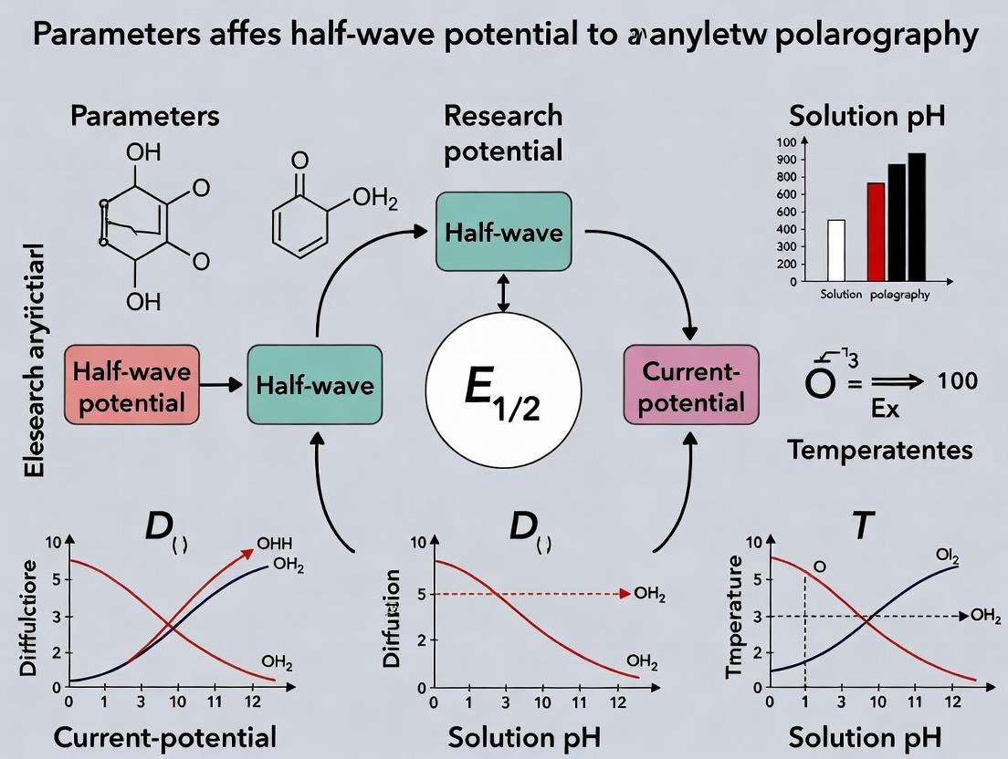
Title: Key Parameters Affecting the Half-Wave Potential
The Scientist's Toolkit: Essential Research Reagent Solutions
| Reagent/Material | Function in Polarographic Analysis |
|---|---|
| High-Purity Mercury | The working electrode material for the DME. Must be purified (e.g., acid-washed) to eliminate trace metal contaminants. |
| Inert Supporting Electrolyte (e.g., KCl, KNO₃, HClO₄) | Eliminates migration current, controls ionic strength, and defines the electrochemical window. Choice can influence E₁/₂. |
| Oxygen Scavenger (Nitrogen/Argon Gas) | Removes dissolved O₂, which produces interfering reduction waves (~ -0.05 V and -0.9 V vs. SCE). |
| Maximum Suppressor (e.g., Triton X-100) | A surface-active agent added at low concentration to suppress the polarographic maximum—anomalous current peaks that distort waves. |
| Standard Buffer Solutions | To systematically study the effect of pH on E₁/₂ for processes involving proton transfer. |
| Complexing Agent (e.g., NH₃, EDTA, Cl⁻) | Used intentionally to shift E₁/₂ via complex formation, enabling the resolution of overlapping waves or qualitative identification. |
| Internal Standard Solution (e.g., Tl⁺) | A known redox species with a stable E₁/₂ used to verify instrument calibration and electrode performance. |
Troubleshooting Guide & FAQ
Q1: During a polarographic experiment, my measured half-wave potential (E₁/₂) shows a positive shift compared to the theoretical value. What are the primary causes and solutions?
A: A positive shift in E₁/₂ often indicates kinetic or adsorption complications affecting reversibility.
- Cause 1: Slow Electron Transfer Kinetics. The system is not electrochemically reversible. The Nernst equation, which strictly applies at equilibrium, is not fully applicable. The process becomes quasi-reversible or irreversible.
- Solution: Increase temperature to improve kinetics or change supporting electrolyte to reduce activation energy. Verify reversibility via cyclic voltammetry (peak separation ~59/n mV at 298 K).
- Cause 2: Specific Adsorption of Reactants or Products. Adsorption of the reduced form onto the mercury electrode shifts E₁/₂ positively.
- Solution: Change the supporting electrolyte composition or ionic strength. Use surfactants (e.g., Triton X-100) to test for adsorption effects, but note they may suppress the polarographic maximum.
- Cause 3: Uncompensated Solution Resistance (iR Drop). This is common in non-aqueous or low-ionic-strength solutions.
- Solution: Use a supporting electrolyte at sufficient concentration (typically ≥0.1 M). Employ positive feedback iR compensation if available on your potentiostat.
Q2: The polarographic wave is drawn-out and not sigmoidal, making E₁/₂ determination difficult. How can I fix this?
A: A non-ideal wave shape suggests non-Nernstian behavior.
- Cause 1: The redox couple is not thermodynamically reversible. Coupled chemical reactions (EC, CE mechanisms) distort the wave.
- Solution: Perform a DC polarography scan at different drop times. If the wave shape is drop-time dependent, a coupled chemical reaction is likely. Conduct controlled-potential electrolysis to identify unstable products.
- Cause 2: The electrode process is complicated by preceding or following chemical reactions. Common in drug research with metal complexes or organic molecules undergoing protonation.
- Solution: Run experiments at varying pH (see Table 1). A shift in E₁/₂ with pH indicates proton involvement. Also, vary concentration of complexing agents.
- Cause 3: Polarographic Maximum. An abnormal current peak at the start of the wave.
- Solution: Add a maximum suppressor like gelatin or Triton X-100 at low concentration (0.001-0.01%).
Q3: How do I experimentally confirm that my system is reversible and thus that the Nernst equation correctly describes the E₁/₂?
A: Perform the following diagnostic tests:
- Log Plot Analysis (Ilkovič Equation): Plot log[(id - i)/i] vs. E(dme). A linear plot with a slope of (nF/2.303RT) confirms Nernstian reversibility. Deviation indicates irreversibility.
- Temperature Dependence: For a reversible process, E₁/₂ shifts slightly with temperature (based on entropy change). For an irreversible process, the shift is more pronounced and relates to activation energy.
- Drop Time Dependence: For a diffusion-controlled, reversible process, the limiting current (i_d) is proportional to the square root of the drop time. Kinetic control shows a different dependence.
Q4: In drug development, how do ligand binding and protonation states affect the half-wave potential of a metal complex?
A: These factors directly alter the standard potential (E°) in the Nernst equation. E₁/₂ ≈ E° when activity coefficients are similar.
- Ligand Binding: Stronger binding to the reduced form shifts E₁/₂ negatively. The shift is quantified by the stability constant difference.
- Protonation: If H⁺ participates in the reduction (e.g., M^(n+) + e⁻ + H⁺ → MH^(n-1)+), E₁/₂ becomes pH-dependent with a slope of -0.059 V/ph unit at 25°C.
- Protocol: To study this, run polarography in buffered solutions across a wide pH range. Keep ionic strength constant with inert electrolyte (e.g., KCl). Deoxygenate all solutions with inert gas (N₂ or Ar) for 15-20 minutes before measurement.
Data Presentation
Table 1: Effect of Experimental Parameters on Half-Wave Potential (E₁/₂)
| Parameter | Reversible System (Nernstian) | Irreversible/Quasi-Reversible System | Diagnostic Test |
|---|---|---|---|
| Variation of [Ox] | E₁/₂ independent of concentration. | E₁/₂ may shift with concentration. | DC polarography at different analyte conc. (0.1-5 mM). |
| Variation of pH | No shift unless H⁺ is involved in the electrode reaction. | Shift may occur if kinetics are pH-dependent. | Polarography in a series of buffers (pH 3-10). |
| Slope of log plot | (59/n) mV at 25°C. | > (59/n) mV. | Plot log[(i_d-i)/i] vs. E. |
| Effect of Temperature | Small, predictable shift (∂E/∂T based on ΔS). | Larger, unpredictable shift. | Measure E₁/₂ at 15°C, 25°C, 35°C. |
| Effect of Drop Time (t) | i_d ∝ t^(1/6) (diffusion current constant independent). | Abnormal dependence. | Measure i_d at varied controlled drop times. |
Table 2: Research Reagent Solutions Toolkit
| Reagent/Solution | Function in Polarography |
|---|---|
| Supporting Electrolyte (e.g., 0.1 M KCl, TBAPF₆) | Eliminates migration current, maintains constant ionic strength, defines electrical field. |
| Maximum Suppressor (e.g., 0.005% Triton X-100) | Suppresses streaming artifacts at the Hg drop to produce smooth polarographic waves. |
| Redox Standard (e.g., 1 mM [Fe(CN)₆]³⁻/⁴⁻) | Validates electrode and potentiostat performance for reversible systems. |
| Buffer Solution (e.g., BR buffer, phosphate) | Controls pH to study proton-coupled electron transfers and stabilize analyte. |
| Oxygen Scavenger (e.g., Nitrogen/Argon gas) | Removes dissolved O₂, which produces interfering reduction waves (~ -0.05 V & -0.9 V vs. SCE). |
| Hg Pool or Ag/AgCl/KCl(sat'd) Reference | Provides a stable, known reference potential for the working electrode (DME). |
Experimental Protocols
Protocol 1: Verifying Reversibility via Log Plot Analysis
- Prepare a 1.0 mM solution of your analyte in a suitable supporting electrolyte (e.g., 0.1 M KCl). Deoxygenate for 15 min.
- Record a DC polarogram from a potential positive of the reduction wave to well past the limiting current plateau. Use a standard drop time (e.g., 2 s).
- From the polarogram, record the current (i) at regular potential intervals (e.g., every 10 mV) across the rising part of the wave. Record the limiting current (i_d).
- For each data point, calculate log[(i_d - i)/i].
- Plot this value (y-axis) against the applied potential E (x-axis).
- Fit a linear regression to the central 60% of the data. A linear fit with a slope close to (59.2/n) mV at 25°C confirms a reversible, Nernstian process.
Protocol 2: Investigating pH Dependence of E₁/₂
- Prepare a stock solution of your analyte (e.g., 10 mM in organic solvent if needed).
- Prepare a series of buffered supporting electrolytes (e.g., Britton-Robinson buffer, pH 2-9), all with the same final ionic strength (adjusted with KCl).
- Spike each buffer with the stock to a final analyte concentration of 0.5 mM. Deoxygenate each.
- Record a DC polarogram for each pH solution.
- Determine E₁/₂ for each wave (potential at i = i_d/2).
- Plot E₁/₂ vs. pH. A slope of ~ -0.059 V/ph unit indicates an equal number of protons and electrons transferred in the rate-determining step.
Visualizations
Diagnosing Nernstian Behavior in Polarography Workflow
Thermodynamic & Kinetic Foundations of E1/2
Troubleshooting & FAQs: Technical Support Center
FAQ 1: Why do I observe an irreversible polarographic wave even with a theoretically reversible quinone moiety?
- Answer: This is often due to a slow electron transfer rate constant (k⁰). While the quinone/hydroquinone couple is inherently reversible, its kinetics on the electrode surface are modulated by adjacent functional groups. Bulky substituents or steric hindrance near the redox-active center can impede the necessary conformational change for electron transfer, leading to quasi-reversible or irreversible behavior. Check for alkyl chains, aryl rings, or heteroatoms adjacent to the quinone.
FAQ 2: How does changing the pH of my supporting electrolyte cause a shift in my compound's half-wave potential (E₁/₂)?
- Answer: A pH-dependent shift in E₁/₂ indicates proton-coupled electron transfer (PCET). The number of protons (m) and electrons (n) involved dictates the shift. For many organic functionalities (e.g., carbonyls, azo groups), reduction is easier (more positive E₁/₂) at lower pH as protons are readily available. Use the Nernst equation derivation: ΔE₁/₂/ΔpH = −0.059(m/n) V at 25°C. A deviation from this slope suggests a change in the PCET mechanism.
FAQ 3: My homologous series of compounds shows an unexpected, non-linear trend in E₁/₂ with increasing chain length. What could be the cause?
- Answer: Non-linear effects often arise from through-space vs. through-bond electronic effects. Initial alkyl chain elongation exhibits a small, linear inductive effect. A sudden deviation may occur when the chain length allows for intramolecular folding or interaction (e.g., a long chain folds back to shield the redox center, altering its solvation or double-layer effects). Consider conformational analysis via molecular modeling.
FAQ 4: Why does the introduction of an electron-withdrawing group (EWG) sometimes make reduction more difficult (shift E₁/₂ more negative)?
- Answer: This counter-intuitive result typically occurs when the EWG stabilizes the reduced form more than the oxidized form. For example, if reduction adds negative charge to a moiety, an adjacent EWG (like a nitro group) can stabilize that anionic product so effectively that it becomes thermodynamically less accessible, requiring more negative potential. Analyze the resonance and inductive stabilization of both redox states.
FAQ 5: I am getting poor reproducibility in drop time and current in my manual polarography setup. What are the critical checkpoints?
- Answer: Follow this checklist:
- Capillary: Ensure it is clean, vertical, and has a consistent, unchanging tip. Measure drop time in your electrolyte without potential applied; it should be constant.
- Mercury Column Height (h): Maintain a fixed, measured height. Current is proportional to √h.
- Deaeration: Inadequate oxygen removal is the most common cause of noisy, drifting baselines. Extend purging time with high-purity inert gas (N₂/Ar).
- Solution Resistance: Use a supporting electrolyte at sufficient concentration (typically 0.1 M minimum) to minimize iR drop, especially for non-aqueous studies.
Table 1: Influence of Substituents on the Half-Wave Potential of Benzene Derivatives (Reduction at DME, vs. SCE)
| Core Structure | Substituent | E₁/₂ (V) | Solvent/Supporting Electrolyte | Effect & Mechanism |
|---|---|---|---|---|
| Nitrobenzene | -H | -0.48 V | 50% EtOH, 0.1 M KCl | Reference value |
| Nitrobenzene | -p-OH | -0.33 V | 50% EtOH, 0.1 M KCl | Positive shift. -OH donates electrons, destabilizes the anion radical product. |
| Nitrobenzene | -p-NO₂ | -0.62 V | 50% EtOH, 0.1 M KCl | Negative shift. Additional -NO₂ withdraws electrons, stabilizes the anion radical product. |
| Benzaldehyde | -H | -1.32 V | DMF, 0.1 M TBAP | Reference value |
| Benzaldehyde | -p-CH₃ | -1.38 V | DMF, 0.1 M TBAP | Slight negative shift. +I effect of -CH₃ stabilizes the reactant carbonyl. |
| Benzaldehyde | -p-CN | -1.24 V | DMF, 0.1 M TBAP | Positive shift. Strong -I/-M effect of -CN destabilizes the reactant carbonyl. |
Table 2: Redox Potential Trends for Common Organic Functional Groups (Approximate Ranges)
| Functional Group | Typical E₁/₂ Range (Reduction, V vs. SCE) | Key Structural Dependencies |
|---|---|---|
| Azo (-N=N-) | -0.2 to -0.9 V | Heavily dependent on aryl substitution and pH (PCET). |
| Quinone | 0.0 to -0.5 V | Conjugation, aromaticity, and hydrogen bonding in hydroquinone. |
| Nitro (-NO₂) | -0.3 to -1.0 V | Conjugation with aromatic system; undergoes 4e⁻/4H⁺ stepwise reduction. |
| Carbonyl (>C=O) | -1.2 to -2.5 V | Adjacent π-systems (conjugation shifts positively), solvent polarity. |
| Alkene (isolated) | < -2.2 V (in DMF) | Significantly positively shifted by conjugation with EWGs or aromatics. |
Experimental Protocols
Protocol 1: Determining the Proton-Coupled Electron Transfer (PCET) Mechanism via pH Variation Objective: To establish the number of protons (m) and electrons (n) involved in the redox process of an analyte. Method:
- Prepare a stock solution of the analyte (~1 mM) in a mixed solvent (e.g., 30% water/70% DMF for poor solubility) with a constant ionic strength buffer (e.g., 0.1 M Britton-Robinson buffer adjusted from pH 2 to 12).
- For each pH, transfer 10 mL to the polarographic cell. Deoxygenate with nitrogen for 10 minutes.
- Record DC polarograms from a starting potential positive of the expected wave to a final negative potential, using a controlled drop time (e.g., 2 s).
- Measure the E₁/₂ for each wave at each pH. Plot E₁/₂ vs. pH.
- Analysis: Linear segments of the plot will have a slope ΔE₁/₂/ΔpH. Calculate m/n = -(slope / 0.059) at 25°C. A break in the plot indicates a pKₐ change in either the reactant or product.
Protocol 2: Assessing Electronic Effects in a Homologous Series Objective: To quantify the Hammett-type relationship between substituent constant (σ) and E₁/₂. Method:
- Synthesize or obtain a series of compounds with the same redox core (e.g., para-substituted nitrobenzenes).
- Prepare identical experimental conditions: e.g., 0.1 M KCl in 50% aqueous ethanol, 25°C.
- Record polarograms for each compound. Use a standard internal reference (e.g., 0.5 mM dimethylformamide) if absolute potential alignment is critical.
- Measure ΔE₁/₂ relative to the unsubstituted parent compound.
- Analysis: Plot ΔE₁/₂ vs. the substituent's Hammett σ constant (σₚ). A linear correlation (ρ is the slope) confirms the dominance of through-bond electronic effects.
Visualizations
Title: Polarographic Reduction Pathway at the Dropping Mercury Electrode
Title: How Molecular Factors Dictate the Measured Half-Wave Potential
The Scientist's Toolkit: Key Research Reagent Solutions
Table 3: Essential Materials for Polarographic Studies of Redox Potentials
| Item | Function & Rationale |
|---|---|
| High-Purity Mercury | The working electrode material for the Dropping Mercury Electrode (DME). Purity is critical to prevent alloy formation and ensure a reproducible, renewable surface. |
| Inert Gas Supply (N₂/Ar) | Used for deaeration of solutions to remove dissolved O₂, which interferes with reduction waves of organic analytes. Must be high-purity with an O₂ trap. |
| Tetraalkylammonium Salts (e.g., TBAP, TBAPF₆) | Common supporting electrolytes for non-aqueous polarography (e.g., in DMF, ACN). Provide conductivity without introducing reactive protons. |
| Aprotic Solvents (DMF, DMSO, Acetonitrile) | Allow access to very negative potentials and study of reduction processes without interference from protons, revealing "true" electron affinity. |
| Buffering Systems (Britton-Robinson, Phosphate) | For controlled aqueous pH studies to investigate Proton-Coupled Electron Transfer (PCET) mechanisms. Must be non-complexing. |
| Internal Potential Reference (e.g., Ferrocene/Ferrocenium) | Added to non-aqueous solutions to calibrate and report potentials against a known, solvent-independent standard (Fc/Fc⁺). |
| Capillary for DME | Glass capillary with precise internal diameter and length to control mercury flow and drop time, the foundation of reproducible current measurement. |
Troubleshooting Guides & FAQs
FAQ 1: Why does my half-wave potential (E₁/₂) shift when I change the supporting electrolyte?
Answer: The half-wave potential is sensitive to the ionic strength and composition of the supporting electrolyte. Changes in ionic strength alter the activity coefficients of the electroactive species, shifting the potential via the Nernst equation. Specific ion interactions (e.g., complexation) can also stabilize the reduced/oxidized form, causing a more significant shift. Always report the exact electrolyte composition and concentration.
FAQ 2: How do I choose a supporting electrolyte to minimize migration current?
Answer: The primary rule is that the concentration of the supporting electrolyte should be at least 100 times greater than the concentration of the analyte. This ensures the migration current is negligible (<<1%) compared to the diffusion current. Use electrolytes with high conductivity (e.g., KCl, LiClO₄, TBAP) to lower solution resistance.
FAQ 3: My polarographic wave is distorted or not well-defined. What could be the issue?
Answer: This is often related to improper supporting electrolyte conditions.
- Check Ionic Strength: Too low ionic strength increases solution resistance and causes poorly shaped waves.
- Check for Adsorption: Some electrolytes or their impurities adsorb on the mercury electrode. Try purifying the salt or switching to a different type (e.g., from tetraalkylammonium to alkali metal salts).
- Check for Overlapping H⁺ Reduction: In aqueous solutions at insufficient pH buffering, hydrogen ion reduction can distort the wave. Use a suitable buffer system relevant to your pH range.
FAQ 4: How can I confirm if my analyte is complexing with the supporting electrolyte?
Answer: Perform a systematic study. Measure the half-wave potential (E₁/₂) of your analyte across a range of concentrations of a suspected complexing agent (e.g., Cl⁻, CN⁻, citrate) while keeping ionic strength constant with a non-complexing salt (e.g., KClO₄). A linear plot of E₁/₂ vs. log[ligand] with a slope near the theoretical value for the complex stoichiometry confirms complexation.
Guide 1: Troubleshooting Unstable Current Readings
| Symptom | Possible Cause (Related to Supporting Electrolyte) | Solution |
|---|---|---|
| Current drifting upwards | Impurities in electrolyte undergoing slow redox reactions. | Purify the supporting electrolyte by recrystallization. Use high-purity salts. Deoxygenate solution thoroughly. |
| Noisy/fluctuating current | Solution resistance too high due to low ionic strength. | Increase concentration of supporting electrolyte. Use a more conductive salt. |
| Irregular drop time | Surfactant impurities in electrolyte adsorbing on Hg. | Clean glassware meticulously. Use a different source or batch of electrolyte. |
Guide 2: Addressing Irreversible or Broad Waves
| Observation | Diagnostic Test | Corrective Action |
|---|---|---|
| Wave is broad, ΔE₃/₄-₄/₁ > 56/n mV | Test at different concentrations of supporting electrolyte (constant ionic strength). | If broadening decreases with higher [electrolyte], increase concentration to improve kinetics. Switch to an electrolyte with different cation (e.g., Li⁺ vs. TBA⁺) which can affect double layer structure. |
| Wave is irreversible (non-Nernstian) | Perform a scan rate study. | Use a supporting electrolyte that provides a more favorable reaction environment (e.g., non-aqueous media with TBAP for organics). Ensure electrolyte does not participate in chemical steps following electron transfer. |
Key Experimental Protocols
Protocol 1: Determining the Effect of Ionic Strength on E₁/₂
Objective: To isolate and quantify the shift in half-wave potential due solely to changes in ionic strength (I), avoiding complexation effects. Methodology:
- Prepare a series of 10 solutions containing a fixed, low concentration of your analyte (e.g., 0.5 mM Cd²⁺).
- Vary the concentration of a non-complexing, inert supporting electrolyte (e.g., KClO₄) from 0.01 M to 1.0 M.
- For each solution, record the DC polarogram.
- Measure the E₁/₂ for the reduction wave.
- Plot E₁/₂ vs. √I (or log I). The slope relates to the change in activity coefficient. Key Consideration: Use an electrolyte like KClO₄ or LiNO₃ that minimally interacts with most metal ions. Maintain all other conditions (pH, temperature) constant.
Protocol 2: Investigating Specific Complexation Effects
Objective: To determine the stoichiometry and stability constant of a complex formed between the analyte and a ligand present in the supporting electrolyte. Methodology (DeFord-Hume Method):
- Prepare a set of solutions with constant ionic strength (I) maintained by a high concentration of inert salt (e.g., 1.0 M NaNO₃).
- In each solution, keep the analyte concentration constant and vary the concentration of the complexing ligand (L) (e.g., Cl⁻, NH₃).
- Record polarograms for each solution.
- For each ligand concentration, calculate the function F₀(L) = antilog[(nF/2.303RT) * (E₁/₂(simple) - E₁/₂(complex)) + log(I₄/Is)].
- Plot F₀(L) vs. [L]. The form of the plot (linear, parabolic) indicates the highest complex number. Successive extrapolations yield overall stability constants (β).
Data Presentation
Table 1: Effect of Supporting Electrolyte Cation on Half-Wave Potential of 1.0 mM Pb²⁺ in 0.1 M XCl Media
| Supporting Electrolyte (0.1 M) | E₁/₂ vs. SCE (V) | ΔE₁/₂ from KCl (V) | Notes |
|---|---|---|---|
| LiCl | -0.405 | +0.007 | Smaller cation, stronger hydration, weaker interaction. |
| NaCl | -0.410 | +0.002 | -- |
| KCl | -0.412 | 0.000 | Reference. |
| Tetramethylammonium Cl | -0.428 | -0.016 | Large organic cation adsorbs, shifts potential negatively. |
Table 2: Shift in E₁/₂ for Zn²⁺ due to Complexation in Different Electrolyte Compositions (I=0.5 M)
| Electrolyte Composition | E₁/₂ vs. SCE (V) | Ligand | Apparent Shift (V) | Inferred Complex |
|---|---|---|---|---|
| 0.5 M KNO₃ (Inert) | -1.010 | -- | 0.000 | [Zn(H₂O)₆]²⁺ |
| 0.5 M KCl | -1.040 | Cl⁻ | -0.030 | [ZnClₙ]²⁻ⁿ |
| 0.5 M KCN | -1.420 | CN⁻ | -0.410 | [Zn(CN)₄]²⁻ |
| 0.1 M NH₃ / 0.4 M KNO₃ | -1.340 | NH₃ | -0.330 | [Zn(NH₃)₄]²⁺ |
Visualizations
Experimental Workflow for Electrolyte Studies
Factors Influencing Half-Wave Potential
The Scientist's Toolkit: Key Research Reagent Solutions
| Item | Function in Polarography | Key Considerations |
|---|---|---|
| Inert Salts (KNO₃, KClO₄) | Maintain constant high ionic strength to suppress migration current and control activity coefficients. | KClO₄ is oxidant; avoid with strong reductants. Pre-purify to remove electroactive impurities. |
| Tetraalkylammonium Salts (TBAP, TBABF₄) | Supporting electrolyte for non-aqueous (aprotic) polarography. Large cations minimize specific adsorption. | Hydroscopic; must be stored dry. Often requires recrystallization from ethyl acetate. |
| pH Buffers (Acetate, Phosphate, Ammonia) | Control proton activity, crucial for analytes involving H⁺ in electrode reaction. | Buffer component must not complex the analyte (e.g., phosphate complexes many metals). |
| Complexing Agents (KCN, KCl, EDTA) | Deliberately added to shift E₁/₂, resolve overlapping waves, or determine stability constants. | Safety: Handle KCN with extreme care in basic solution to prevent HCN gas. |
| Oxygen Scavenger (Nitrogen/Argon Gas) | Remove dissolved O₂, which reduces in two steps (-0.05V & -0.9V vs. SCE) and interferes with analyses. | Use high-purity grade. Sparge for 10-15 minutes before measurement. |
| Electrode Cleaning Solution (Dil. HNO₃, Ethanol) | Clean glassware and cell to prevent surfactant contamination which adsorbs on Hg and affects E₁/₂. | Use trace-metal grade acids. Rinse extensively with high-purity water (18.2 MΩ·cm). |
Technical Support Center
Frequently Asked Questions (FAQs) & Troubleshooting Guides
Q1: During polarographic analysis of a novel pharmaceutical compound, my measured half-wave potential (E₁/₂) shifts significantly between different solvent systems. Why does this happen, and which solvent property is most responsible? A: This is a direct manifestation of solvent influence on electron transfer thermodynamics. The primary property is the solvent's dielectric constant (ε). A higher ε increases the solvent's ability to stabilize charged species (reactants, products, transition states) through dielectric polarization. The solvation energy change (ΔGsolv) between the oxidized and reduced states dictates the E₁/₂ shift. Use the Born approximation as a starting model: ΔE₁/₂ ∝ (1/ε) * (1/rred - 1/r_ox), where r are the ionic radii. A shift to more negative E₁/₂ in a high-ε solvent often indicates better stabilization of the reduced (anionic) state.
Q2: How can I quantitatively correct my experimental half-wave potential for solvent effects to compare data obtained in different media? A: You can reference potentials to a solvent-independent redox couple. The detailed protocol is below.
- Protocol: Referencing to the Ferrocene/Ferrocenium (Fc/Fc⁺) Couple
- Preparation: In your test solvent (e.g., DMSO, acetonitrile, DMF), prepare a 1 mM solution of your analyte with 0.1 M supporting electrolyte (e.g., TBAPF₆).
- Measurement: Record the polarogram (or cyclic voltammogram) of your analyte. Note the E₁/₂ (midpoint potential).
- Internal Standard Addition: Add a small amount of solid ferrocene (Fc) to the same cell to achieve ~0.5 mM concentration. Mix thoroughly.
- Re-measure: Record a new polarogram. You will observe your analyte wave and the new Fc/Fc⁺ wave.
- Calculation: Measure the E₁/₂ of the Fc/Fc⁺ couple in your solvent. Report your analyte's potential as E₁/₂ vs. Fc/Fc⁺. For reporting vs. Standard Hydrogen Electrode (SHE), add the known conversion factor (e.g., Fc/Fc⁺ in acetonitrile is +0.641 V vs. SHE).
Q3: My voltammetric peaks are broad and poorly resolved in a low-dielectric constant solvent (ε < 10). What is the cause and solution? A: This is likely due to high solution resistance (R_u) and improper uncompensated iR drop. Low-ε solvents have lower ionic conductivity, leading to significant potential distortion across the cell.
- Troubleshooting Steps:
- Increase Electrolyte Concentration: Use a higher concentration (e.g., 0.3-0.5 M) of a suitable supporting electrolyte to enhance conductivity.
- Employ Positive Feedback iR Compensation: If your potentiostat has this feature, enable and carefully adjust the compensation. Avoid over-compensation.
- Reduce Scan Rate: Lower your polarographic scan rate or drop time to decrease current (i), thereby minimizing the iR error.
- Use a Microelectrode: Switch to a smaller working electrode (e.g., Pt microdisk) which reduces current and minimizes iR effects.
Q4: I suspect specific solute-solvent interactions (beyond dielectric effects) are influencing my electron transfer kinetics. How can I investigate this? A: You are likely dealing with specific solvation (e.g., hydrogen bonding, Lewis acidity/basicity). To investigate:
- Solvent Parameter Correlation: Measure E₁/₂ in a series of solvents with similar ε but different donor/acceptor numbers (DN, AN). Plot E₁/₂ vs. DN or AN. A strong correlation indicates specific interactions.
- Kinetic Measurement: Use fast-scan cyclic voltammetry to measure the standard heterogeneous electron transfer rate constant (k⁰) in different solvents. A change in k⁰ that doesn't correlate with ε may indicate specific interactions affecting the transition state solvation.
- Spectroscopic Validation: Use techniques like FT-IR or NMR to detect shifts in analyte vibrational or chemical shift frequencies indicative of specific solvent coordination.
Summary of Quantitative Solvent Effects
Table 1: Common Solvent Properties and Their Impact on Polarographic Measurements
| Solvent | Dielectric Constant (ε) | Viscosity (cP) | Donor Number | Typical ΔE₁/₂ (vs. Fc/Fc⁺) Shift for Anionic Reduction* |
|---|---|---|---|---|
| Water | 80.1 | 0.89 | 18 | Reference (by definition for SHE) |
| N,N-Dimethylformamide (DMF) | 36.7 | 0.92 | 26.6 | Shifts negative |
| Acetonitrile (MeCN) | 37.5 | 0.34 | 14.1 | Shifts negative |
| Dimethyl Sulfoxide (DMSO) | 46.7 | 2.00 | 29.8 | Shifts more negative |
| Dichloromethane (DCM) | 8.93 | 0.41 | 1.0 | Shifts positive; iR issues likely |
| Tetrahydrofuran (THF) | 7.58 | 0.46 | 20.0 | Shifts positive; iR issues likely |
*General trend for reduction to an anionic product. Magnitude depends on ionic radii.
Table 2: Troubleshooting Chart for Solvent-Related Issues
| Symptom | Probable Cause | Recommended Action |
|---|---|---|
| Irreversible or broad waves | Slow electron transfer due to poor reactant solvation | Increase temperature; use a more polar solvent; check for coupled chemical reactions. |
| Inconsistent E₁/₂ between replicates | Variable water content (hygroscopic solvents) | Rigorously dry solvent and electrolyte; use molecular sieves; employ inert atmosphere glovebox. |
| High background current | Solvent or electrolyte impurity | Purify solvent (distillation); recrystallize supporting electrolyte; use higher purity grade. |
| Multiple, unclear reduction waves | Solvent-assisted side reactions (e.g., protonation) | Switch to aprotic, non-coordinating solvent with high purity; use a weaker acid supporting electrolyte. |
Experimental Protocols
Protocol: Determining the Dielectric Constant Dependence of E₁/₂ Objective: To empirically measure the relationship between solvent polarity (ε) and the half-wave potential for a model compound. Materials: See "Research Reagent Solutions" below. Procedure:
- Prepare a 1.0 mM solution of quinone (e.g., 1,4-benzoquinone) in each of the following dry, deaerated solvents: DMF, MeCN, DMSO, and a DMF:DCM mixture series (e.g., 100:0, 75:25, 50:50, 25:75 v/v). Maintain 0.1 M TBAPF₆ as supporting electrolyte.
- Using a standard three-electrode polarographic setup (DME working electrode), record the DC polarogram for each solution.
- For each polarogram, determine the E₁/₂ precisely.
- Add internal ferrocene and re-measure to obtain E₁/₂ vs. Fc/Fc⁺.
- Plot E₁/₂ (vs. Fc/Fc⁺) against the inverse of the solvent's dielectric constant (1/ε). Analyze the linearity of the relationship.
Protocol: Assessing Solvation Energy via Computational Methods Objective: To complement experimental data with calculated solvation energies. Procedure:
- Geometry Optimization: Using computational chemistry software (e.g., Gaussian, ORCA), optimize the geometry of both the oxidized and reduced states of your analyte in the gas phase at the DFT level (e.g., B3LYP/6-31+G(d)).
- Single Point Energy in Solvent: Perform a single-point energy calculation on the optimized structures using a Polarizable Continuum Model (PCM) specifying your experimental solvent (e.g., ε = 46.7 for DMSO).
- Calculation: The solvation energy (ΔGsolv) for each species is output by the software. Calculate the difference: ΔΔGsolv = ΔGsolv(red) - ΔGsolv(ox).
- Correlation: Relate the calculated ΔΔGsolv to the experimentally observed shift in E₁/₂. A more negative ΔΔGsolv correlates with a more negative (easier) reduction potential.
Diagrams
Title: Logical Flow of Solvent Impact on E₁/₂
Title: Experimental Workflow for Solvent Effect Study
The Scientist's Toolkit
Table 3: Essential Research Reagent Solutions for Solvent Effect Studies
| Reagent / Material | Function & Importance |
|---|---|
| Tetrabutylammonium Hexafluorophosphate (TBAPF₆) | Standard supporting electrolyte for non-aqueous electrochemistry. Large ions minimize ion-pairing, and PF₆⁻ is relatively inert and non-coordinating. |
| Ferrocene (Fc) | Internal redox standard for potential referencing across different solvents. Its E₁/₂ is relatively insensitive to specific solvation. |
| Distilled & Dried Solvents (DMF, MeCN, DMSO) | High-purity, anhydrous solvents are critical. Trace water can act as a proton source, drastically altering reduction mechanisms. |
| 3Å or 4Å Molecular Sieves | Used to maintain anhydrous conditions in solvent and electrolyte stock solutions. |
| Dropping Mercury Electrode (DME) | The classic working electrode for polarography. Provides a renewable, smooth surface ideal for reproducible diffusion-controlled currents. |
| Nonaqueous Reference Electrode (e.g., Ag/Ag⁺) | Provides a stable potential in organic solvents. Often filled with matching solvent/electrolyte and referenced to Fc/Fc⁺ for reporting. |
| Polarographic Cell with Gas Inlet/Outlet | Allows for deaeration of solutions with inert gas (N₂, Ar) to remove dissolved O₂, which is electroactive. |
Technical Support Center: Troubleshooting Polarography Experiments
FAQs & Troubleshooting Guides
Q1: Why does my measured half-wave potential (E₁/₂) shift significantly when I change the experimental temperature?
A: A shift in E₁/₂ with temperature indicates that the electrode reaction has a measurable entropy change (ΔS). This is a thermodynamic contribution. An increase in E₁/₂ with temperature suggests a positive ΔS for the reduction (favorable disorder increase). To isolate this effect, ensure your cell is thermally equilibrated for at least 15 minutes, use a calibrated thermometer in the solution (not just the bath), and verify that your reference electrode potential is temperature-corrected.
Q2: My polarographic wave becomes broad and poorly defined at higher temperatures. What is the cause and solution?
A: Wave broadening is typically a kinetic issue. Increased temperature accelerates the electron transfer rate (increasing the standard rate constant, k⁰), but it can also exacerbate problems like inadequate buffering (pH shifts), increased convection, or faster follow-up chemical reactions (EC mechanisms). Ensure your supporting electrolyte concentration is high (≥0.1 M) to maintain a constant ionic strength and pH. Use a three-electrode system with minimal uncompensated resistance.
Q3: How do I determine if an observed temperature effect on E₁/₂ is due to thermodynamics or kinetics?
A: You must perform a series of experiments at different temperatures and analyze the data systematically.
- Protocol: Record polarograms (or cyclic voltammograms) at 5°C intervals from 10°C to 40°C.
- Analysis:
- Plot E₁/₂ vs. T (K). A linear relationship with a significant slope (∂E₁/₂/∂T) confirms a thermodynamic contribution (ΔS° = nF(∂E₁/₂/∂T)).
- Plot the wave width (e.g., E₃/₄ - E₁/�) vs. T. A change indicates a kinetic contribution. For a reversible system, the width should be constant (~56.4/n mV at 25°C).
- For quasi-reversible systems, use the variation of peak separation (ΔEp) in cyclic voltammetry with temperature to calculate the activation energy (Ea) via an Arrhenius plot of ln(k⁰) vs. 1/T.
Q4: I suspect a temperature-induced chemical decomposition of my drug analyte. How can I confirm this during my polarographic study?
A: This is a common issue in drug development.
- Protocol for Stability Check:
- Prepare your drug solution and place it in the thermostated cell at your experimental temperature.
- Record successive polarograms every 2-5 minutes over 30-60 minutes.
- Plot limiting current (iₗ) and E₁/₂ vs. time.
- Diagnosis: A steady decrease in iₗ (diffusion-controlled) indicates loss of electroactive species. A shift in E₁/₂ suggests a change in speciation (e.g., hydrolysis). Perform this test at each key temperature. Always use freshly prepared, degassed solutions.
Table 1: Thermodynamic Parameters from Temperature-Dependent E₁/₂ Shifts for Model Compounds
| Compound / System | ∂E₁/₂/∂T (mV/K) | ΔS° of Reduction (J/mol·K) | n (electrons) | Temperature Range (°C) |
|---|---|---|---|---|
| [Fe(CN)₆]³⁻/⁴⁻ (Reversible) | ~0.0 | ~0 | 1 | 10-40 |
| Quinone/Hydroquinone | +1.2 | +116 | 2 | 15-35 |
| Cd²⁺ in 1M KCl | -0.3 | -29 | 2 | 10-40 |
| Model Drug A (Proton-Coupled) | +0.8 | +77 | 2 | 20-40 |
Table 2: Kinetic Parameters from Temperature-Dependent Voltammetry
| System | Standard Rate Constant, k⁰ @ 25°C (cm/s) | Activation Energy, Ea (kJ/mol) | Method |
|---|---|---|---|
| [Fe(CN)₆]³⁻/⁴⁻ (1M KCl) | 0.05 | 20-30 | CV (ΔEp) |
| [Ru(NH₃)₆]³⁺/²⁺ | > 0.1 | < 20 | CV (ΔEp) |
| Model Drug B (Irreversible) | 3.2 x 10⁻⁴ | 65 | Polarography (Wave Analysis) |
Experimental Protocol: Determining Thermodynamic & Kinetic Parameters
Objective: To separately determine the entropy change (ΔS°) and the electron transfer activation energy (Ea) for a reversible, one-electron reduction.
Materials: Polarograph/Voltammeter, Hanging Mercury Drop Electrode (HMDE) or DME, Pt counter electrode, Ag/AgCl reference electrode, Thermostated cell (±0.1°C), Analyte, High-purity supporting electrolyte, N₂ gas for degassing.
Methodology:
- Prepare 10 mL of a 1.0 mM analyte solution in 0.1 M supporting electrolyte. Degas with N₂ for 10 minutes.
- Set cell temperature to 10°C. Allow thermal equilibration for 15 min with gentle stirring.
- Record 3 consecutive DC polarograms (or cyclic voltammograms at slow scan rate, e.g., 50 mV/s) from a potential positive of the wave to negative of the limiting current.
- Precisely measure the half-wave potential (E₁/₂) and the limiting current (iₗ) for each polarogram. Average the values.
- Increase the temperature to 15°C. Repeat steps 2-4. Continue in 5°C increments up to 40°C.
- For Thermodynamics: Plot average E₁/₂ (in Volts) vs. Absolute Temperature (T in Kelvin). Perform a linear regression. ∂E₁/₂/∂T = slope. Calculate ΔS° = nF*(slope).
- For Kinetics (from CV): At each temperature, measure the peak separation (ΔEp). Use digital simulation software or Nicholson's method to estimate the standard rate constant (k⁰) from ΔEp. Plot ln(k⁰) vs. 1/T. The slope of the linear fit = -Ea/R.
Visualizations
Title: How Temperature Affects Polarographic Signals
Title: Temperature-Dependence Experimental Workflow
The Scientist's Toolkit: Key Research Reagent Solutions
Table 3: Essential Materials for Temperature-Controlled Polarography
| Item | Function & Specification | Critical Note for Temperature Studies |
|---|---|---|
| Thermostated Electrochemical Cell | Maintains solution temperature within ±0.1°C. Jacketed cells connected to a circulator are ideal. | Ensure the reference electrode compartment is also temperature-controlled to avoid thermal junction potentials. |
| Supporting Electrolyte (e.g., 1.0 M KCl or Phosphate Buffer) | Provides ionic conductivity, fixes ionic strength, and can control pH. | Use a high buffer capacity. Check for temperature-dependent pH shifts (especially for phosphate/amine buffers) and adjust calculations. |
| Redox Potential Reference Standard (e.g., Ferrocene/ Ferrocenium or [Ru(bpy)₃]²⁺/³⁺) | Internal reference for potential calibration, correcting for reference electrode drift with T. | Add a small amount post-experiment. Confirm its E₁/₂ is temperature-independent or well-characterized. |
| High-Purity Mercury (Triple Distilled) | For the working electrode (DME or HMDE). | Mercury resistivity changes with T; ensure potentiostat compensation is adequate for precise iR drop correction. |
| Inert Gas Supply (N₂ or Ar, Oxygen-Free) | Removes dissolved O₂, which interferes with reduction waves. | Gas solubility decreases with T. Degas after thermal equilibration at each new temperature for a consistent time. |
| Calibrated Thermometer or Probe | Directly measures solution temperature. | Do not rely on the circulator readout. Use a certified NIST-traceable probe inserted into the cell. |
Precision in Practice: Methodological Control for Accurate E₁/₂ Measurement
Technical Support Center
Troubleshooting Guides & FAQs
Q1: My polarographic wave is poorly defined and not sigmoidal. What could be wrong with my DME setup? A: This is often due to incorrect DME characteristics. Ensure:
- Capillary Clogging: The capillary tip may be partially blocked. Clean according to protocol.
- Incorrect Mercury Column Height (h): This controls drop time (
t_d). Measure and adjusthto achieve at_dbetween 3-6 seconds for a clean, renewable surface. A too-shortt_dyields an ill-defined wave. - Dirty Mercury: Use triple-distilled mercury. Oxidized mercury forms a film that distorts the current.
Experimental Protocol: Calibrating DME Characteristics
- Fill the mercury reservoir with clean Hg.
- In a quiet, vibration-free environment, suspend a clean DME capillary in air over a catch basin.
- Measure the height (
h) from the tip to the meniscus in the reservoir using a ruler. - Using a stopwatch, record the time (
t_d) for 10 drops to fall. Calculate averaget_d. - Adjust
hand repeat untilt_dis in the optimal range. Record the exacthand correspondingt_dand mercury flow rate (m).
Q2: I observe excessive noise and unstable current in my cell. How should I troubleshoot the cell design? A: This typically stems from improper cell design or setup.
- Electrical Shielding: Ensure the entire cell is housed in a grounded Faraday cage to block external electromagnetic interference.
- Vibration: Place the apparatus on a vibration-damping table. Mechanical disturbance of the DME causes current fluctuations.
- Cell Geometry & Placement: The DME, reference electrode (RE), and counter electrode (CE) must be positioned correctly. The RE Luggin capillary should be close (~2 mm) to the DME to minimize uncompensated resistance (
R_u), but not so close as to disturb diffusion. - Deaeration: Inadequate removal of dissolved oxygen is a prime noise source. Purge with high-purity nitrogen or argon for at least 15 minutes before measurement and maintain a blanket over the solution.
Q3: My measured half-wave potential (E₁/₂) shifts between experiments. Could this be related to my reference electrode? A: Yes. E₁/₂ is referenced against the potential of the RE. Instability indicates a faulty RE.
- Contamination: The RE frit may be contaminated by sample components. Soak and rinse the RE according to manufacturer guidelines.
- Filled/Leaking Electrolyte: Ensure the RE is filled to the correct level with fresh filling solution (e.g., saturated KCl for SCE). Check for crystals clogging the junction.
- Inappropriate RE Choice: Using an RE with a liquid junction potential sensitive to your solution composition (e.g., SCE in non-aqueous media) causes shifts. Select a compatible RE (see Table 2).
Experimental Protocol: Validating Reference Electrode Stability
- Measure the open-circuit potential of your RE against a second, freshly prepared, identical RE in a 0.1 M KCl solution.
- The potential should be stable and read < ±2 mV. Drift or a larger offset indicates the RE needs maintenance or replacement.
- Record this validation check before each experimental series.
Data Presentation
Table 1: Impact of DME Parameters on Polarographic Wave and E₁/₂
| Parameter | Typical Optimal Value | Effect if Too Low | Effect if Too High | Primary Impact on E₁/₂? |
|---|---|---|---|---|
Drop Time (t_d) |
3 - 6 s | Ill-defined wave, high capacitive current | Increased diffusion layer, time-dependent analysis | Minor indirect effect via current shape |
Mercury Column Height (h) |
40 - 80 cm | Short t_d, erratic drops |
Long t_d, prolonged analysis time |
No direct effect |
Mercury Flow Rate (m) |
1 - 3 mg/s | - | - | No direct effect |
Capillary Radius (r) |
25 - 50 µm | - | - | No direct effect |
Table 2: Common Reference Electrodes for Polarography
| Electrode | Full Name | Potential vs. SHE (25°C) | Typical Use Case | Advantage | Disadvantage |
|---|---|---|---|---|---|
| SCE | Saturated Calomel Electrode | +0.241 V | Aqueous, non-complexing media. | Stable, widely used. | KCl leakage; unstable in non-aq. media. |
| Ag/AgCl (sat. KCl) | Silver/Silver Chloride | +0.197 V | General aqueous work. | Simple, robust. | Ag+ may interfere in some systems. |
| Ag/Ag+ | Silver/Silver Ion | User-defined | Non-aqueous solvents (e.g., DMF, MeCN). | Compatible with organic media. | Requires careful preparation, less stable. |
| TMS | Thallium Amalgam | -0.557 V (in sat. KCl) | Used as internal ref. in some cells. | Minimizes liquid junction. | Toxic, preparation complexity. |
Mandatory Visualizations
Troubleshooting Path for Polarographic Setup Optimization
Key Parameters Influencing Measured Half-Wave Potential
The Scientist's Toolkit: Research Reagent Solutions
| Item | Function in Polarographic Setup |
|---|---|
| Triple-Distilled Mercury | High-purity working electrode material for the DME. Essential for a clean, reproducible surface. |
| DME Capillary (Glass) | Precisely controls the formation and fall of the mercury drop. The inner diameter and cleanliness are critical. |
| Saturated Calomel Electrode (SCE) | Common reference electrode for aqueous studies, providing a stable potential to measure E₁/₂ against. |
| Tetraalkylammonium Salt (e.g., TBAPF₆) | Supporting electrolyte at high concentration (~0.1 M). Carries current and fixes ionic strength, minimizing migration current. |
| High-Purity Inert Gas (N₂/Ar) | Used to deoxygenate the analyte solution by bubbling, removing O₂ which interferes by reducing in the same potential window. |
| Faraday Cage | Metallic enclosure that shields the sensitive electrochemical cell from external electromagnetic noise. |
| Vibration-Damping Table | Isolates the DME from building vibrations, preventing premature drop dislodgement and current noise. |
| Luggin Capillary | A thin tube extending from the reference electrode compartment. Positions the RE close to the DME to minimize uncompensated resistance (iR drop). |
Technical Support Center: Troubleshooting & FAQs
Q1: During polarographic analysis, my polarogram shows erratic and unstable currents, with sharp spikes or noise. What could be the cause and how do I resolve it? A: This is a classic symptom of insufficient solution deaeration. Residual oxygen reacts at the dropping mercury electrode (DME), producing overlapping reduction waves and noisy baselines. Follow this protocol:
- Protocol: Sparge the analyte solution with high-purity nitrogen (N₂) or argon (Ar) for a minimum of 15-20 minutes prior to measurement. During the scan, maintain a gentle blanket of inert gas over the solution surface. For very sensitive measurements, consider pre-saturating the purge gas by bubbling it through an electrolyte solution identical to your analyte cell to prevent solvent evaporation.
- Thesis Context: Uncontrolled oxygen reduction interferes with the accurate measurement of the half-wave potential (E₁/₂), shifting it and introducing irreproducibility due to fluctuating background currents.
Q2: I am observing an unexpected shift in the half-wave potential (E₁/₂) for my target analyte between experimental runs. How can I ensure solution purity is not the culprit? A: Contaminants, especially heavy metal ions, can complex with analytes or adsorb onto the electrode, altering the electrode process kinetics. Implement this purity check:
- Protocol: Use the highest grade chemicals available (e.g., ACS Reagent Grade or better). Utilize high-purity deionized water (resistivity ≥18.2 MΩ·cm). For critical experiments, pre-clean electrochemical cells by soaking in 50% (v/v) nitric acid overnight, followed by copious rinsing with pure water. Run a blank polarogram of your supporting electrolyte to check for impurity waves.
- Thesis Context: Impurities that complex with the analyte will directly change the standard potential of the redox couple, thereby shifting the measured E₁/₂, a core parameter in your thesis.
Q3: My polarographic wave is poorly defined or not detectable. What concentration range should my analyte be in, and how does supporting electrolyte concentration affect this? A: The analyte concentration must be within the optimal linear range of the polarographic method, and the supporting electrolyte must be in sufficient excess.
- Protocol: For classical DC polarography with DME, the typical analyte concentration range is 10⁻⁵ M to 10⁻² M. The concentration of the supporting electrolyte (e.g., KCl, HCl, buffer salts) should be at least 100 times greater than the analyte concentration to maintain a constant ionic strength and eliminate migration current.
- Thesis Context: At very low concentrations, the diffusion current (i_d) becomes too small to measure accurately, affecting the determination of E₁/₂. An insufficient supporting electrolyte concentration alters the electrical double layer, which can lead to shifts in E₁/₂.
Data Presentation: Key Parameters for Solution Preparation
Table 1: Recommended Concentration Ranges and Purity Standards
| Component | Purpose | Recommended Concentration Range | Purity Grade | Critical Parameter |
|---|---|---|---|---|
| Analyte | Species of interest for redox study | 10⁻⁵ M to 10⁻² M | ≥99.0% (or highest available) | Must be soluble and stable in the chosen solvent/electrolyte. |
| Supporting Electrolyte | Eliminates migration current, controls pH & ionic strength | 0.1 M to 1.0 M (≥100x [Analyte]) | ACS Reagent Grade, low heavy metals | Must be electroinactive in the potential window of interest. |
| Solvent | Dissolves analyte and electrolyte | N/A (bulk medium) | HPLC/Chromatography Grade | Low water content for non-aqueous work; degassed. |
| Water | Aqueous medium preparation | N/A | Type I (18.2 MΩ·cm) | Resistivity ≥18.2 MΩ·cm, TOC <5 ppb. |
| Purge Gas | Solution deaeration | N/A | High-Purity Grade (≥99.999%) | Oxygen content <1 ppm; use with in-line gas scrubber. |
Table 2: Common Issues & Systematic Troubleshooting Guide
| Symptom | Likely Cause | Immediate Action | Preventive Protocol |
|---|---|---|---|
| Noisy, unstable baseline | Inadequate deaeration (O₂ present) | Extend purging time to 25 mins; check gas lines for leaks. | Standardize a 20-min pre-purge and continuous surface blanket. |
| Shift in E₁/₂ between runs | Contaminated cell or reagents | Clean cell with acid; prepare fresh electrolyte from new stock. | Dedicate cells to specific analytes; use aliquots, not from stock bottles. |
| Poorly formed wave, low current | Analyte concentration too low | Concentrate sample or use a more sensitive technique (e.g., DP polarography). | Verify stock solution molarity; confirm solubility limits. |
| Broad, drawn-out wave | Unbuffered solution or incorrect ionic strength | Adjust pH with buffer; increase supporting electrolyte concentration. | Always use a buffer at adequate concentration (>0.05 M). |
Experimental Protocols
Protocol 1: Standard Solution Deaeration for Aqueous Polarography
- Preparation: Fill the electrochemical cell with the prepared solution containing analyte and supporting electrolyte.
- Purging: Insert a clean glass or Teflon gas dispersion tube (frit) connected to an N₂/Ar line. Sparge the solution vigorously for 15-20 minutes.
- Blanketing: After purging, raise the gas tube above the solution level but within the cell neck. Maintain a slow, continuous inert gas flow over the solution surface for the duration of the experiment.
- Verification: Record a polarogram of the blank supporting electrolyte from 0.0 V to -1.0 V vs. SCE. A flat, featureless baseline confirms successful deaeration.
Protocol 2: Preparation of a Standard Series for Calibration
- Prepare a stock solution of the analyte at a precisely known concentration (e.g., 1.00 x 10⁻³ M).
- Prepare a master batch of supporting electrolyte/buffer solution.
- Using volumetric glassware, perform serial dilutions of the analyte stock into the supporting electrolyte to create a standard series (e.g., 5 μM, 10 μM, 25 μM, 50 μM, 100 μM).
- Deaerate each standard individually following Protocol 1 before measurement.
- Plot diffusion current (i_d) vs. concentration. The linear range defines the usable concentration window for your system.
Visualization of Workflows
Standard Solution Prep & Troubleshooting Workflow
How Solution Parameters Affect Half-Wave Potential
The Scientist's Toolkit: Key Research Reagent Solutions
Table 3: Essential Materials for Polarographic Solution Preparation
| Item | Function / Rationale | Specification Notes |
|---|---|---|
| Supporting Electrolyte Salts | Provides ionic conductivity, suppresses migration current, fixes ionic strength. | Potassium chloride (KCl), Sodium perchlorate (NaClO₄), Tetraalkylammonium salts (for non-aqueous). Must be electroinactive. |
| Buffer Components | Maintains constant pH, critical as E₁/₂ for many species is pH-dependent. | Phosphate, acetate, borate, or ammonium buffers. Use at ≥0.05 M concentration. |
| High-Purity Deionized Water | Solvent for aqueous studies; contaminants can introduce spurious redox waves. | Type I water, resistivity ≥18.2 MΩ·cm at 25°C. |
| Inert Purge Gas | Removes interfering dissolved oxygen from solution. | Ultra-high purity Nitrogen (N₂) or Argon (Ar) with oxygen trap. |
| Mercury | For the Dropping Mercury Electrode (DME). Must be clean for reproducible drops. | Triple-distilled, ACS Grade. |
| Standard Reference Solutions | For calibration of current and potential axes. | Certified standard solutions of known analytes (e.g., 1.00 mM Cd²⁺). |
| Cell Cleaning Solution | To remove adsorbed contaminants from glassware and electrodes. | 50% (v/v) Nitric Acid (HNO₃), followed by pure water rinse. |
Technical Support Center
Troubleshooting Guide: Common Polarographic Issues
Issue 1: Unstable or Drifting Half-Wave Potential (E₁/₂)
- Q: Why is my measured half-wave potential drifting between repeated scans?
- A: This is often due to a contaminated mercury electrode or an unstable reference electrode potential.
- Actionable Steps:
- Clean the Dropping Mercury Electrode (DME): Rinse the capillary with dilute nitric acid (0.1 M) followed by copious amounts of deionized water.
- Check the Reference Electrode: Ensure the reference electrode (e.g., Ag/AgCl, SCE) is filled with the correct electrolyte and that the junction is not clogged. Measure its potential against a second, fresh reference electrode.
- Degas the Solution: Dissolved oxygen is a common interferent. Purge the analyte solution with an inert gas (N₂ or Ar) for at least 10 minutes before measurement and maintain a blanket gas during the run.
- Control Temperature: Use a thermostated cell. E₁/₂ can shift -0.5 to -2 mV/°C for many metal ions.
Issue 2: Poorly Defined or Broad Waves
- Q: My polarographic wave is very broad, making E₁/₂ difficult to determine accurately. What causes this?
- A: Broad waves indicate slow electrode kinetics or an uncompensated solution resistance (iR drop).
- Actionable Steps:
- Add a Supporting Electrolyte: Increase the concentration of the inert electrolyte (e.g., KCl, KNO₃) to at least 0.1 M to minimize migration current and reduce iR drop.
- Check for Unwanted Chemical Reactions: The electroactive species may be involved in a preceding or following chemical reaction (e.g., complexation). Review solution chemistry.
- Verify Instrument Settings: Ensure the correct voltage scan rate is selected. For conventional DC polarography, a slow scan rate (e.g., 0.5-2 mV/s) is typical.
Issue 3: Non-Linear Ilkovič Plot (Limiting Current vs. √Hg Height)
- Q: My plot of limiting current (iₗ) versus the square root of the mercury column height is not linear, suggesting the current is not purely diffusion-controlled.
- A: Contributions from kinetic or catalytic currents are likely.
- Actionable Steps:
- Systematic Variation of Hg Height: Measure iₗ at a minimum of five different, precisely controlled mercury heights. Plot iₗ vs. √h. A linear plot confirms diffusion control.
- Vary Ligand Concentration: If studying a metal complex, perform experiments with systematically varied ligand concentrations while keeping metal ion concentration constant. This helps isolate kinetic parameters.
- Change pH Methodically: For pH-dependent systems, perform a detailed pH study in increments of 0.2-0.5 pH units to identify proton-coupled electron transfer steps.
Frequently Asked Questions (FAQs)
Q1: How do I systematically determine if an observed shift in E₁/₂ is due to complexation or a change in pH? A: You must isolate the parameters. First, fix the pH in a well-buffered system and vary the ligand concentration in a stepwise manner. Record E₁/₂ for each step. In a separate experiment, fix the ligand concentration at zero (or a constant value) and systematically vary the pH. Compare the magnitude and direction of the shifts. Data should be organized as in Table 1.
Q2: What is the most effective way to confirm the reversibility of an electrode reaction in my polarographic experiment? A: Use cyclic voltammetry (CV) on a static mercury drop electrode (SMDE) as a complementary technique. A peak separation (ΔEp) close to 59/n mV at slow scan rates indicates reversibility. Alternatively, within DC polarography, a logarithmic analysis of the wave (plot of log[(iₗ-i)/i] vs. E) yielding a slope of 59/n mV is indicative of a reversible process.
Q3: My drug compound is poorly soluble in aqueous buffer. Can I still perform polarography? A: Yes, but you must systematically vary the solvent composition. Start with a minimal amount of a co-solvent like methanol, acetonitrile, or DMSO (e.g., 1% v/v). Ensure the supporting electrolyte is soluble. Record your baseline and then incrementally increase the co-solvent percentage (e.g., 1%, 2%, 5%) while monitoring the consistency of control measurements (e.g., standard ferrocene redox couple) to account for solvent effects on the reference potential.
Table 1: Systematic Variation of Ligand Concentration on Cd²⁺ Half-Wave Potential Supporting Electrolyte: 0.1 M KNO₃, pH = 7.0 (HEPES buffer), T = 25°C
| [CN⁻] (mM) | E₁/₂ vs. SCE (V) | ΔE₁/₂ (V) | Wave Slope (mV/log) | Interpretation |
|---|---|---|---|---|
| 0.0 | -0.601 | 0.000 | 61 | Free Cd²⁺ |
| 0.5 | -0.723 | -0.122 | 63 | Complexation |
| 1.0 | -0.782 | -0.181 | 62 | Complexation |
| 2.0 | -0.845 | -0.244 | 64 | Complexation |
| 5.0 | -0.942 | -0.341 | 60 | Complexation |
Table 2: Systematic Variation of pH on 8-Hydroxyquinoline Half-Wave Potential [Compound] = 0.1 mM, Supporting Electrolyte: 0.1 M KCl, T = 25°C
| pH (Buffer) | E₁/₂ vs. Ag/AgCl (V) | Limiting Current (nA) | Wave Character |
|---|---|---|---|
| 3.0 (Acetate) | -0.45 | 48 | Single, broad |
| 5.0 (Acetate) | -0.62 | 52 | Single |
| 7.0 (Phosphate) | -0.89 | 105 | Well-defined |
| 9.0 (Borate) | -1.15 | 108 | Well-defined |
| 11.0 (Carbonate) | -1.32 | 52 | Broadened |
Experimental Protocols
Protocol 1: Baseline Establishment for Reversible System Title: Calibration of Polarographic System Using Standard Cd²⁺ Solution.
- Preparation: Prepare 50 mL of a 0.1 mM Cd(NO₃)₂ solution in 0.1 M KNO₃. Adjust pH to 3.0 using HNO₃ to prevent hydrolysis.
- Degassing: Transfer solution to the polarographic cell. Bubble high-purity nitrogen gas through the solution for 12 minutes. Maintain a nitrogen atmosphere above the solution during runs.
- Instrument Setup: Set DME parameters (drop time: 2 s, scan rate: 1 mV/s). Set initial potential to 0.0 V vs. SCE and final potential to -1.0 V vs. SCE.
- Measurement: Record three consecutive polarograms. The average E₁/₂ for Cd²⁺ under these conditions should be -0.58 ± 0.01 V vs. SCE.
Protocol 2: Isolating the Effect of Complexation Title: Stepwise Variation of Ligand (Cyanide) Concentration.
- Stock Solutions: Prepare 100 mL of 0.1 mM Cd(NO₃)₂ in 0.1 M KNO₃. Prepare a 10 mM KCN stock solution in 0.1 M KNO₃. Caution: Use in fume hood.
- Systematic Addition: To the polarographic cell containing 10 mL of the Cd²⁺ solution, add aliquots of the KCN stock solution using a micropipette to achieve the final concentrations listed in Table 1. Stir thoroughly after each addition.
- Measurement: After each addition and degassing (2 min N₂), record the polarogram from 0.0 V to -1.2 V vs. SCE.
- Analysis: For each wave, determine E₁/₂ and the limiting current. Plot E₁/₂ vs. log[CN⁻] to determine complex stoichiometry and stability constant.
Visualizations
Title: Systematic Parameter Isolation Workflow
Title: Oxygen Interference on Polarographic Wave
The Scientist's Toolkit: Key Research Reagent Solutions
| Item | Function in Polarography | Key Consideration |
|---|---|---|
| High-Purity Mercury | Forms the working electrode (DME or SMDE). Must be triple-distilled to eliminate trace metal contaminants that can create interfering redox waves. | |
| Inert Salts (KCl, KNO₃, NaClO₄) | Supporting electrolyte at high concentration (≥0.1 M). Carries current to eliminate migration effects, defines ionic strength, and minimizes iR drop. | Use highest purity to avoid reducible impurities. |
| pH Buffer Solutions | Controls and stabilizes pH, which is critical for studying proton-coupled reactions or analyte stability. Must be non-complexing and electro-inactive in the potential window (e.g., phosphate, acetate, borate). | |
| Oxygen Scavengers | Removes dissolved O₂ which produces two irreversible reduction waves (-0.05 V & -0.9 V vs. SCE) that can mask analyte waves. | Pre-purge with N₂/Ar is standard. Sodium sulfite can be used in alkaline media. |
| Standard Redox Couples | Used for system calibration and verification of reference electrode potential (e.g., Ferrocene/Ferrocenium in non-aqueous media, K₃Fe(CN)₆/K₄Fe(CN)₆ in aqueous). | |
| Complexing Agents (e.g., CN⁻, EDTA, NTA) | Systematically varied to study metal speciation, stability constants, and the effect of complexation on E₁/₂. | Concentration must be known precisely; stability in the electrochemical window is required. |
Technical Support Center
Troubleshooting Guides & FAQs
Q1: My polarographic wave is distorted and not sigmoidal. What could be the cause? A: A non-ideal waveform often stems from uncompensated resistance (iR drop) or adsorption of the electroactive species or supporting electrolyte onto the dropping mercury electrode (DME). First, ensure your supporting electrolyte concentration is sufficiently high (typically ≥0.1 M) to minimize solution resistance. Verify that your reference electrode is placed correctly in the Luggin capillary to reduce iR drop. If the problem persists, consider changing the supporting electrolyte composition, as certain ions may specifically adsorb.
Q2: The measured half-wave potential (E₁/₂) shifts between experimental runs. How can I improve reproducibility? A: E₁/₂ drift is commonly linked to reference electrode potential instability or changes in liquid junction potential. Calibrate your reference electrode (e.g., Ag/AgCl, SCE) before each set of experiments using a known redox couple. Ensure a consistent and stable temperature, as E₁/₂ is temperature-dependent. Use a fresh, deoxygenated solution for each run and maintain a constant, pure inert gas (N₂ or Ar) purge flow rate.
Q3: I observe excessive noise in the sampled DC polarogram, obscuring the wave. How do I resolve this? A: High-frequency noise usually indicates electrical interference. Use a high-quality Faraday cage to enclose the electrochemical cell and grounding wires properly. Ensure all connections are secure. Low-frequency drift can be due to temperature fluctuations or mechanical vibration; isolate the setup from drafts and equipment vibration. Employ digital filtering (e.g., low-pass Savitzky-Golay) during post-processing, but be cautious not to distort the wave shape.
Q4: The Ilkovič equation predicts a different diffusion current (i_d) than I measure. What parameters should I verify? A: The Ilkovič constant is sensitive to DME characteristics and analyte properties. Precisely measure the mercury column height (h) and the drop time (t) in your experimental solution. Confirm the analyte's diffusion coefficient (D) value for your specific solvent/electrolyte system and temperature. Common causes for discrepancy include incorrect capillary m and t values, partial electrode blockage, or non-diffusion-controlled currents (e.g., kinetic currents).
Q5: How do I accurately correct for the charging (capacitive) current to achieve a true diffusion-controlled wave? A: Use pulse polarographic techniques (e.g., Normal Pulse or Differential Pulse) which are designed to minimize charging current contributions. If using DC polarography, you can perform a background subtraction by recording a polarogram of the supporting electrolyte alone under identical conditions and subtracting it from the analyte polarogram. Advanced software tools often include this subtraction function.
Table 1: Key Solution Parameters Affecting E₁/₂
| Parameter | Effect on E₁/₂ | Typical Experimental Range | Rationale |
|---|---|---|---|
| Supporting Electrolyte | Can shift by 10-100 mV | 0.1 M to 1.0 M | Changes in ionic strength and specific anion/cation adsorption alter the double-layer structure and activity coefficients. |
| pH | Significant shift for H⁺-coupled reactions | pH 2 - 12 | Direct involvement of H⁺ in redox reaction (e.g., E₁/₂ ∝ -0.059 pH for equal H⁺/e⁻ ratio). |
| Complexing Agents | Large negative shifts (up to -1 V+) | Ligand excess ≥ 10:1 | Formation of stable complexes with the oxidized form makes reduction easier (e.g., Cd²⁺ in NH₃ vs. KCl). |
| Solvent Composition | Moderate shifts (10-50 mV) | 0-50% organic modifier | Changes solvation energy, dielectric constant, and junction potentials. |
| Temperature | Minor shift (~1-2 mV/°C) | 25 ± 5 °C | Affects diffusion coefficients, equilibrium constants, and electrode kinetics. |
Table 2: Common Instrumental & Measurement Errors
| Error Source | Effect on Apparent E₁/₂ | Diagnostic Check | Correction Protocol |
|---|---|---|---|
| Uncompensated iR Drop | Shifts to more negative (reduction) | Plot i vs. E; deviation from symmetry. | Use positive feedback iR compensation or a high [supporting electrolyte]. |
| Reference Electrode Drift | Random shift between runs | Measure potential vs. a second, stable reference. | Use fresh filling solution, maintain stable T, re-calibrate frequently. |
| Incorrect Potential Scan Rate | Can shift E₁/₂ for kinetically slow systems | Vary scan rate; E₁/₂ should be constant for reversible systems. | Use standard, slow scan rates (e.g., 1-5 mV/s for DC). |
| Oxygen Interference | Overlapping waves mask E₁/₂ | Run blank solution; look for O₂ reduction waves at ~ -0.1V & -0.9V. | Purge with N₂/Ar for ≥ 15 min before scan, maintain blanket during. |
Experimental Protocols
Protocol 1: Standard DC Polarography for Reversible System E₁/₂ Determination
- Solution Preparation: Prepare a 1.0 mM solution of analyte (e.g., Cd²⁺) in a high-purity, non-complexing supporting electrolyte (e.g., 1.0 M KNO₃). Adjust pH if necessary.
- Deaeration: Transfer solution to polarographic cell. Sparge with high-purity nitrogen gas for 15 minutes to remove dissolved oxygen. Maintain a gentle N₂ blanket over the solution during measurement.
- Electrode Setup: Assemble the three-electrode system: DME as Working Electrode, Saturated Calomel Electrode (SCE) as Reference, and Platinum wire as Auxiliary.
- Instrument Settings: Set initial potential to 0.0 V vs. SCE. Set final potential to -1.4 V vs. SCE. Set scan rate to 2 mV/s. Set drop time to 2-5 seconds (use a mechanical drop knocker if integrated).
- Data Acquisition: Start the potential scan. Record the full current (i) vs. potential (E) polarogram.
- Background Subtraction: Repeat the scan on the supporting electrolyte alone and subtract this background current from the analyte polarogram.
- E₁/₂ Determination: On the corrected polarogram, identify the limiting current (id). E₁/₂ is the potential at which i = id/2.
Protocol 2: Evaluation of Complexation via E₁/₂ Shift
- Perform Protocol 1 on the metal ion in a non-complexing medium (e.g., 1 M HClO₄). Record E₁/₂(1).
- Prepare a new solution with identical metal ion concentration but in a complexing medium (e.g., 1 M NH₃ / 1 M NH₄Cl).
- Perform Protocol 1 on this new solution. Record E₁/₂(2).
- Analysis: Use the shift ΔE₁/₂ = E₁/₂(2) - E₁/₂(1) to calculate the formation constant (β) of the complex using the Lingane equation: ΔE₁/₂ = (0.059/n) log β + (0.059/n) p log [Ligand], at 25°C.
Visualizations
Title: Workflow for Precise Polarographic E₁/₂ Determination
Title: Logical Tree of Parameters Affecting E₁/₂ Measurement
The Scientist's Toolkit: Key Research Reagent Solutions
Table 3: Essential Materials for Polarographic Experiments
| Item | Function | Example & Notes |
|---|---|---|
| High-Purity Supporting Electrolyte | Minimizes migration current, provides constant ionic strength, defines electrical field. | 1.0 M KCl, KNO₃, HClO₄. Must be inert and highly soluble. |
| Oxygen Scavenging Gas | Removes interfering O₂ reduction waves from the analytical window. | Ultra-high purity (≥99.998%) Nitrogen or Argon with in-line oxygen trap. |
| Reference Electrode | Provides a stable, known potential reference point. | Saturated Calomel Electrode (SCE) or Ag/AgCl (sat'd KCl). Requires regular maintenance. |
| Dropping Mercury Electrode (DME) Capillary | The working electrode; provides a renewable, reproducible Hg surface. | Glass capillary with precise bore length & diameter. Must be kept clean and vertical. |
| Redox Standard Solution | Validates instrument calibration and reference electrode stability. | 1.0 mM Potassium Ferricyanide in 1.0 M KCl (E₁/₂ ~ +0.22V vs. SCE). |
| Complexing Buffer | For studying metal speciation and determining formation constants. | 1.0 M NH₃/NH₄Cl buffer (for Cd, Zn, Cu), Acetate buffer, Citrate buffer. |
| Maximum Suppressor | Prevents polarographic maxima (spurious current peaks) distorting the wave. | 0.005% Triton X-100 or gelatin. Use minimal concentration required. |
Technical Support Center
Troubleshooting Guides & FAQs
Q1: During potentiometric pKa determination, my titration curve shows excessive buffering and an indistinct inflection point. What could be wrong? A: This is often caused by interference from atmospheric CO₂ dissolving in your aqueous solvent and forming carbonic acid. It acts as an additional buffer. Ensure solutions are prepared with freshly boiled and cooled deionized water to remove CO₂, and maintain an inert atmosphere (N₂ or Ar) over the titration cell throughout the experiment. Low compound solubility can also cause this; consider using a mixed solvent (e.g., water-methanol) and apply appropriate correction (e.g., Yasuda-Shedlovsky extrapolation).
Q2: My measured Log P (octanol-water) value is inconsistent between replicates. What are the key sources of error? A: The primary sources are: 1) Incomplete phase separation – Centrifuge samples to ensure clear phase separation before sampling. 2) Volatile solvent loss – Ensure vials are tightly sealed during shaking. 3) Impurity interference – Use HPLC-UV or LC-MS for analysis instead of direct spectrophotometry to specifically quantify the compound of interest. 4) pH control – For ionizable compounds, use a buffer sufficiently far from the pKa (typically ±2 units) to ensure the neutral form dominates, or report the apparent Log D at a specific pH.
Q3: In competitive UV-Vis titrations for metal-binding constant determination, the data fitting yields a poor fit. How can I improve it? A: Poor fits commonly arise from: 1) Incorrect stoichiometry assumption – Perform Job's plot (method of continuous variation) first to confirm the binding stoichiometry (M:L ratio). 2) Wavelength selection – Choose an analyte wavelength where the absorbance change (ΔA) upon binding is maximal. 3) Metal hydrolysis – Use buffers (e.g., MES, HEPES) that control pH but do not complex the metal ion significantly. Keep total metal concentration low to minimize polynuclear complex formation. 4) Software constraints – Ensure your fitting model accounts for both metal-ligand and possible proton-ligand equilibria if working at non-ideal pH.
Q4: How do these physicochemical parameter measurements relate to polarographic half-wave potential (E½) studies in my thesis research? A: The half-wave potential (E½) in polarography for a reducible organic compound is directly modulated by these parameters. pKa influences the protonation state of the molecule at the working pH, changing the energy required for electron transfer. Log P relates to the compound's hydrophobicity, affecting its adsorption onto the mercury electrode surface, which can shift E½. Metal-Binding Constants are crucial if the electroactive species is a metal complex; ligand binding alters the electron density and stability of the metal center, causing a significant shift in E½. Systematic measurement of these parameters allows for the construction of quantitative structure-electroactivity relationships (QSeRs) in drug development.
Table 1: Typical pKa Ranges for Common Drug Functional Groups
| Functional Group | Acidic/Basic | Typical pKa Range (in water) | Common Analytical Method |
|---|---|---|---|
| Carboxylic acid | Acidic | 3.0 - 5.0 | Potentiometric Titration |
| Aromatic amine | Basic | 4.0 - 6.0 | Potentiometric/UV-Vis |
| Aliphatic amine | Basic | 9.0 - 11.0 | Potentiometric Titration |
| Phenol | Acidic | 9.5 - 10.5 | Potentiometric/UV-Vis |
| Imidazole | Basic | ~6.0 - 7.0 | Potentiometric Titration |
Table 2: Log P Classification and Drug Relevance
| Log P Value | Hydrophobicity Classification | Relevance to Drug Development |
|---|---|---|
| < 0 | Very Hydrophilic | Often poor membrane permeability |
| 0 - 3 | Moderate (Optimal range) | Good balance of solubility & permeability |
| 3 - 5 | Hydrophobic | Potential solubility & metabolic issues |
| > 5 | Very Hydrophobic | Likely poor aqueous solubility |
Table 3: Stability Constant (log β) Interpretation for Metal Complexes
| log β (for 1:1 complex) | Binding Affinity | Typical Application Context |
|---|---|---|
| < 2 | Very Weak | Nonspecific, transient interaction |
| 2 - 6 | Moderate | Physiological chelators, transport |
| 6 - 12 | Strong | Therapeutic chelation, metalloenzyme mimics |
| > 12 | Very Strong | Decorporation agents, diagnostic radiopharmaceuticals |
Experimental Protocols
Protocol 1: Potentiometric pKa Determination using a GLpKa Instrument
- Preparation: Dissolve 1-5 mg of compound in 20 mL of 0.15 M KCl (ionic strength adjuster). Use a mixed solvent if needed (e.g., water:methanol 50:50). Degas with inert gas (N₂) for 10 minutes.
- Acidification: Titrate the solution with 0.5 M HCl to reach pH 2.0 using the automated titrator.
- Titration: Titrate with standardized 0.5 M KOH at a controlled rate (e.g., 0.01 mL/min) while monitoring pH and temperature. Maintain N₂ atmosphere.
- Data Analysis: Use the instrument software (e.g, pKa LOGP) to analyze the pH vs. volume curve, applying the appropriate fitting algorithm (e.g., Bjerrum, Yasuda-Shedlovsky for cosolvents) to determine pKa values.
Protocol 2: Shake-Flask Log P Determination with HPLC-UV Analysis
- Saturation: Pre-saturate n-octanol and aqueous buffer (e.g., pH 7.4 phosphate) by mutually shaking for 24 hours. Separate.
- Partitioning: Add a known mass of compound to a vial containing 1 mL of each pre-saturated phase. Seal tightly.
- Equilibration: Shake vigorously for 1 hour at constant temperature (25°C). Centrifuge at 3000 rpm for 15 minutes for complete phase separation.
- Analysis: Carefully sample from each layer. Dilute as necessary. Analyze using an HPLC-UV method validated for the compound.
- Calculation: Log P = log10( [Compound]octanol / [Compound]aqueous ). Report the mean of at least 5 replicates.
Protocol 3: Competitive UV-Vis Titration for Metal-Binding Constant (Job's Plot + Titration)
- Job's Plot (Stoichiometry): Prepare a series of 10 solutions where the total concentration of metal (M) and ligand (L) is constant (e.g., 1 mM), but the mole fraction (XL = [L]/([M]+[L])) varies from 0.1 to 0.9. Hold pH constant with non-complexing buffer. Measure UV-Vis spectra. Plot ΔA * XL vs. X_L. The peak indicates the M:L stoichiometry (e.g., peak at 0.5 = 1:1 complex).
- Titration (Binding Constant): Prepare a ligand solution (e.g., 50 µM) in buffer. Record reference spectrum. Titrate with small aliquots of a concentrated metal stock solution. After each addition, mix, wait for equilibrium, and record the full spectrum.
- Data Fitting: At a selected wavelength with strong absorbance change, fit the absorbance vs. [Metal]total data using specialized software (e.g., HypSpec, SPECFIT) with a model for the determined stoichiometry to calculate the stability constant (β).
Visualizations
Diagram 1: Relationship of Physicochemical Parameters to Polarographic E½
Diagram 2: Workflow for Integrated Physicochemical Profiling in Drug Dev
The Scientist's Toolkit: Research Reagent Solutions
| Item | Function in Experiments |
|---|---|
| Ionic Strength Adjuster (e.g., 0.15 M KCl) | Maintains constant ionic strength during pKa titrations, ensuring activity coefficients are stable for accurate calculation. |
| Pre-Saturated n-Octanol & Buffer | Prevents volume changes during Log P shaking by mutually saturating the phases, leading to more accurate concentration measurements. |
| Non-Complexing Buffer (e.g., MES, TRIS) | Maintains constant pH in metal-binding studies without competitively binding to the metal ion, which would interfere with the measured constant. |
| Competitive Chelator (e.g., EDTA, NTA) | Used in competitive metal-binding assays to calibrate or determine very strong binding constants by competing for the metal ion. |
| Inert Atmosphere Setup (N₂/Ar Gas) | Excludes O₂ and CO₂ during pKa and sensitive metal-binding experiments to prevent oxidation, reduction, or carbonation side reactions. |
| Mercury Electrode & Supporting Electrolyte | The core components in polarography; the dropping mercury electrode (DME) provides a renewable surface, and the inert electrolyte (e.g., TBAP) carries current. |
| Spectrophotometric Grade Solvents | Ensure low UV absorbance background for accurate measurements in UV-Vis based pKa and metal-binding studies. |
| pH Calibration Buffer Set (pH 4, 7, 10) | Essential for accurate calibration of the glass electrode before and after any potentiometric measurement. |
Technical Support Center
Troubleshooting Guides & FAQs
Q1: During polarographic measurement, my half-wave potential (E₁/₂) values are inconsistent between replicates. What could be the cause? A: Inconsistent E₁/₂ values typically stem from three primary issues. First, check the reference electrode; a clogged junction or depleted filling solution causes drift. Rinse and refill according to protocol. Second, oxygen contamination can distort the wave. Ensure deaeration with high-purity nitrogen or argon for a minimum of 10 minutes before scanning and maintain a blanket during measurement. Third, a contaminated or poorly renewed working electrode (e.g., DME drop irregularity, solid electrode fouling) is a common culprit. Clean the electrode thoroughly and establish a stable drop time. Finally, verify the concentration of your supporting electrolyte is sufficient (typically ≥0.1 M) to maintain constant ionic strength.
Q2: How does a shift in E₁/₂ to a more positive value relate to the antioxidant activity of a drug compound? A: A positive shift in E₁/₂ indicates a lower Gibbs free energy change for the reduction, meaning the compound is more easily reduced. In the context of antioxidant activity, this often correlates with a higher propensity to donate an electron, a key mechanism in neutralizing reactive oxygen species (ROS). A more easily reduced compound (more positive E₁/₂) is typically a stronger reducing agent (antioxidant). However, note that thermodynamic ease (E₁/₂) must be considered alongside kinetic factors (rate of electron transfer).
Q3: When correlating E₁/₂ with in vitro cytotoxicity, what experimental parameters are most critical to control? A: For a valid correlation, polarographic and cell-based experiments must share consistent conditions. Most critical is the pH of the buffer, as E₁/₂ for many organic pharmaceuticals is pH-dependent. Use the identical buffer system (type, ionic strength, pH) in both electrochemical and cell culture media for drug exposure. Second, control temperature (e.g., 25°C for polarography, 37°C for cell assays) and account for its known effect on E₁/₂ in your analysis. Third, ensure the drug is in the same redox state and formulation (solubilized in DMSO/buffer identically) for both experiments.
Q4: What does a poorly defined or broad polarographic wave suggest about my pharmaceutical compound? A: A broad, ill-defined wave often indicates slow electron transfer kinetics or adsorption phenomena at the electrode surface. This can occur with complex molecules undergoing multi-step reduction or those that adsorb onto the mercury electrode, inhibiting the reaction. Troubleshoot by: 1) Verifying sufficient deaeration, 2) Increasing the concentration of supporting electrolyte to reduce migration current, 3) Trying a different buffer/pH where the compound may be more soluble, or 4) Using techniques like cyclic voltammetry to diagnose adsorption (look for peaks in both forward and reverse scans).
Q5: Can E₁/₂ be used to predict drug toxicity mechanisms like redox cycling? A: Yes, E₁/₂ is a key parameter. Compounds with an E₁/₂ that lies within a specific "redox cycling window" (approximately between the reduction potential of biological dioxygen/O₂ to superoxide and the reduction of flavoproteins) are prone to undergo redox cycling. They can be reduced by cellular enzymes (e.g., P450 reductases) and then auto-oxidize, generating superoxide and causing oxidative stress. Measuring a compound's E₁/₂ relative to known biological redox couples provides an initial risk assessment for this toxicity pathway.
Data Presentation
Table 1: Correlation of E₁/₂ with Antioxidant (ORAC) and Cytotoxicity (IC₅₀) Data for Model Compounds
| Compound | E₁/₂ (V vs. SCE) | pH | ORAC Value (μM TE) | IC₅₀ (μM) in HepG2 Cells | Proposed Redox Activity |
|---|---|---|---|---|---|
| Trolox | +0.480 | 7.4 | 1.00 (Reference) | >1000 | Direct radical scavenger |
| Compound A | +0.520 | 7.4 | 1.85 | 125 | Potent antioxidant/pro-oxidant |
| Compound B | -0.120 | 7.4 | 0.45 | 45 | Mild antioxidant, high toxicity |
| Compound C | -0.350 | 7.4 | 0.10 | >500 | Low redox activity |
Table 2: Effect of Key Experimental Parameters on Measured Half-Wave Potential (E₁/₂)
| Parameter | Typical Range | Effect on E₁/₂ | Control Recommendation |
|---|---|---|---|
| pH | 3.0 - 10.0 | Linear shift (~59 mV/pH for H⁺ involved processes) | Use high-capacity buffer (e.g., 0.1 M phosphate). |
| Temperature | 20°C - 40°C | Minor shift (~0.5 - 2 mV/°C) | Thermostat cell at 25.0 ± 0.2°C. |
| Supporting Electrolyte Conc. | 0.01 - 0.5 M | Negligible if >0.1 M; shifts at low conc. | Maintain at ≥0.1 M. |
| Solvent System | Aqueous / Mixed | Can shift dramatically | Use consistent % of co-solvent (e.g., <2% DMSO). |
| Reference Electrode Calibration | - | Absolute value offset | Calibrate vs. known standard (e.g., 1 mM K₃Fe(CN)₆). |
Experimental Protocols
Protocol 1: Determination of Half-Wave Potential (E₁/₂) via DC Polarography Objective: To obtain a reproducible E₁/₂ value for a pharmaceutical compound in a physiologically relevant buffer. Materials: Polarograph/ potentiostat, three-electrode cell (Dropping Mercury Electrode (DME) as WE, Saturated Calomel Electrode (SCE) as RE, Platinum wire as CE), pH meter, high-purity N₂ gas. Procedure:
- Prepare a 0.1 M phosphate buffer solution (PBS), pH 7.4.
- Dissolve the pharmaceutical compound in minimal DMSO (<0.5% final v/v) and dilute with PBS to a final concentration of 0.1 - 0.5 mM.
- Add KCl as an inert supporting electrolyte to a final concentration of 0.1 M.
- Transfer 10 mL of the solution to the polarographic cell.
- Purge the solution with N₂ gas for 10 minutes to remove dissolved oxygen. Maintain a slight N₂ overpressure during the run.
- Set the DME parameters: drop time = 2 s, scan rate = 5 mV/s.
- Record the DC polarogram from 0.0 V to -1.5 V (vs. SCE) or the appropriate range.
- Identify the plateau of the sigmoidal wave. E₁/₂ is the potential at which the current is exactly half of the limiting current (Iₗ).
- Repeat in triplicate using a freshly prepared solution and a clean cell.
Protocol 2: Validating Redox-Based Cytotoxicity Using an NAC Rescue Assay Objective: To determine if a compound's toxicity is mediated by oxidative stress. Materials: HepG2 cell line, DMEM culture media, drug compound, N-Acetylcysteine (NAC), MTT assay kit, CO₂ incubator, microplate reader. Procedure:
- Seed HepG2 cells in a 96-well plate at 10,000 cells/well in 100 μL complete media. Incubate for 24 h.
- Prepare two sets of drug dilutions in serum-free media covering a range around the suspected IC₅₀.
- For the "rescue" set, supplement each drug dilution with 5 mM NAC (a precursor to glutathione, an intracellular antioxidant).
- Aspirate media from cells and add 100 μL of either: drug-only media, drug+NAC media, or NAC-only control media.
- Incubate cells for 24 h.
- Perform MTT assay per manufacturer's instructions to determine cell viability.
- Interpretation: A significant rightward shift in the IC₅₀ curve (reduced toxicity) in the presence of NAC strongly suggests the toxicity is redox-mediated and linked to the compound's E₁/₂.
Mandatory Visualization
Title: Experimental Workflow Linking E₁/₂ to Bioactivity
Title: Redox Cycling Toxicity Mechanism
The Scientist's Toolkit
Table 3: Essential Research Reagent Solutions for E₁/₂ Studies
| Reagent/Solution | Function & Importance | Typical Specification |
|---|---|---|
| High-Purity Inert Salt (e.g., KCl, KNO₃) | Serves as supporting electrolyte to carry current and minimize migration effects. Ensures ionic strength is constant. | ACS grade, ≥99.0%, low heavy metal content. |
| Deoxygenation Gas (N₂ or Ar) | Removes dissolved O₂, which produces interfering reduction waves (~-0.05V and ~-0.9V vs. SCE). Critical for clear data. | Ultra-high purity (≥99.999%), equipped with O₂ scrubber. |
| pH Buffer (e.g., Phosphate, Britton-Robinson) | Maintains physiologically relevant and stable pH, which directly impacts E₁/₂ for many compounds. | 0.1 M concentration, prepared with Milli-Q water. |
| Internal Standard (e.g., 1 mM K₃Fe(CN)₆) | Used to verify and calibrate the reference electrode potential before/after measurements. | Freshly prepared in the same supporting electrolyte. |
| Mercury (for DME) | The working electrode material for traditional polarography. Provides a renewable, smooth surface ideal for reduction studies. | Triple-distilled, high purity. Handle with strict safety protocols. |
| Standard Solution for Electrode Cleaning (e.g., 0.1 M HNO₃, Ethanol) | For cleaning reference electrode junctions and solid working electrodes to prevent contamination and carryover. | Trace metal grade for acids. |
Diagnosing Drift and Distortion: Troubleshooting Common E₁/₂ Challenges
Technical Support Center: Troubleshooting Polarographic Analysis
Thesis Context: This support center addresses common experimental issues that impact the reproducibility and accuracy of half-wave potential (E₁/₂) measurements in polarography, a critical parameter in electroanalytical chemistry for drug development and molecular characterization.
Troubleshooting Guides
Issue 1: Gradual Negative or Positive Drift in E₁/₂ Over Time
- Potential Cause: Reference electrode potential instability due to clogged junction, depleted filling solution, or contamination.
- Mitigation Protocol:
- Regularly refurbish or replace the reference electrode (e.g., Ag/AgCl, SCE).
- Use a double-junction reference electrode to prevent analyte contamination.
- Verify reference potential daily against a certified redox standard.
- Ensure stable temperature; use a thermostated cell.
Issue 2: Erratic or Noisy Current Baselines
- Potential Cause: Uncompensated solution resistance (iR drop) or electrical interference.
- Mitigation Protocol:
- Enable iR Compensation: Use your potentiostat's positive feedback or current interrupt function. Apply cautiously to avoid oscillation.
- Shielding: Use a grounded Faraday cage around the electrochemical cell.
- Grounding: Ensure all instruments share a common, clean earth ground.
- Purge Gases: Use high-purity, oxygen-free inert gas (N₂, Ar) and extend purging time.
Issue 3: Unreproducible Wave Shapes and E₁/₂ Between Replicates
- Potential Cause: Working electrode surface fouling or inconsistent renewal.
- Mitigation Protocol (for Dropping Mercury Electrode - DME):
- Ensure constant mercury column height and clean capillary.
- Use a mechanical knocker for consistent drop dislodgement.
- For solid electrodes, implement a strict cleaning/polishing/activation regimen between runs.
- Filter and degas all solutions immediately before analysis.
Frequently Asked Questions (FAQs)
Q1: Why does my half-wave potential shift when I change the supporting electrolyte concentration or type? A: E₁/₂ is sensitive to the ionic strength and composition of the solution, which affects the double-layer structure and activity coefficients. This is expected. For valid comparisons, always use the same high-purity supporting electrolyte at a consistent, high concentration (typically 0.1 M minimum) to maintain a constant ionic strength and mask unwanted migration currents.
Q2: How do I know if my observed E₁/₂ drift is from the instrument or the chemical system? A: Perform a diagnostic test with a known, stable redox couple (e.g., 1.0 mM K₃Fe(CN)₆ in 1.0 M KCl). If the E₁/₂ of this standard is stable over time, the issue is likely chemical (e.g., analyte decomposition, electrode fouling). If it drifts, the issue is instrumental/electrochemical (e.g., reference electrode, temperature fluctuation).
Q3: Can dissolved oxygen really affect my baseline and E₁/₂ that much? A: Yes. Oxygen undergoes a two-step reduction in aqueous solutions, producing significant and irregular currents that distort the baseline, obscure analyte waves, and can shift apparent E₁/₂. Rigorous deoxygenation for 10-15 minutes with inert gas and maintaining a blanket during runs is non-negotiable for precise work.
Q4: What is the single most important step to stabilize my polarographic baseline? A: Meticulous control of temperature. The temperature coefficient for E₁/₂ is approximately -1 to -2 mV/°C. Use a water-jacketed cell connected to a precision circulator (±0.1 °C) and allow ample time for thermal equilibration before measurement.
Table 1: Primary Experimental Parameters Influencing E₁/₂ Stability
| Parameter | Typical Acceptable Range | Effect on E₁/₂ | Recommended Control Measure |
|---|---|---|---|
| Temperature | 25.0 ± 0.2 °C | -1.0 to -2.0 mV / °C shift | Thermostated bath with calibrated thermometer. |
| Supporting Electrolyte Concentration | ≥ 0.1 M | Up to ±30 mV shift per log[M⁺] change | Use high, constant concentration (e.g., 0.1 M to 1.0 M). |
| Solution pH | As required by analyte | 59 mV/pH for H⁺-involved processes | Use high-capacity buffer (≥ 0.05 M). |
| Dissolved O₂ | < 1 ppm | Causes pre-wave, elevates baseline. | Purge with N₂/Ar for >10 min; use gas blanket. |
| Reference Electrode Stability | ±1 mV over 8h | Direct 1:1 impact on measured E₁/₂. | Use fresh filling solution; check vs. standard. |
| iR Drop (Uncompensated) | < 1% of applied E | Causes wave broadening & E₁/₂ shift. | Enable compensation; use smaller electrodes/higher electrolyte. |
Experimental Protocol: Standard Calibration and Diagnostic Run
Objective: To validate polarographic system stability and diagnose sources of drift. Materials: See "The Scientist's Toolkit" below.
- Cell Assembly: Clean all glassware with aqua regia (caution!) and rinse copiously with distilled water. Assemble the three-electrode cell in the thermostated jacket at 25.0°C.
- Solution Preparation: Prepare 50 mL of 1.0 mM Potassium Hexacyanoferrate(III) (K₃Fe(CN)₆) in 1.0 M KCl as supporting electrolyte.
- Deoxygenation: Sparge the solution vigorously with high-purity nitrogen gas for a minimum of 15 minutes. Maintain a gentle nitrogen stream over the solution during measurements.
- Electrode Preparation: Insert freshly filled reference electrode and clean platinum wire counter electrode. Set up DME with specified drop time.
- Instrument Setup: Configure potentiostat for DC polarography. Set scan from +0.6 V to -0.1 V vs. Ag/AgCl at a scan rate of 5 mV/s.
- Diagnostic Run: Record five consecutive polarograms without cleaning or renewing the surface beyond the normal drop cycle.
- Data Analysis: Measure the E₁/₂ for the Fe(CN)₆³⁻/⁴⁻ reduction in each scan. Calculate the mean and standard deviation. A stable system will show E₁/₂ variability < ±2 mV. Drift > 5 mV indicates a problem requiring investigation per the troubleshooting guides.
Visualizations
Diagram Title: Decision Workflow for Diagnosing Polarographic Drift
Diagram Title: Root Cause & Mitigation Map for Unstable Baselines
The Scientist's Toolkit: Essential Research Reagent Solutions
Table 2: Key Materials for Stable Polarographic Experiments
| Item | Function & Importance | Specification Notes |
|---|---|---|
| High-Purity Supporting Electrolyte | Minimizes migration current, fixes ionic strength, determines double-layer structure. | Use salts like KCl, KNO₃, LiClO₄. ACS grade or higher. Dry before use. |
| Inert Purging Gas | Removes electroactive interference from dissolved O₂. | Nitrogen (N₂) or Argon (Ar), 99.998% purity or higher, with O₂ trap. |
| Stable Reference Electrode | Provides a constant potential reference for all measurements. | Ag/AgCl (3M KCl) with Vycor tip. Maintain proper filling level. |
| Working Electrode System | Generates reproducible mercury drops for consistent diffusion. | DME with clean capillary, constant Hg column height, and mechanical knocker. |
| Redox Standard Solution | For system diagnostics and validation. | 1.0-5.0 mM K₃Fe(CN)₆ in 1.0 M KCl. Prepare fresh daily. |
| pH Buffer Solution | Controls proton activity for pH-dependent processes. | Use non-complexing buffers (e.g., acetate, phosphate, TRIS) at ≥ 0.05 M. |
| Temperature Control Bath | Stabilizes temperature-sensitive electrochemical parameters. | Circulating bath with stability of ±0.1 °C and water-jacketed cell. |
Resolving Issues of Irreversibility and Kinetic Control in Polarographic Waves
Within the broader thesis on parameters affecting the half-wave potential ($E{1/2}$) in polarography, the phenomena of irreversibility and kinetic control present significant challenges. These factors distort polarographic waves, shifting $E{1/2}$ and complicating quantitative analysis. This technical support center provides targeted guidance for researchers and drug development professionals encountering these issues in their experiments.
Troubleshooting Guides & FAQs
Q1: My polarographic wave is drawn-out and shifts negatively compared to literature values for a supposedly reversible system. What is the primary cause and solution? A: This indicates electrochemical irreversibility. The electron transfer kinetic constant ($k^0$) is too slow relative to the scan rate, causing a large overpotential. The shift in $E{1/2}$ is governed by: $\Delta E{1/2} = (RT/\alpha nF) \ln(k^0 \sqrt{\frac{RT}{\pi D \nu}})$ where $\nu$ is scan rate, D is diffusion coefficient, and $\alpha$ is transfer coefficient.
Troubleshooting Protocol:
- Reduce Scan Rate ($\nu$): Decrease from e.g., 100 mV/s to 10 mV/s. A reversible wave shape at slower scan rates confirms irreversibility.
- Check for Adsorption: Run a baseline with supporting electrolyte only.
- Modify Electrode Surface: Mechanically polish the working electrode (DME, HDME, or SMDE) to ensure a clean, reproducible surface.
- Change Solution Conditions: Increase temperature or switch to a different supporting electrolyte (e.g., from phosphate to perchlorate buffer) which may improve kinetics.
Q2: The wave height (limiting current) is lower than predicted by the Ilkovic equation and appears to be controlled by a chemical reaction. How do I diagnose and characterize this? A: You are likely observing a kinetic current, where the electroactive species is generated via a preceding chemical reaction (CE mechanism).
Diagnostic Protocol:
- Vary Drop Time ($t{drop}$): For a DME, mechanically vary the drop time. Kinetic currents are relatively independent of $t{drop}$, while diffusion currents scale with $\sqrt{t_{drop}}$.
- Perform pH Dependence Study: If the reaction involves a protonation step, measure limiting current across a pH range. A profile will reveal the $pK_a$ of the controlling step.
- Conduct Temperature Study: The apparent activation energy will differ significantly from that of a purely diffusion-controlled process.
Q3: How can I quantitatively distinguish between irreversibility due to slow electron transfer and irreversibility due to a coupled chemical reaction? A: Systematic variation of experimental time windows is key.
Experimental Distinction Method:
- For Electron Transfer (ET) Kinetics: Use cyclic polarography/voltammetry. The separation between anodic and cathodic peak potentials ($\Delta Ep$) is diagnostic. For a reversible system, $\Delta Ep \approx 59/n$ mV at 25°C. Larger, scan-rate-dependent values indicate slow ET.
- For Chemical Kinetics (CE, EC, etc.): Use Normal Pulse Polarography (NPP) vs. DC Polarography. NPP has a shorter sampling window. Compare currents: $i{NPP}/i{DC}$. A ratio <1 suggests a chemical kinetic limitation.
Data Presentation
Table 1: Diagnostic Tests for Irreversibility & Kinetic Control
| Test | Parameter Varied | Reversible Diffusion Control | Slow Electron Transfer | Coupled Chemical Kinetics (CE) |
|---|---|---|---|---|
| Scan/Drop Time Dependence | $\nu$ or $t_{drop}$ | $id \propto \nu^{1/2}$ or $t{drop}^{1/6}$ | Wave shifts with $\log(\nu)$ | $ik$ independent of $t{drop}$ |
| Cyclic Voltammetry | $\Delta E_p$ | ~59/n mV | >59/n mV, increases with $\nu$ | Distorted, scan-shape dependent |
| Pulse Techniques | $i{NPP}/i{DC}$ | ~1 | ~1 | <1 |
| Temperature Dependence | Activation Energy ($E_a$) | Low (Diffusion ~20 kJ/mol) | Can be high | Very High (>50 kJ/mol) |
Table 2: Common Reagents to Modify Electrode Kinetics
| Reagent/Solution | Typical Concentration | Function in Resolving Issues |
|---|---|---|
| Tetraalkylammonium Salts (e.g., TBAPF6) | 0.1 M | Minimizes specific adsorption; provides inert ionic strength. |
| 1,4-Dithiothreitol (DTT) | 1-10 mM | Reduces disulfide bonds in protein/drug samples, preventing electrode fouling. |
| Urea or Guanidine HCl | 1-6 M | Denatures proteins to expose redox centers, improving electron transfer. |
| Dimethylformamide (DMF) or Acetonitrile | Varies | Changes solvent to improve solubility of organic analytes and shift redox potentials. |
| Brij-35 or Triton X-100 | 0.001-0.01% | Non-ionic surfactant to suppress polarographic maxima without affecting kinetics. |
| Catalase Enzyme | 100-1000 U/mL | Removes dissolved oxygen in biological samples, preventing interfering reduction waves. |
Experimental Protocols
Protocol 1: Determination of Electron Transfer Kinetic Parameters ($\alpha$, $k^0$)
- Setup: Use a three-electrode cell (DME/HDME, SCE reference, Pt counter) with purified $N_2$ saturation.
- Solution: 1 mM analyte in 0.1 M supporting electrolyte (e.g., 0.1 M KCl).
- Measurement: Record DC polarograms at multiple drop times ($t{drop}$ = 0.5, 1, 2, 4 s). For each, measure $E{1/2}$ and the slope of the wave.
- Analysis: Plot $E{de}$ (potential at $i = id/4$) vs. $\log(t_{drop})$. The slope gives $\alpha n$. Use standard $E^0$ if known, or an extrapolation method, to calculate $k^0$.
Protocol 2: Characterizing a CE Mechanism
- Setup: Standard polarographic cell with SMDE in NPP mode.
- pH Study: Prepare a series of 0.05 M Britton-Robinson buffers from pH 2-10, each containing 0.1 M KCl and 0.5 mM analyte.
- Measurement: Record DC and NPP polarograms for each pH. Pre-purge each solution with $N_2$.
- Analysis: Plot limiting current ($il$) vs. pH. Fit to the equation for a CE process: $il / id = \frac{\sqrt{K}}{1+\sqrt{K}}$ where $K$ is the equilibrium constant for the preceding reaction, to determine $pKa$.
The Scientist's Toolkit: Research Reagent Solutions
| Item | Function & Relevance to Irreversibility/Kinetics |
|---|---|
| Hanging Mercury Drop Electrode (HMDE) | Provides a static, renewable Hg surface for precise kinetic studies by controlling drop life. |
| Normal Pulse Polarography (NPP) Module | Applies short voltage pulses to minimize contributions from slow chemical reactions, isolating kinetic information. |
| Oxygen Scrubbing System | (e.g., Gas bubblers with acidic vanadium(II) chloride) Produces ultra-pure $N2$ or $Ar$ to remove $O2$, eliminating its irreversible reduction wave. |
| Digital Potentiostat with IR Compensation | Corrects for ohmic drop ($iR_u$) in non-aqueous or low-ionic-strength solutions, preventing distorted wave shapes. |
| Temperature-Controlled Electrochemical Cell | Allows precise measurement of activation parameters to distinguish diffusion from kinetic control. |
| Ultra-Pure Water System (18.2 MΩ·cm) | Prevents trace metal impurities that can catalyze or interfere with electrode processes. |
| Mercury Ultrapure (Triple Distilled) | Ensures a pristine, reproducible electrode surface free of metallic contaminants that alter $E_{1/2}$. |
Visualizations
Troubleshooting Path for Irreversible/Kinetic Waves
Experimental Workflow for Mechanism Elucidation
Addressing Maxima Suppression and Capacitive Current Interference
Troubleshooting Guides & FAQs
Q1: During a polarographic experiment, I observe distorted, irregular peaks ("maxima") that obscure the half-wave potential. What is this, and how do I fix it? A1: This is a classical polarographic maxima. It is an interference caused by irregular streaming of the solution at the dropping mercury electrode (DME) surface. To suppress it, you must add a maxima suppressor like Triton X-100 to your analyte solution.
- Protocol: Prepare a stock solution of 0.01% (w/v) Triton X-100. Add this to your supporting electrolyte and analyte solution to achieve a final concentration of 0.0005% to 0.001% (w/v). Re-run the polarogram. Excess suppressor can suppress the diffusion current, so titrate carefully.
Q2: My polarographic baseline is not flat; it has a significant sloping background, making it hard to measure the diffusion current plateau. What causes this? A2: A sloping baseline is primarily due to capacitive (charging) current. This is an inherent, non-faradaic current that charges the electrical double layer at the DME surface as the potential changes. It directly interferes with accurate measurement of the faradaic (diffusion) current, impacting the precision of the half-wave potential (E₁/₂) determination.
- Protocol: To minimize its impact, ensure your supporting electrolyte is at a high concentration (typically 0.1 M to 1.0 M) relative to the analyte. This decreases the solution resistance and stabilizes the double layer. Techniques like Differential Pulse Polarography (DPP) are specifically designed to subtract capacitive current.
Q3: How do I choose the right concentration of maxima suppressor? Using too much seems to dampen my signal. A3: Excessive suppressor adsorbs strongly on the mercury drop, inhibiting the electrochemical reaction itself. You must perform an optimization.
- Protocol: Record a series of polarograms for your analyte with increasing suppressor concentration (e.g., 0.0001%, 0.0005%, 0.001%, 0.005%). Plot the measured diffusion current (id) versus suppressor concentration. The optimal point is the lowest concentration that yields a smooth, maxima-free wave without reducing id.
Q4: In my thesis on factors affecting E₁/₂, how do I experimentally isolate the effect of capacitive current on my measurements? A4: You can perform a blank subtraction experiment.
- Protocol:
- Record a detailed polarogram of only your high-purity supporting electrolyte (with optimized suppressor). This is your capacitive current profile.
- Record the polarogram of your analyte in the identical supporting electrolyte.
- Digitally subtract the blank curve (step 1) from the analyte curve (step 2). The resultant waveform more accurately represents the faradaic current, allowing for clearer identification of the half-wave potential.
Table 1: Effect of Triton X-100 Concentration on Polarographic Parameters for 0.1 mM Cd²⁺ in 0.1 M KCl
| Triton X-100 (% w/v) | Observed Maxima? | Diffusion Current (µA) | Half-wave Potential E₁/₂ (V vs. SCE) | Waveform Clarity |
|---|---|---|---|---|
| 0.0000 | Yes (Severe) | 1.85 | -0.605 | Poor |
| 0.0002 | Yes (Mild) | 1.82 | -0.608 | Fair |
| 0.0005 | No | 1.80 | -0.612 | Excellent |
| 0.0020 | No | 1.65 | -0.620 | Good (Suppressed signal) |
Table 2: Impact of Supporting Electrolyte Concentration on Capacitive Current Slope
| Electrolyte (KNO₃) Concentration | Approx. Baseline Slope (µA/V) * | Signal-to-Background Ratio for 0.1 mM Pb²⁺ |
|---|---|---|
| 0.01 M | High (0.15) | 2.1:1 |
| 0.1 M | Moderate (0.05) | 6.5:1 |
| 1.0 M | Low (0.02) | 15.0:1 |
*Measured over the potential range -0.2 to -0.8 V.
Detailed Experimental Protocol: Optimizing Maxima Suppression
Objective: To determine the optimal concentration of Triton X-100 for obtaining a well-defined polarographic wave of a target ion. Materials: See "The Scientist's Toolkit" below. Method:
- Prepare 1.0 L of 0.1 M supporting electrolyte (e.g., KCl, KNO₃) using high-purity water and reagent-grade salt.
- Prepare a 0.01% (w/v) stock solution of Triton X-100 in the supporting electrolyte.
- In five 25 mL volumetric flasks, prepare the following solutions:
- Flask 1-4: Add 2.5 mL of 1.0 mM analyte (e.g., Cd²⁺) standard solution.
- Flask 5 (Blank): No analyte.
- To Flasks 1-4, add 0, 12.5 µL, 62.5 µL, and 125 µL of the Triton X-100 stock solution, respectively. Dilute all flasks to the mark with the supporting electrolyte. This yields final Triton X-100 concentrations of 0, 0.00005%, 0.00025%, and 0.0005%.
- Deoxygenate each solution by purging with high-purity nitrogen or argon for 8-10 minutes.
- Using a polarograph with a DME, SCE, and platinum auxiliary electrode, record the current-voltage curve for each solution from -0.2 V to -0.9 V vs. SCE.
- Record the polarogram for the blank solution (Flask 5) under identical conditions.
- Analyze the waves for the presence of maxima, measure the limiting diffusion current (id), and determine the half-wave potential (E₁/₂).
Visualizations
Title: Maxima Formation and Suppression Workflow
Title: Capacitive vs. Faradaic Current Pathways
The Scientist's Toolkit: Key Research Reagent Solutions
| Item | Function in Experiment |
|---|---|
| High-Purity Supporting Electrolyte (e.g., KCl, KNO₃, HCl) | Minimizes solution resistance, defines ionic strength, and carries current. Suppresses migration current and reduces capacitive current interference. |
| Maxima Suppressor (e.g., Triton X-100, Gelatin) | Surface-active agent that adsorbs at the DME/solution interface to eliminate streaming, producing smooth polarographic waves. |
| Oxygen Scavenging Gas (High-purity N₂ or Ar) | Removes dissolved oxygen from solution, as O₂ is electroactive and produces interfering reduction waves. |
| Standard Analyte Solutions (Certified reference materials) | Used for calibration of diffusion current and precise determination of half-wave potential (E₁/₂). |
| Triple-Distilled Mercury | Essential for the DME. High purity ensures reproducible drop formation and minimizes metallic impurities. |
| pH Buffer Solutions | Critical for studying systems where E₁/₂ is pH-dependent (e.g., organic molecules, metal complexes). |
Technical Support Center: Troubleshooting Guides & FAQs
Thesis Context: All content addresses challenges in polarographic analysis, specifically related to the broader research on parameters affecting half-wave potential (E½), which governs wave position and separation.
FAQ 1: Why are my polarographic waves overlapping, making E½ values impossible to determine?
- Answer: Overlap occurs when the half-wave potentials of two or more electroactive species are too close (typically <0.2V apart). Within the thesis context, this is primarily caused by:
- Insufficient Complexation: Failure to use a complexing agent to sufficiently shift the E½ of one species relative to another.
- Suboptimal Supporting Electrolyte: The pH or composition of the base electrolyte does not optimize the separation of the reduction/oxidation steps.
- Excessive Concentration: High analyte concentrations lead to broader waves that merge.
- Uncompensated Resistance (iR Drop): This can distort and broaden waves, causing artificial overlap.
FAQ 2: What deconvolution strategies are most effective for quantifying individual species from a merged polarographic wave?
- Answer: The choice depends on the nature of the overlap.
- For Moderate Overlap: Use Curve Fitting (Non-Linear Least Squares) assuming the individual waves follow the shape described by the Heyrovský-Ilkovič equation. This is the most common computational approach.
- For Severe Overlap with Known Components: Multivariate Calibration (e.g., Partial Least Squares regression) using a set of standard mixtures can deconvolute the signal.
- Fundamental Approach: Optimize Experimental Conditions First (see Protocol 1) to achieve physical separation before relying solely on mathematical deconvolution.
FAQ 3: My deconvolution results are unstable or non-reproducible. What are the key troubleshooting steps?
- Answer: This indicates poor conditioning of the problem.
- Increase Data Quality: Ensure a high signal-to-noise ratio (S/N). Increase scan duration or use signal averaging.
- Constrain Fitting Parameters: Limit the possible range for E½ and limiting current (iₗ) based on known chemical behavior or prior experiments.
- Validate with Standards: Always test the deconvolution algorithm on synthetic data (known mixtures) to assess its accuracy before applying it to unknown samples.
- Check Instrument Calibration: Verify potentiostat accuracy and reference electrode stability, as E½ drift will ruin deconvolution.
Experimental Protocols
Protocol 1: Systematic Optimization for Wave Separation Aim: To physically separate overlapping polarographic waves by modulating E½ through solution chemistry. Method:
- Prepare a standard mixture of the target analytes (e.g., Cd²⁺ and In³⁺) in a concentration ratio of 1:1 (10⁻⁴ M each).
- Use a standard dropping mercury electrode (DME) and a three-electrode cell.
- Test a series of different supporting electrolytes (e.g., 0.1 M KCl, HCl, NH₄Cl/NH₃ buffer, KCN).
- For each electrolyte, record a DC polarogram from a potential 0.5V more positive than the first reduction wave to 0.5V more negative than the last.
- Identify the electrolyte providing the largest ΔE½ between waves.
- Within the optimal electrolyte, perform a pH titration (if applicable) by adding small volumes of acid/base and recording a polarogram at each step to find the pH that maximizes ΔE½.
- If separation remains poor, introduce a complexing agent (e.g., tartrate, EDTA) at varying concentrations and repeat.
Protocol 2: Digital Deconvolution via Curve Fitting Aim: To mathematically resolve overlapping waves post-data acquisition. Method:
- Acquire a high-quality polarogram (I vs. E) of the overlapping mixture with a well-defined baseline before and after the wave complex. Export the data (E, I).
- In scientific software (e.g., Python/SciPy, MATLAB, Origin), define a model function that is the sum of n individual polarographic waves. For a reversible reduction, each wave can be modeled by:
I(E) = i_{l} / [1 + exp((E - E_{½}) * (nF/RT))] + linear baselinewhere iₗ is the limiting current, E½ is the half-wave potential, n is electrons transferred, F is Faraday's constant, R is the gas constant, and T is temperature. - Use a non-linear least-squares algorithm to fit the model to the experimental data.
- The output parameters (E½, iₗ for each species) provide the resolved quantification. The quality of fit (e.g., R²) indicates success.
Data Presentation
Table 1: Effect of Supporting Electrolyte on ΔE½ for Cd²⁺ and In³⁺ (10⁻⁴ M each)
| Supporting Electrolyte (0.1 M) | pH | E½ for Cd²⁺ (V vs. SCE) | E½ for In³⁺ (V vs. SCE) | ΔE½ (V) | Resolution Quality |
|---|---|---|---|---|---|
| Potassium Chloride (KCl) | 3.0 | -0.60 | -0.56 | 0.04 | Poor (Fused wave) |
| Hydrochloric Acid (HCl) | 1.0 | -0.64 | -0.55 | 0.09 | Poor (Shoulders) |
| Ammonia Buffer (NH₄Cl/NH₃) | 9.2 | -0.81 | -1.00 | 0.19 | Good (Baseline separated) |
| Potassium Cyanide (KCN) | 10.0 | -1.20 | -1.15 | 0.05 | Poor (Fused wave) |
Table 2: Performance of Deconvolution Algorithms on Synthetic Overlapping Waves
| Algorithm | Input ΔE½ | Added Noise (S/N) | Mean Error in E½ | Mean Error in Concentration | Computational Cost |
|---|---|---|---|---|---|
| Non-Linear Least Squares | 0.10 V | 50:1 | ± 0.002 V | ± 1.5% | Low |
| Non-Linear Least Squares | 0.10 V | 20:1 | ± 0.008 V | ± 5.8% | Low |
| Partial Least Squares (PLS) Regression | 0.05 V | 50:1 | ± 0.005 V | ± 2.2% | Medium |
| Fourier Transform Filtering + Derivative | 0.15 V | 50:1 | ± 0.015 V | >10% | Low |
Mandatory Visualization
Title: Decision Workflow for Resolving Overlapping Waves
Title: Key Parameters Affecting Wave Separation
The Scientist's Toolkit: Research Reagent Solutions
Table 3: Essential Materials for Wave Separation & Deconvolution Experiments
| Item | Function in Context of Thesis |
|---|---|
| Dropping Mercury Electrode (DME) | The working electrode. Provides a renewable, reproducible surface for reduction. Its characteristics directly affect wave shape and reproducibility of E½. |
| Saturated Calomel Electrode (SCE) | Stable reference electrode. Critical for accurate, reproducible measurement of the half-wave potential (E½). |
| High-Purity Supporting Electrolytes (e.g., KCl, NH₄Cl, KNO₃) | Eliminates migration current and controls ionic strength. Its chemical nature is the primary tool for shifting E½ via complexation or pH. |
| Complexing Agents (e.g., Ammonia, Tartrate, EDTA, Cyanide) | Selectively bind to metal ions, altering their standard potential and thus their E½. The main lever for separating waves of similar ions. |
| pH Buffers (e.g., Acetate, Phosphate, Ammonia) | Control solution pH, which critically affects the E½ of H⁺-involved reactions and many metal complexes. |
| Oxygen-Free Nitrogen (or Argon) Gas | For deaeration of solutions. Dissolved oxygen produces two large, overlapping reduction waves that interfere with analysis of other species. |
| Potentiostat with High-Resolution DAC/ADC | The voltage source and current measurer. High digital resolution is essential for capturing subtle wave features needed for successful deconvolution. |
| Non-Linear Curve Fitting Software (e.g., custom Python/Matlab scripts, Origin) | Required for implementing mathematical deconvolution strategies to extract E½ and iₗ from overlapping signals. |
Troubleshooting Guides & FAQs
FAQ 1: Why is my polarographic half-wave potential (E₁/₂) shifting unpredictably in serum samples, and how can I stabilize it? Answer: Unpredictable shifts in E₁/₂ in complex matrices like serum are primarily caused by competitive adsorption of proteins and surfactants onto the mercury electrode surface. This alters the double-layer structure and the effective potential required for reduction. To stabilize it:
- Use a Supporting Electrolyte with a Complexing Agent: This can shift the analyte's reduction potential away from the region where matrix components adsorb.
- Implement a Cleaning Pulse: In modern polarographic techniques (e.g., Differential Pulse), apply a high-potential cleaning pulse (+0.1V to +0.5V vs. Ag/AgCl) at the end of each drop cycle to oxidatively desorb foulants.
- Apply a Pre-treatment Protocol: Dilute the serum sample with a supporting electrolyte containing a protective agent like Triton X-100 (0.001-0.01% w/v).
Experimental Protocol for Serum Analysis:
- Prepare a 0.1 M ammonium acetate buffer (pH 9.2) as supporting electrolyte.
- Add Triton X-100 to a final concentration of 0.005% w/v.
- Mix serum sample 1:10 with the prepared electrolyte.
- Deoxygenate with pure nitrogen for 300 seconds.
- Run Differential Pulse Polarography with the following parameters: drop time = 1 s, pulse amplitude = 50 mV, scan rate = 5 mV/s. Include a cleaning pulse at +0.3V for 100 ms.
FAQ 2: My current response is decreasing over successive scans. What are the most effective electrode surface regeneration techniques? Answer: This indicates progressive electrode fouling. The regeneration method depends on your electrode material.
Table 1: Electrode Regeneration Protocols
| Electrode Type | Fouling Type | Regeneration Protocol | Expected Outcome |
|---|---|---|---|
| Static Mercury Drop Electrode (SMDE) | Polymer/Protein Film | Mechanical drop dislodgment + potential cycling (-0.1V to -1.5V, 10 cycles) in clean electrolyte. | >95% signal recovery. |
| Glassy Carbon (for modified electrodes) | Strong Adsorbates | Chemical polishing with 0.05µm alumina slurry, followed by sonication in 50:50 ethanol/water for 60s. | Requires re-calibration. 90-98% activity restored. |
| Gold Electrode | Thiol-based foulants | Electrochemical cleaning in 0.5 M H₂SO₄ via cyclic voltammetry (0V to +1.5V, 20 cycles). | Clean, reproducible surface voltammogram achieved. |
FAQ 3: How do I choose between a protective agent and a competitive displacer for my specific matrix? Answer: The choice is based on the primary fouling mechanism and the analyte's properties.
Diagram Title: Decision Workflow: Protective Agent vs. Competitive Displacer
FAQ 4: What are the quantitative effects of common buffer components on E₁/₂ and fouling? Answer: Ionic strength, pH, and specific ion interactions significantly influence both E₁/₂ and the rate of fouling.
Table 2: Effect of Buffer Parameters on E₁/₂ and Fouling
| Parameter | Typical Range Tested | Effect on E₁/₂ (for a model nitroaromatic) | Impact on Fouling in Protein Matrices | Recommended for Complex Matrices |
|---|---|---|---|---|
| Ionic Strength (NaCl) | 0.01 - 0.5 M | Shift of -28 mV per 10-fold increase (Double-layer effect). | High ionic strength (>0.1M) promotes protein precipitation and fouling. | Moderate (0.05 - 0.1 M). |
| pH (Phosphate Buffer) | 3.0 - 9.0 | Shift of -59 mV/pH unit for H⁺-involved reductions. | Near protein isoelectric point (pI) fouling is maximized. | Choose pH 2-3 units away from sample pI. |
| Ca²⁺/Mg²⁺ ions | 1 - 10 mM | Can cause positive shift up to +15 mV via complexation. | Greatly enhances adsorption of anionic proteins and lipids. | Chelate with 1-5 mM EDTA. |
| Chaotropic Ion (ClO₄⁻) | 0.1 M | Minimal direct shift. | Reduces protein adsorption by 60-70% compared to Cl⁻. | Preferred anion in supporting electrolyte. |
The Scientist's Toolkit: Key Research Reagent Solutions
Table 3: Essential Reagents for Fouling Prevention Studies
| Reagent/Material | Function & Rationale |
|---|---|
| Triton X-100 (0.001-0.01% w/v) | Non-ionic surfactant. Forms a protective monolayer on Hg electrode, preventing macromolecular adsorption without inhibiting most analyte diffusion. |
| Polyvinyl Alcohol (PVA, 50 ppm) | Polymer protective agent. Creates a physical diffusion barrier against foulants; useful in flow analysis systems. |
| Camphor (2-5 mM) | Classic competitive displacer. Strongly adsorbs on Hg, displacing other organic molecules; can also shift E₁/₂. |
| EDTA Disodium Salt (1-5 mM) | Chelating agent. Binds polyvalent cations (Ca²⁺, Mg²⁺) that bridge anionic foulants to the electrode surface. |
| Ammonium Buffer with Perchlorate | Provides pH control and ionic strength. NH₄⁺ and ClO₄⁻ are chaotropic, reducing structural order of water and protein adhesion. |
| Alumina Slurry (0.05 µm) | Abrasive polishing agent for solid electrodes. Removes irreversibly adsorbed layers to regenerate a fresh surface. |
| Nafion Solution (0.1-0.5%) | Cation-exchange polymer coating. Can be applied to electrodes to repel anionic foulants (e.g., proteins at pH > pI). |
Experimental Protocol: Systematic Evaluation of a Protective Additive Objective: Quantify the efficacy of Triton X-100 in preventing fouling from Bovine Serum Albumin (BSA).
- Prepare a 10 µM solution of your target analyte (e.g., cadmium ion) in 0.1 M KCl.
- Record a standard DP polarogram (pulse amp: 50 mV, drop time: 1 s). Note the peak height (I_p1).
- To the cell, add BSA to a final concentration of 50 mg/L. Stir and let adsorb for 120 s.
- Record a new polarogram for the same analyte. Note the decreased peak height (I_p2).
- Regenerate the electrode: Form a new mercury drop. Add Triton X-100 to the cell (final conc. 0.005%).
- Let the system equilibrate for 60 s. Record the polarogram again (I_p3).
- Calculate Protection Efficacy: % Efficacy = [(Ip3 - Ip2) / (Ip1 - Ip2)] * 100. Results typically show >85% efficacy for Triton X-100 under these conditions.
Best Practices for Calibration, Validation, and Reproducibility in E₁/₂ Measurements
Troubleshooting Guides and FAQs
Q1: Why is my measured half-wave potential (E₁/₂) shifting between runs on the same solution? A: This is often due to a non-reproducible reference electrode junction potential or electrode contamination. Ensure the reference electrode (e.g., Ag/AgCl) is freshly filled with the correct electrolyte, the frit is not clogged, and it is placed consistently. Clean the working electrode (e.g., Hg drop) meticulously between runs.
Q2: How can I validate that my polarographic system is correctly calibrated? A: Use a well-characterized internal standard. Routinely measure the E₁/₂ of a known redox couple, such as 1.0 mM potassium ferricyanide in 1.0 M KCl, which has a known potential vs. common references. Compare your measured value to the literature value. A deviation > ±10 mV indicates a need for system calibration.
Q3: What are the primary factors causing poor reproducibility of E₁/₂ in drug compound analysis? A: The main factors are: (1) Oxygen contamination: Residual O₂ can interfere with the redox wave. Deaerate with inert gas (N₂ or Ar) for a consistent time before each scan. (2) Solvent/pH effects: Small changes in buffer pH or ionic strength significantly shift E₁/₂. Use high-purity buffers and measure pH post-experiment. (3) Adsorption: The drug or its reduction product may adsorb to the electrode, shifting E₁/₂. Perform multiple scans and note trends.
Q4: How do I differentiate between a reversible and irreversible wave, and why does it matter for reproducibility? A: For a reversible system, E₁/₂ is independent of scan rate and concentration. For an irreversible system, E₁/₂ shifts with scan rate. Determine this by running cyclic polarography at different scan rates (e.g., 20, 50, 100 mV/s). Irreversible systems require stricter control of experimental parameters for reproducible E₁/₂.
Table 1: Expected E₁/₂ Shifts with Key Experimental Parameters (for a reversible system)
| Parameter | Change | Approximate E₁/₂ Shift | Reason |
|---|---|---|---|
| Solution pH | Increase of 1 unit | -59 mV (for H⁺-coupled) | Nernstian dependence on [H⁺] |
| Ligand Concentration | 10-fold increase | Variable (can be >100 mV) | Change in formal potential of complex |
| Ionic Strength (I) | Increase from 0.01 M to 0.1 M | ±5-15 mV | Alteration in activity coefficients |
| Temperature | Increase of 10°C | Slight (theoretical shift) | Changes in diffusion coefficients & kinetics |
| Reference Electrode | Saturated Calomel (SCE) to Ag/AgCl (3M KCl) | +42 mV | Difference in reference potential |
Table 2: Calibration Standards for Common Reference Electrodes
| Redox Couple | Concentration/Medium | Expected E₁/₂ vs. SCE | Expected E₁/₂ vs. Ag/AgCl (3M KCl) |
|---|---|---|---|
| Potassium Ferricyanide | 1 mM in 1 M KCl | +0.218 V | +0.260 V |
| Ferrocene Carboxylic Acid | 1 mM in Phosphate Buffer | +0.320 V | +0.362 V |
| Quinhydrone | Saturated in pH 7.0 Buffer | +0.228 V | +0.270 V |
Experimental Protocols
Protocol 1: Daily System Calibration and Validation
- Preparation: Prepare 10 mL of 1.0 mM potassium ferricyanide in 1.0 M KCl deionized water solution.
- Deaeration: Transfer to polarographic cell. Sparge with high-purity nitrogen for 10 minutes.
- Measurement: Record a DC polarogram from +0.6 V to 0.0 V vs. your reference electrode. Use a drop time of 2 s.
- Analysis: Determine the E₁/₂ from the wave's inflection point. Compare to Table 2.
- Acceptance Criterion: Measured E₁/₂ must be within ±10 mV of the literature value for your reference electrode.
Protocol 2: Assessing Reversibility for a Novel Drug Compound
- Setup: Prepare a 0.5 mM solution of the drug in appropriate supporting electrolyte (e.g., pH 7.4 buffer).
- Variable Scan Rate: Using cyclic polarography mode, record voltammograms at scan rates of 10, 25, 50, and 100 mV/s over a range encompassing the reduction wave.
- Analysis: Plot E₁/₂ vs. log(scan rate). A near-zero slope indicates a reversible process. A significant slope (>30 mV per log unit) indicates irreversibility.
- Documentation: Report the slope and correlation coefficient. For irreversible systems, report E₁/₂ at a standard scan rate (e.g., 50 mV/s).
Workflow Diagram
Title: Workflow for Reliable E₁/₂ Measurement
Diagram 2: Key Factors Affecting E₁/₂ in Polarography
Title: Primary Parameters Affecting E₁/₂
The Scientist's Toolkit: Research Reagent Solutions
Table 3: Essential Materials for Reliable E₁/₂ Measurements
| Item | Function & Specification | Critical for: |
|---|---|---|
| High-Purity Mercury | Triply distilled; used for dropping mercury electrode (DME). Must be clean to avoid contamination of the drop. | All polarographic measurements with Hg electrodes. |
| Supporting Electrolyte | Inert, high-purity salt (e.g., KCl, TBAPF₆) at ≥0.1 M concentration. Minimizes migration current and defines ionic strength. | Creating a well-defined diffusion layer. |
| pH Buffer Solutions | Non-complexing buffers (e.g., phosphate, acetate) at known, stable pH. Must be electroinactive in potential window. | Studies of H⁺-coupled reactions; reproducibility. |
| Internal Redox Standard | Stable, reversible couple (e.g., ferrocene, ferricyanide) with known potential. Used for in-situ validation. | Daily system calibration and validation. |
| Oxygen Scavenging Gas | Ultra-high purity Nitrogen or Argon (≥99.999%) with in-line oxygen scrubber. | Solution deaeration to remove O₂ interference. |
| Stable Reference Electrode | Double-junction Ag/AgCl electrode with matching electrolyte bridge. Prevents contamination of cell. | Providing a stable, known reference potential. |
| Faradaic Cage / Shielding | Electrically grounded metal enclosure surrounding the cell. | Minimizing electrical noise for clean waveforms. |
Beyond Classical Polarography: Validating E₁/₂ with Modern Electroanalytical Techniques
Technical Support Center
Troubleshooting Guides & FAQs
Q1: Why is my measured half-wave potential (E₁/₂) consistently shifting negatively in Differential Pulse Polarography (DPP) compared to literature values? A: This is often due to an uncompensated solution resistance (iR drop). DPP is sensitive to resistive effects because of its small, constant amplitude pulse.
- Check/Adjust: Ensure your supporting electrolyte concentration is sufficiently high (typically ≥0.1 M). For non-aqueous solvents, add a tetraalkylammonium salt. Use a smaller working electrode or position the reference electrode closer via a Luggin capillary.
- Protocol: To diagnose, run a cyclic voltammetry (CV) experiment at a slow scan rate (e.g., 20 mV/s). A large separation (>59 mV for a reversible system) between anodic and cathodic peaks indicates significant iR drop.
Q2: In Cyclic Voltammetry (CV), the E₁/₂ calculated from (Eₚₐ + Eₚ꜀)/2 varies with scan rate. Which scan rate should I report? A: This indicates quasi-reversible or irreversible electrode kinetics. The thermodynamic E₁/₂ is ideally obtained under reversible (diffusion-controlled) conditions.
- Check/Adjust: Perform a scan rate study from 10 mV/s to 1000 mV/s. Plot ΔEₚ (Eₚₐ - Eₚ꜀) vs. scan rate. Use the E₁/₂ value from the slowest scan rate where ΔEₚ is constant and close to 59/n mV.
- Protocol: For a 1 mM ferrocene in 0.1 M Bu₄NPF₆/ACN solution, record CVs at 10, 20, 50, 100, 200, 500 mV/s. The 10-50 mV/s range typically yields the most reliable E₁/₂ for reversible systems.
Q3: My Square Wave Voltammetry (SWV) peaks are broad or asymmetric, making E₁/₂ determination difficult. A: This is commonly caused by incorrect frequency or step potential settings relative to the electron transfer kinetics.
- Check/Adjust: Optimize SWV parameters. For a reversible system, use a square wave frequency (f) of 15-25 Hz and a step potential (Eₛ) of 5-10 mV. For slower kinetics, reduce frequency to 5-10 Hz.
- Protocol: Hold analyte concentration constant. Acquire SWV voltammograms at frequencies: 5, 10, 15, 25, 50 Hz. Select the highest frequency that still yields a symmetric peak. The peak potential (Eₚ) in SWV for a reversible system is a direct measure of E₁/₂.
Q4: How do I validate that my measured E₁/₂ is independent of the technique used? A: Perform a cross-method validation on the same solution under identical chemical conditions.
- Protocol:
- Prepare a single, deaerated solution of your analyte (e.g., 1.0 mM drug candidate) in pH 7.0 phosphate buffer with 0.1 M KCl.
- Using the same three-electrode setup, perform DPP (pulse amplitude 50 mV, pulse width 50 ms), CV (scan rate 50 mV/s), and SWV (frequency 15 Hz, amplitude 25 mV, step potential 5 mV).
- Determine E₁/₂ from each method (peak potential for DPP/SWV, midpoint of peak potentials for CV).
- Compare values. Agreement within ±10 mV confirms a robust, technique-independent measurement.
Quantitative Data Comparison
Table 1: Comparison of E₁/₂ Determination Methods for Model Compounds (vs. Ag/AgCl, 3M KCl)
| Compound (1.0 mM) | Supporting Electrolyte | DPP E₁/₂ (mV) | CV E₁/₂ (mV) | SWV E₁/₂ (mV) | Ideal for Kinetics? |
|---|---|---|---|---|---|
| Potassium ferricyanide | 0.1 M KCl (aq) | +215 ± 2 | +218 ± 3 | +216 ± 1 | Reversible Standard |
| Dopamine | 0.1 M PBS, pH 7.0 | +175 ± 5 | +168 ± 8 | +172 ± 3 | Quasi-reversible |
| Chlorpromazine | 0.1 M Acetate, pH 4.5 | +650 ± 10 | +655 ± 15 | +652 ± 5 | Irreversible Analysis |
| Ciprofloxacin | 0.1 M BR buffer, pH 9.0 | -1350 ± 25 | -1360 ± 30 | -1355 ± 20 | Adsorption Studies |
Table 2: Key Operational Parameters for Each Voltammetric Technique
| Parameter | DPP | CV | SWV |
|---|---|---|---|
| Measured Signal | Difference current (Δi) | Net current (i) | Forward/Reverse current difference (Δi) |
| E₁/₂ Determination | Peak potential (Eₚ) | Midpoint potential (Eₚₐ+Eₚ꜀)/2 | Peak potential (Eₚ) |
| Typical Pulse/Step Amp | 25-50 mV | N/A (Sweep) | 10-50 mV |
| Effective Scan Rate | 2-10 mV/s (modulated) | 10-1000 mV/s | Very fast (f × Eₛ) |
| Concentration Sensitivity | High (∼10⁻⁸ M) | Moderate (∼10⁻⁵ M) | Very High (∼10⁻⁹ M) |
| Kinetic Info. Access | Limited | Excellent (ΔEₚ vs. scan rate) | Good (Peak width, f dependence) |
Experimental Protocols
Protocol 1: Standardized Setup for Cross-Method Validation
- Cell Preparation: Use a three-electrode cell: Glassy Carbon working electrode (3 mm diameter), Pt wire counter electrode, Ag/AgCl (3M KCl) reference electrode.
- Electrode Polishing: Polish GCE sequentially with 1.0, 0.3, and 0.05 μm alumina slurry on a microcloth. Rinse thoroughly with deionized water.
- Solution Preparation: Dissolve analyte to 1.0 mM final concentration in a supporting electrolyte (e.g., 0.1 M phosphate buffer with 0.1 M KCl). Purge with inert gas (N₂ or Ar) for 10 minutes before analysis.
- Instrument Settings: Set initial potential 500 mV positive of expected E₁/₂. Allow 10 sec equilibration.
Protocol 2: Optimized Parameters for Each Technique
- DPP: Scan increment = 4 mV. Pulse amplitude = 50 mV. Pulse period = 0.1 s. Scan rate (effective) = 40 mV/s.
- CV: Initial potential = +0.5 V vs. E₁/₂. Switch potential = -0.5 V vs. E₁/₂. Scan rates = 20, 50, 100, 200 mV/s.
- SWV: Frequency = 15 Hz. Amplitude = 25 mV. Step potential = 5 mV. Corresponds to effective scan rate of 75 mV/s.
Visualization: Workflow & Relationships
Workflow for Cross-Method E½ Determination
Key Parameters Affecting Measured E½
The Scientist's Toolkit: Research Reagent Solutions
Table 3: Essential Materials for Reliable E₁/₂ Determination
| Item | Function & Specification | Example/Typical Use |
|---|---|---|
| Supporting Electrolyte | Minimizes migration current and iR drop. Must be inert in potential window. | 0.1 M Tetrabutylammonium hexafluorophosphate (TBAPF₆) for organic solvents; 0.1 M KCl for aqueous. |
| Redox Potential Standard | Validates reference electrode potential and setup accuracy. | Ferrocene/Ferrocenium (Fc/Fc⁺) in organic media; Potassium ferricyanide in aqueous. |
| Buffer System | Controls pH, critical for analytes with proton-coupled electron transfer. | 0.05 M Britton-Robinson buffer (pH 2-12 range); 0.1 M Phosphate buffer (pH 6-8). |
| Electrode Polishing Kit | Ensines reproducible, clean electrode surface for consistent kinetics. | Alumina or diamond polishing slurry (1.0, 0.3, 0.05 μm) on microcloth pads. |
| Oxygen Scavenger | Removes dissolved O₂ which causes interfering reduction waves. | High-purity Nitrogen or Argon gas with bubbling/deaeration for 10-15 min. |
| Reference Electrode | Provides stable, known reference potential. | Ag/AgCl (3M KCl) - check frequently with standard. Double-junction for non-aqueous. |
| Ultra-Pure Solvent | Reduces background current from impurities. | HPLC-grade acetonitrile (dry) for non-aqueous; Milli-Q water (18.2 MΩ·cm). |
Technical Support Center: Troubleshooting & FAQs
This support center is framed within a thesis research context investigating parameters affecting the half-wave potential (E₁/₂) in polarography. The following guides address common experimental issues when comparing techniques for reversible redox systems.
FAQ 1: My polarographic half-wave potential (E₁/₂) is shifting negatively compared to literature values. What are the primary culprits?
Answer: A negative shift in E₁/₂ for a reversible system often points to issues with the reference electrode or solution composition.
- Troubleshooting Guide:
- Check Reference Electrode: Ensure your reference electrode (e.g., SCE, Ag/AgCl) is properly filled and not contaminated. Measure its potential against a known standard.
- Verify Supporting Electrolyte Concentration: Insufficient ionic strength can alter the diffusion layer and affect E₁/₂. Increase concentration to ≥0.1 M.
- Confirm Reversibility: Use cyclic voltammetry (CV) to check the system is truly reversible (ΔEp ≈ 59/n mV at 25°C). Irreversible kinetics can distort E₁/₂.
- Check for Junction Potentials: Ensure stable liquid junctions in your cell setup. Re-make salt bridges if unsure.
FAQ 2: In cyclic voltammetry, my peak separation (ΔEp) is much larger than the theoretical 59 mV for a reversible, one-electron process. What does this indicate and how can I fix it?
Answer: An enlarged ΔEp indicates non-ideal reversibility. This can be due to instrumental, resistive, or kinetic factors.
- Troubleshooting Guide:
- Measure Uncompensated Resistance (Ru): High solution resistance causes iR drop, distorting peaks and increasing ΔEp. Use a smaller electrode, add more supporting electrolyte, or apply positive feedback iR compensation if available on your potentiostat.
- Check Scan Rate: At very high scan rates, electron transfer kinetics may appear quasi-reversible. Verify behavior at slow scan rates (e.g., 20-100 mV/s).
- Clean Working Electrode: Contamination can slow electron transfer. Re-polish glassy carbon or re-hang a new mercury drop.
- Verify Counter Electrode: Ensure your counter electrode (Pt wire) is not fouled and has sufficient surface area.
FAQ 3: My polarographic limiting current (iₗ) is not proportional to concentration or the square root of the mercury column height. What is wrong?
Answer: This deviation suggests the current is not purely diffusion-controlled, which is a core assumption for reversible wave analysis.
- Troubleshooting Guide:
- Purge Solutions: Dissolved oxygen is a common redox interferent. Purge with inert gas (N₂, Ar) for at least 10-15 minutes before measurement and maintain a blanket during the run.
- Check Capillary Characteristics: The capillary may be partially blocked. Measure drop time and mass flow rate (m) of Hg.
- Test for Adsorption: Analyte adsorption on the Hg drop can alter currents. Run experiments at different concentrations to see if iₗ/concentration ratio changes.
- Temperature Control: Ensure thermostatic control of the cell, as temperature affects diffusion coefficients.
Table 1: Advantages and Limitations for Reversible Systems
| Feature | Polarography (DME) | Cyclic Voltammetry (Stationary Electrode) |
|---|---|---|
| Key Output | Sigmoidal wave (i vs. E). | Peaked waveform (i vs. E). |
| Primary Analytical Parameter | Half-wave potential (E₁/₂), limiting current (iₗ). | Peak potentials (Epa, Epc), peak currents (ipa, ipc). |
| Advantage: E₁/₂ Determination | E₁/₂ is clearly defined and independent of concentration for a reversible system. | Formal potential (E⁰') ≈ (Epa+Epc)/2, but requires well-defined peaks. |
| Advantage: Mass Transport | Renewed electrode surface eliminates fouling; steady-state diffusion. | Fast experiment; rapid assessment of reversibility. |
| Limitation: Speed & Scan Rate | Slow (several minutes per voltammogram). | Fast (seconds per cycle), but high scan rates can induce quasi-reversibility. |
| Limitation: Data Interpretation | Primarily for reversible/quasi-reversible systems. Complex for coupled chemistry. | Excellent for diagnosing reaction mechanisms (e.g., EC, CE). |
| Key Parameter Affecting E₁/₂ | Support electrolyte composition, complexation, junction potentials. | Scan rate, uncompensated resistance (iR drop), double-layer effects. |
Table 2: Key Experimental Parameters & Their Impact
| Parameter | Effect on Polarographic E₁/₂ | Effect on Cyclic Voltammetry ΔEp |
|---|---|---|
| Supporting Electrolyte | High conc. stabilizes E₁/₂; specific ion interactions can shift it. | Reduces uncompensated resistance (Ru), minimizing ΔEp distortion. |
| Temperature (±1°C) | Minor direct shift. Affects diffusion coefficient, altering iₗ. | Can affect kinetics, changing ΔEp for near-reversible systems. |
| Electrode Geometry/Freshness | DME drop time & mass flow critical for iₗ. | Stationary electrode surface cleanliness is paramount for kinetics. |
| Oxygen Presence | Causes interfering reduction waves, distorting the baseline. | Obscures redox peaks, can be mistaken for an analyte signal. |
Experimental Protocols
Protocol A: Standard Polarographic Determination of E₁/₂ for a Reversible System
- Objective: Obtain a polarogram for potassium ferricyanide in 1 M KCl and determine its E₁/₂.
- Materials: See "The Scientist's Toolkit" below.
- Method:
- Prepare a 1.0 mM solution of K₃[Fe(CN)₆] in 1.0 M KCl (supporting electrolyte).
- Transfer 10 mL to the polarographic cell. Insert the DME (working), SCE reference, and Pt wire counter electrode.
- Sparge the solution with nitrogen gas for 15 minutes to remove oxygen. Maintain a N₂ blanket above the solution during the run.
- Set the potentiostat parameters: Initial potential = 0.0 V vs. SCE, final potential = -0.8 V vs. SCE, scan rate = 5 mV/s, drop time = 0.5 s.
- Start the linear sweep scan. Record the current-potential waveform.
- Locate the limiting current plateau (iₗ). The E₁/₂ is the potential at which i = iₗ/2.
- Validate by checking the plot of E vs. log[(iₗ-i)/i] is linear with a slope of ~59/n mV.
Protocol B: Cyclic Voltammetry Assessment of Reversibility
- Objective: Confirm the reversibility of the ferricyanide/ferrocyanide couple and determine ΔEp.
- Materials: See "The Scientist's Toolkit" below.
- Method:
- Prepare the same solution as in Protocol A.
- Use a clean, polished 3 mm diameter glassy carbon working electrode, SCE reference, and Pt counter.
- Purge with N₂ as in Step A.3.
- Set CV parameters: Initial potential = +0.5 V, first vertex = -0.1 V, second vertex = +0.5 V, scan rate = 100 mV/s.
- Run the CV cycle.
- Measure the anodic peak potential (Epa) and cathodic peak potential (Epc). Calculate ΔEp = Epa - Epc.
- For a reversible 1-electron process, ΔEp should be approximately 59 mV at 25°C. Verify that ipa/ipc ≈ 1.
Visualizations
Diagram 1: Workflow for Selecting Electroanalytical Technique
Diagram 2: Key Parameters Affecting Half-Wave Potential (E1/2)
The Scientist's Toolkit: Essential Research Reagents & Materials
| Item | Function in Experiment |
|---|---|
| Dropping Mercury Electrode (DME) | The working electrode in polarography; provides a renewable, reproducible Hg surface for reduction reactions. |
| Saturated Calomel Electrode (SCE) | Common reference electrode providing a stable, known potential for measuring E₁/₂ and Ep. |
| High-Purity Potassium Chloride (KCl) | Used as a high-concentration supporting electrolyte to minimize solution resistance and migrate effects. |
| Potassium Ferricyanide (K₃[Fe(CN)₆]) | Standard reversible redox probe for validating both polarographic and CV setups (1e⁻ transfer). |
| Nitrogen or Argon Gas (O₂-free) | Used to purge dissolved oxygen from solutions, eliminating its interfering reduction waves. |
| Glassy Carbon Working Electrode | Common stationary electrode for CV; requires polishing for reproducible kinetics. |
| Potentiostat/Galvanostat | Instrument for applying controlled potentials and measuring resulting currents. |
Correlating Polarographic E₁/₂ with Computational Chemistry (DFT) Predictions
Technical Support & Troubleshooting Center
FAQ: Common Issues in Correlating E₁/₂ with DFT Predictions
Q1: My experimentally measured E₁/₂ values show poor correlation with DFT-calculated HOMO/LUMO energies. What are the main culprits? A: This is a common issue. The primary factors are:
- Solvent & Electrolyte Effects: DFT calculations are often performed in vacuo or with an implicit solvation model (e.g., PCM, SMD), which may not fully capture specific ion-pairing, hydrogen bonding, or double-layer effects present in your experimental electrolyte (e.g., 0.1 M TBAP in DMF). The half-wave potential is heavily influenced by the medium.
- Reference Electrode Inconsistency: DFT output (e.g., orbital energies) is referenced to an absolute vacuum scale. Your experimental E₁/₂ is relative to a reference electrode (Ag/AgCl, SCE). Ensure a consistent and correct conversion to a common scale (e.g., Fc/Fc+ or SHE).
- Irreversible Electrode Kinetics: DFT typically predicts thermodynamic redox potentials. If your polarographic wave is electrochemically irreversible (slow electron transfer), the measured E₁/₂ will be shifted from the thermodynamic potential. Check your wave shape.
Q2: How do I accurately account for solvation in my DFT protocol for better prediction? A: Follow this protocol:
- Geometry Optimization: Optimize the geometry of both the oxidized and reduced forms of your molecule using a functional like B3LYP and a basis set like 6-31+G(d) with an implicit solvation model (e.g., SMD or PCM) parameterized for your experimental solvent (e.g., water, acetonitrile).
- Single-Point Energy Calculation: Perform a higher-level single-point energy calculation on the optimized geometries using a larger basis set (e.g., 6-311++G(2d,p)) and the same solvation model.
- Free Energy Correction: Calculate the Gibbs free energy in solution (Gsol) for each species. The predicted redox potential is derived from ΔGsol for the redox couple.
Q3: My polarographic wave is broad or has unusual shape, making E₁/₂ hard to determine. What should I do? A: This suggests non-ideal behavior.
- Check for Adsorption: The electroactive species may adsorb onto the mercury electrode. Try varying the concentration; if the wave height is not proportional to concentration, adsorption is likely.
- Verify Supporting Electrolyte: Ensure your background electrolyte (e.g., KCl, TBAP) is at a sufficiently high concentration (≥ 0.1 M) to minimize migration effects and has a wide potential window. Purge it thoroughly with inert gas (N₂, Ar) to remove dissolved O₂.
- Assess Electrode Kinetics: Run experiments at different drop times. If E₁/₂ shifts with drop time, the process is likely irreversible. Use the equation for irreversible waves to extract E₁/₂.
Q4: What is the step-by-step workflow for a combined polarographic and DFT study? A: Follow this integrated experimental-computational workflow.
Title: Integrated Workflow for E1/2-DFT Correlation Study
Q5: How do I convert my DFT-calculated energy to a potential vs. a standard reference electrode?
A: Use a thermodynamic cycle. A common approximation for a one-electron reduction is:
E_pred ≈ - (E_HOMO / e) - ΔG_solv(SHE) + Constant
Where the constant aligns the scale. The most reliable method is to use an internal reference, like ferrocene. Calculate the redox potential of your molecule and ferrocene at the same theoretical level. The relative difference can be compared directly to your experimental ΔE versus Fc/Fc+.
Experimental Protocols
Protocol 1: Determination of Reversible Half-Wave Potential (E₁/₂) by DC Polarography Objective: To obtain a clean, well-defined polarogram for a reversible redox couple. Materials: See "Scientist's Toolkit" below. Procedure:
- Prepare a 1.0 mM stock solution of the analyte in purified solvent (e.g., DMF, acetonitrile).
- In the polarographic cell, combine 9.0 mL of 0.1 M supporting electrolyte (e.g., Tetrabutylammonium perchlorate, TBAP) and 1.0 mL of the analyte stock. Final concentration: 0.1 mM analyte in 0.1 M TBAP.
- Purge the solution with argon or nitrogen for a minimum of 10 minutes to remove dissolved oxygen.
- Set the polarograph parameters: Drop time = 2 s, scan rate = 5 mV/s, initial potential = 0 V, final potential = -2.0 V (vs. Ag/AgCl reference).
- Record the current-potential (I-E) curve.
- Determine E₁/₂ graphically as the potential at I = Id/2, where Id is the limiting diffusion current (the plateau).
Protocol 2: DFT Calculation of Redox Potential (Implicit Solvation) Objective: To compute the Gibbs free energy change for reduction/oxidation in solution. Software: Gaussian, ORCA, or similar. Method:
- Input Generation: Build molecular structures for the oxidized (Ox) and reduced (Red) species.
- Geometry Optimization & Frequency: Optimize both structures using B3LYP/6-31+G(d) with the SMD solvation model for your solvent. Run a frequency calculation to confirm a minimum (no imaginary frequencies) and obtain thermal corrections to Gibbs free energy at 298.15 K.
- High-Energy Calculation: Perform a single-point energy calculation on the optimized geometry using a higher-level method (e.g., ωB97XD/def2-TZVP) with the SMD model.
- Compute ΔGsol: ΔGsol = Gsol(Red) - Gsol(Ox), where G_sol includes electronic energy, thermal correction, and solvation free energy from the single-point calculation.
- Calculate Epred: For a reduction: Epred (vs. SHE) = -ΔG_sol / nF, where n is number of electrons and F is Faraday's constant. Convert to your experimental reference scale (e.g., Fc/Fc+).
Data Presentation
Table 1: Comparison of Experimental E₁/₂ and DFT-Predicted Redox Potentials for Selected Quinones (All potentials in Volts vs. Fc/Fc+)
| Compound | Experimental E₁/₂ (Polarography) | DFT-Predicted E_redox (SMD/CH₃CN) | Absolute Error (V) | Key Experimental Condition |
|---|---|---|---|---|
| 1,4-Benzoquinone | -0.51 | -0.48 | 0.03 | 0.1 M TBAP, DME, 25°C |
| 1,4-Naphthoquinone | -0.67 | -0.71 | 0.04 | 0.1 M TBAP, DME, 25°C |
| 2-Methyl-1,4-naphthoquinone | -0.70 | -0.75 | 0.05 | 0.1 M TBAP, DME, 25°C |
| Anthraquinone | -1.03 | -0.95 | 0.08 | 0.1 M TBAP, DMF, 25°C |
Table 2: Parameters Affecting Half-Wave Potential in Polarography
| Parameter | Effect on E₁/₂ for a Reduction | Rationale & DFT Relevance |
|---|---|---|
| Electron-Withdrawing Groups | Makes E₁/₂ more positive (easier reduction) | Stabilizes the reduced form (anion). Correlates with lower LUMO energy in DFT. |
| Solvent Polarity | Shift depends on charge type. For neutral→anion: more polar solvent makes E₁/₂ more positive. | Better solvation of the anion. DFT solvation models (PCM/SMD) aim to capture this. |
| Supporting Electrolyte (Ion Pairing) | Cation (e.g., Na⁺ vs. TBA⁺) can shift E₁/₂ positively if it stabilizes the reduced species. | Hard to model with standard DFT. Requires explicit solvent/ion models. |
| pH (for protic systems) | Significant shift (∼59 mV per pH unit) if H⁺ involvement precedes or follows e⁻ transfer. | Requires calculation of protonation states (ΔG of deprotonation) combined with redox steps. |
| Irreversible Kinetics | E₁/₂ shifts negatively (harder reduction) as electron transfer slows. | DFT gives thermodynamic potential. Extra experimental analysis (e.g., scan rate) needed. |
The Scientist's Toolkit: Key Research Reagent Solutions
| Item & Common Example | Function in Experiment |
|---|---|
| Supporting Electrolyte (e.g., Tetrabutylammonium perchlorate, TBAP) | Minimizes solution resistance, eliminates migration current, and defines the ionic medium. The large TBA⁺ cation minimizes specific ion-pairing effects with reduced anions. |
| Solvent (HPLC/Polarographic Grade) (e.g., Dimethylformamide (DMF), Acetonitrile (MeCN)) | Provides the medium for dissolution. Must have a wide potential window, low water content, and be thoroughly degassed to remove O₂, which is electroactive. |
| Internal Reference (e.g., Ferrocene/Ferrocenium (Fc/Fc⁺) couple) | Provides a reliable, well-behaved redox standard to calibrate both the experimental potential scale and align the computational absolute potential scale. |
| Purification Agents (e.g., Molecular Sieves (3Å), Alumina for solvent drying) | Ensures solvents and electrolytes are free of water and impurities that can react, adsorb, or shift background currents. |
| DFT Solvation Model (e.g., SMD, PCM) | An implicit model within the computational software that approximates the electrostatic and non-electrostatic effects of the solvent on the molecule's electronic structure and energy. |
Technical Support Center: Troubleshooting & FAQs
Q1: During polarographic analysis in human serum, we observe an unpredictable positive shift in half-wave potential (E1/2) and signal suppression. What is the cause and solution?
A: This is typically caused by nonspecific protein adsorption and biofouling on the working electrode surface. Serum proteins (e.g., albumin) form an insulating layer, increasing charge transfer resistance and altering E1/2.
- Troubleshooting Guide:
- Implement a Cleaning Protocol: Between analyses, clean the electrode with a 30-second polish on a microcloth with 0.05 µm alumina slurry, followed by sonication in distilled water for 60 seconds.
- Use a Modified Electrode: Apply a permselective membrane coating (e.g., Nafion) or a self-assembled monolayer (e.g., 6-mercapto-1-hexanol) to prevent fouling.
- Optimize Sample Dilution: Dilute serum 1:1 with supporting electrolyte (e.g., 0.1 M phosphate buffer, pH 7.4) to reduce protein concentration while maintaining detectable analyte levels.
- Standard Addition Method: Use the method of standard additions to the serum sample itself to calibrate and account for matrix effects.
Q2: How do ionic strength and pH differences between a standard buffer and a simulated biological fluid (like simulated synovial fluid) affect E1/2, and how can we validate method transfer?
A: E1/2 is Nernstian and depends on the activity of the electroactive species, which is influenced by ionic strength (via the activity coefficient) and pH (if H+ is involved in the electrode reaction). A change from a simple buffer to a complex, high-ionic-strength fluid will shift E1/2.
- Validation Protocol:
- Prepare Calibration Standards: Spike your analyte into the actual simulated fluid (not just buffer) across the validation range.
- Measure Shift Quantitatively: Record the ΔE1/2 between the buffer and simulated fluid matrices for the same analyte concentration.
- Assess Key Parameters: Determine accuracy (recovery %), precision (%RSD), and the limit of detection (LOD) directly in the simulated fluid.
Q3: What are the critical validation parameters when adapting a polarographic method from a controlled buffer to a complex matrix like simulated gastric fluid (SGF)?
A: The following parameters must be rigorously assessed. Quantitative acceptance criteria should be defined per ICH Q2(R1) guidelines.
Table 1: Key Validation Parameters for Complex Matrices
| Parameter | Objective | Typical Acceptance Criteria (Example) |
|---|---|---|
| Accuracy (Recovery) | Measure of closeness to true value. | 98-102% recovery in buffer; 90-110% in complex matrix. |
| Precision (Repeatability) | Closeness of repeated measurements. | Intra-day %RSD ≤ 2.0%. |
| Linearity & Range | Proportionality of signal to concentration. | R² ≥ 0.998 over specified range. |
| Limit of Detection (LOD) | Lowest detectable concentration. | Signal/Noise ≥ 3. |
| Limit of Quantification (LOQ) | Lowest quantifiable concentration. | Signal/Noise ≥ 10, Accuracy 80-120%, RSD ≤ 10%. |
| Robustness (to pH, IS) | Reliability despite deliberate variations. | E1/2 shift < ±10 mV with ±0.2 pH unit change. |
| Matrix Effect (ΔE1/2) | Shift due to matrix components. | Report absolute shift (mV) versus buffer standard. |
Q4: Can you provide a detailed protocol for validating the impact of serum proteins on the E1/2 of a novel drug candidate?
A: Experimental Protocol: Serum Protein Binding Analysis via Polarography
- Objective: To determine the binding constant (K) and free drug fraction using E1/2 shift.
- Materials: Drug candidate stock solution, Human Serum Albumin (HSA) stock solution (600 µM), 0.1 M phosphate buffer (pH 7.4), Oxygen-free Nitrogen gas.
- Method:
- Deoxygenate all solutions with N₂ for 10 min prior to analysis.
- Prepare a series of 10 mL solutions with constant drug concentration (e.g., 50 µM) and varying [HSA] (0, 10, 20, 40, 60, 100 µM) in phosphate buffer.
- Record differential pulse polarograms for each solution.
- Measure the E1/2 for the drug reduction wave in each solution.
- Data Analysis: Plot ΔE1/2 (vs. [HSA]=0) against [HSA]. Fit data to a binding isotherm model (e.g., Langmuir) to calculate the binding constant K.
Diagram: Workflow for Validating Matrix Effects
Title: Troubleshooting Matrix Effects Workflow
The Scientist's Toolkit: Key Research Reagent Solutions
Table 2: Essential Materials for Polarography in Biological Matrices
| Item | Function & Rationale |
|---|---|
| High-Purity Inert Salt (e.g., KCl, NaClO₄) | Provides supporting electrolyte to maintain constant ionic strength and minimize migration current. |
| Phosphate Buffered Saline (PBS), 0.1 M, pH 7.4 | Standard physiological buffer for initial method development and calibration. |
| Human Serum Albumin (HSA) or Fetal Bovine Serum (FBS) | Model proteins/fluids for studying nonspecific binding and fouling in blood/serum matrices. |
| Simulated Biological Fluids (SGF, SIF, SF) | Validated surrogate matrices mimicking the chemical composition of gastric, intestinal, or synovial fluid. |
| Nafion Perfluorinated Resin Solution | A cation-exchange polymer used to coat electrodes, providing charge selectivity and antifouling properties. |
| Alumina Polishing Slurries (0.05 µm & 0.3 µm) | For sequential mechanical polishing of solid working electrodes to ensure a reproducible, clean surface. |
| Oxygen-Free Nitrogen or Argon Gas | To deoxygenate solutions prior to analysis, removing dissolved O₂ which interferes via reduction waves. |
| Internal Standard (e.g., Ti⁴⁺, [Fe(CN)₆]³⁻) | A redox compound with a well-defined E1/2 added to all samples to correct for potential drift and matrix variations. |
Diagram: Factors Affecting Half-Wave Potential in Matrices
Title: Key Factors Influencing Half-Wave Potential
Benchmarking Against Spectroscopic and Chromatographic Data for Redox-Active Drugs
Technical Support Center
Troubleshooting Guides & FAQs
Q1: Why is the polarographic half-wave potential (E½) for my drug compound shifting between experiments?
A: Shifts in E½ are commonly due to variations in experimental parameters. Ensure the following are consistent:
- Supporting Electrolyte & Ionic Strength: Use a high-purity electrolyte at a consistent concentration (e.g., 0.1 M KCl or phosphate buffer) to maintain a constant ionic strength. Contaminants can complex with your analyte.
- pH: The E½ for many organic drugs is pH-dependent. Use a well-buffered system and verify pH before and after degassing.
- Dissolved Oxygen: Incomplete degassing (with N₂ or Ar) is a primary culprit. Extend degassing time to at least 10-15 minutes and maintain an inert atmosphere blanket during measurement.
- Reference Electrode: Check the calibration of your reference electrode (e.g., Ag/AgCl, SCE) using a standard redox couple like potassium ferricyanide.
- Drug Concentration & Solvent: Work within the ideal polarographic concentration range (10⁻⁵ to 10⁻³ M). Ensure the drug is fully dissolved and the solvent composition (e.g., % of methanol/ethanol in aqueous buffer) is identical.
Q2: My chromatographic (HPLC/LC-MS) data shows multiple peaks for a supposedly pure redox-active drug. How do I interpret this against a single polarographic wave?
A: A single polarographic wave with multiple chromatographic peaks suggests the presence of non-electroactive isomers, dimers, or degradation products that are chromatographically resolved but reducible/oxidizable at a similar potential. To troubleshoot:
- Collect Fractions: Isolate individual HPLC peaks.
- Re-analyze by Polarography: Perform DC polarography or differential pulse polarography (DPP) on each fraction. If all fractions show a similar E½, it confirms the redox center is identical across species.
- Cross-Validate with UV-Vis: Compare the UV-Vis spectrum of each fraction. Differences will confirm structural variations not affecting the redox potential.
Q3: How do I resolve discrepancies between the number of electrons (n) calculated from polarography vs. spectroscopic data?
A: Discrepancies often arise from coupled chemical reactions (EC mechanisms).
- Polarographic 'n': Calculated from the Ilković equation using diffusion current (i_d). This measures all electrons transferred in the electrochemical step.
- Spectroscopic 'n': For a UV-Vis titration, it measures electrons transferred in the overall reaction, which may include follow-up chemistry.
- Protocol to Investigate: Perform cyclic voltammetry at varying scan rates. An increase in cathodic-to-anodic peak separation (ΔEp) with scan rate indicates a coupled chemical reaction that consumes the electrogenerated product, explaining the spectroscopic discrepancy.
Q4: What are the critical steps for validating a polarographic method against a standard chromatographic assay for drug quantification?
A: Follow this validation protocol:
- Prepare Standard Solutions: Drug concentrations from 5 µM to 100 µM in supporting electrolyte.
- Generate Calibration Curves: In triplicate, for both DPP (peak height vs. conc.) and HPLC (peak area vs. conc.).
- Statistical Comparison: Use a t-test to compare slopes and intercepts. The methods are considered concordant if there is no significant difference (p > 0.05).
- Spike-and-Recovery Test: Spike known amounts of drug into a complex matrix (e.g., simulated biological fluid). Compare % recovery between the two methods.
Table 1: Method Comparison for Model Drug "Quinone A"
| Parameter | Differential Pulse Polarography (DPP) | High-Performance Liquid Chromatography (HPLC) | Acceptable Criteria |
|---|---|---|---|
| Linear Range | 2.0 – 80.0 µM | 1.0 – 100.0 µM | - |
| Limit of Detection (LOD) | 0.6 µM | 0.3 µM | - |
| Slope of Calibration (Mean ± SD) | 12.5 ± 0.3 nA/µM | 15500 ± 450 AU/µM | - |
| Intercept (Mean ± SD) | 1.8 ± 1.5 nA | -520 ± 1800 AU | - |
| % Recovery in Matrix (50 µM spike) | 98.7 ± 2.1% | 99.4 ± 1.8% | 95-105% |
| p-value (slope comparison) | 0.32 | > 0.05 |
The Scientist's Toolkit: Research Reagent Solutions
Table 2: Essential Materials for Polarographic-Chromatographic Benchmarking
| Item | Function & Specification |
|---|---|
| High-Purity Supporting Electrolyte (e.g., KCl, Phosphate Buffer) | Provides ionic conductivity, controls pH, and minimizes migration current. Use 99.99% purity to avoid metal ion contamination. |
| Internal Standard for Chromatography (e.g., 4-Nitrophenol for HPLC-UV) | A non-interfering compound with a known retention time to monitor for instrument drift and validate integration. |
| Redox Standard for Potential Calibration (e.g., Potassium Ferricyanide [Fe(CN)₆³⁻/⁴⁻]) | Used to verify the potential accuracy of the reference electrode and the three-electrode polarographic cell. |
| Oxygen Scavenger / Degassing Agent | Ultra-high purity Nitrogen (N₂) or Argon (Ar) gas for deaeration of solutions to remove electroactive oxygen. |
| Standard pH Buffers (pH 4.01, 7.00, 10.01) | For precise calibration of the pH meter, which is critical for pH-dependent E½ studies. |
| Mercury Drop Electrode (DME) or HMDE | The working electrode for classical polarography. Requires high-purity triple-distilled mercury. |
| HPLC-Grade Organic Modifiers (e.g., Acetonitrile, Methanol) | For preparing drug stock solutions and mobile phases. Low UV cutoff and minimal electrochemical background are essential. |
Experimental Protocols
Protocol 1: Determining Half-Wave Potential (E½) and Diffusion Current Constant (I)
- Solution Preparation: Prepare a 1.0 mM stock solution of the drug in appropriate solvent. Dilute to 0.1 mM in the chosen supporting electrolyte (e.g., 0.1 M phosphate buffer, pH 7.4).
- Degassing: Transfer 25 mL to the polarographic cell. Bubble oxygen-free N₂ through the solution for 15 minutes. Maintain a N₂ blanket above the solution during measurement.
- Instrument Setup: Configure the polarograph for DC mode. Set parameters: DME drop time = 2 s, scan rate = 5 mV/s, scan range = +0.2 to -1.2 V vs. Ag/AgCl.
- Measurement: Record the current-voltage (i-E) curve. Repeat in triplicate.
- Analysis: Determine E½ as the potential at half the limiting current plateau. Calculate the diffusion current constant (I = i_d / (C * m^(2/3) * t^(1/6))).
Protocol 2: Cross-Validation Using Spectroscopic Titration (UV-Vis)
- Baseline Scan: Fill a cuvette with the supporting electrolyte buffer. Record a UV-Vis spectrum from 800 nm to 250 nm.
- Reductive Titration: Add a known aliquot of a strong reducing agent (e.g., sodium dithionite solution) to the cuvette. Mix thoroughly and record the spectrum after each addition until no further spectral changes occur.
- Oxidative Titration (Reverse): Starting with the fully reduced sample, add aliquots of an oxidizing agent (e.g., potassium ferricyanide).
- Data Analysis: Plot absorbance change at a characteristic wavelength (λ_max) vs. added equivalents of reductant/oxidant. The inflection point gives the stoichiometry of electrons involved. Compare the redox potential estimated from the mid-point of the titration to the polarographic E½.
Visualizations
Diagram 1: Thesis Context: Key Parameter Effects on E½
Diagram 2: Experimental Workflow for Benchmarking
The Evolving Role of E₁/₂ in the Era of Sensor Arrays and High-Throughput Screening
Technical Support & Troubleshooting Center
Frequently Asked Questions (FAQs)
Q1: Why am I observing a significant positive shift in E₁/₂ when using a carbon nanotube-modified sensor array compared to my traditional dropping mercury electrode (DME)? A: This is a common observation. Nanomaterial-modified sensors, like CNT arrays, increase the effective electrode surface area and electron transfer kinetics. This often leads to a shift in E₁/₂ due to altered double-layer capacitance and reduced polarization. Corrective Action: Run a standard, such as 1.0 mM Cd²⁺ in 0.1 M KCl, on both systems. Calibrate your reported E₁/₂ values against this internal reference for each specific sensor platform. Do not directly compare absolute E₁/₂ values between fundamentally different electrode materials without a rigorous reference system.
Q2: My high-throughput screening (HTS) polarographic data shows high variance in E₁/₂ for replicate samples. What could be the cause? A: In HTS configurations, inconsistent E₁/₂ is frequently traced to:
- Microfluidic Flow Effects: Laminar flow variations can cause inconsistent analyte delivery to the sensor surface.
- Sensor Fouling: Rapid sequential samples can lead to carryover or biofilm formation.
- Reference Electrode Stability: Miniaturized reference electrodes in array formats can show potential drift. Troubleshooting Guide:
- Check: Introduce a dye to visualize flow uniformity across all array cells.
- Clean: Implement an aggressive electrochemical cleaning protocol (e.g., cyclic voltammetry in 0.5 M H₂SO₄) between sample batches.
- Validate: Use a well-characterized redox couple (e.g., Ferrocene carboxylic acid, E₁/₂ ~ 0.45 V vs. SCE) in your background electrolyte to confirm system stability.
Q3: How does the ionic strength of my drug discovery buffer affect the measured E₁/₂ of my target compound? A: Ionic strength directly impacts the diffusion layer and the activity coefficients of electroactive species. According to the Gouy-Chapman-Stern model, an increase in supporting electrolyte concentration (>0.1 M) generally stabilizes E₁/₂ by compressing the double layer. Low ionic strength can cause broadening of the polarographic wave and an unstable E₁/₂. Protocol: Always maintain a consistent, high concentration (e.g., 0.1 M) of inert supporting electrolyte (e.g., PBS, phosphate buffer) across all samples in a screening campaign to ensure E₁/₂ comparability.
Q4: When integrating a polarographic sensor array with an automated liquid handler, why does my baseline current drift upwards over a screening run? A: This is typically an artifact of temperature increase. The activity of the liquid handler's enclosure, combined with continuous operation of potentiostat electronics, can raise the ambient temperature. Since diffusion current is temperature-dependent (approx. 1.5-2% per °C), this causes drift. Solution: Ensure the HTS platform is in a temperature-controlled environment (e.g., 25.0 ± 0.5 °C). Allow the system to thermally equilibrate for 30 minutes before initiating a screening protocol.
Key Parameter Reference Tables
Table 1: Factors Influencing Half-Wave Potential (E₁/₂) in Modern Polarography
| Parameter | Typical Effect on E₁/₂ | Mechanism | Recommended Control for HTS |
|---|---|---|---|
| Electrode Material | Shift of ± 50-300 mV | Altered work function & catalytic properties | Use platform-specific internal standard. |
| Solution pH | Shift of -59 mV/pH (for H⁺ involved reactions) | Change in protonation state of analyte | Buffer at pH relevant to biological target. |
| Complexing Agent | Negative shift (often large) | Changes formal potential of metal ion redox couple | Standardize ligand concentration across all assays. |
| Ionic Strength (I) | Minor shift, stabilizes wave | Modifies double-layer structure | Use high, constant I (≥0.1 M). |
| Temperature | Minor shift (< 0.1 mV/°C for non-kinetic), affects current | Alters diffusion coefficients & kinetics | Thermostat system to ± 0.5 °C. |
Table 2: Troubleshooting Matrix for E₁/₂ Anomalies
| Symptom | Possible Cause | Diagnostic Experiment | Solution |
|---|---|---|---|
| Irreproducible E₁/₂ | Unstable reference electrode | Measure open circuit potential vs. a fresh external reference. | Replace reference electrolyte or entire electrode. |
| Broad, ill-defined wave | Slow electron transfer kinetics | Perform cyclic voltammetry; large ΔEp indicates kinetic limitation. | Consider a surface-modified electrode to catalyze reaction. |
| Unexpected peak instead of wave | Adsorption of analyte to electrode | Vary analyte concentration; peak current may saturate. | Change electrode material or add surfactant to buffer. |
| Continuous negative drift | Formation of metal amalgam (on Hg) | Inspect electrode surface microscopically. | Implement regular electrode polishing/renewal step. |
Experimental Protocol: Standardized E₁/₂ Determination for HTS Validation
Title: Protocol for Calibrating a 96-Well Polarographic Sensor Array.
Objective: To establish a consistent baseline for E₁/₂ measurements across all sensors in a high-throughput array, accounting for inter-electrode variability.
Materials & Reagents:
- Decxygenated Phosphate Buffer (0.1 M, pH 7.4): Serves as the inert supporting electrolyte and biological mimic.
- 1.0 mM Potassium Hexacyanoferrate(III) (K₃[Fe(CN)₆]) Standard: Provides a well-known, reversible one-electron redox couple (E₁/₂ ~ +0.22 V vs. Ag/AgCl in PBS).
- Test Drug Candidate Solution (10 µM in buffer): The target analyte for screening.
- 96-Well Polarographic Sensor Array: Integrated with a multi-channel potentiostat.
- Automated Liquid Handling System.
Procedure:
- System Preparation: Thermostat the entire platform to 25.0 °C. Electrochemically clean all sensor electrodes using a predefined cleaning script (e.g., 10 cycles from -0.1 to +0.6 V at 500 mV/s in blank buffer).
- Array Calibration: Dispense 200 µL of the K₃[Fe(CN)₆] standard solution into all 96 wells. Record differential pulse polarograms (DPP) for each well simultaneously. Parameters: Step potential 5 mV, pulse amplitude 50 mV, pulse duration 50 ms.
- Data Alignment: For each sensor (i), calculate the observed E₁/₂(i) for the ferricyanide reduction. Compute the correction factor ΔE(i) = 0.22 V - E₁/₂(i). This ΔE(i) is applied to all subsequent measurements on that specific sensor.
- Sample Analysis: Replace the standard with the test drug candidate solution across the plate. Record DPP scans.
- Reporting: Report the corrected E₁/₂ for the drug candidate as: E₁/₂(corrected) = E₁/₂(observed) + ΔE(i).
The Scientist's Toolkit: Research Reagent Solutions
Table 3: Essential Reagents for Reliable E₁/₂ Research
| Reagent / Material | Function & Importance |
|---|---|
| High-Purity Supporting Electrolyte (e.g., Tetraalkylammonium salts, PBS) | Minimizes residual current, defines ionic strength, and eliminates migration current. |
| External Ag/AgCl (3M KCl) Reference Electrode | Provides a stable, non-polarizable potential reference for validating integrated miniaturized references. |
| Redox Standard Solutions (e.g., Ferrocene derivatives, Ru(NH₃)₆³⁺) | Used for potential scale calibration and confirming electrode kinetics. |
| Ultra-High Purity (UHP) Nitrogen or Argon Gas | For decxygenation of solutions, as O₂ causes interfering reduction waves. |
| Electrode Cleaning Solution (e.g., 0.5 M H₂SO₄, Alumina Slurry (0.05 µm)) | Essential for maintaining reproducible electrode surfaces and preventing fouling. |
| Complexing Buffer (e.g., Tris, acetate buffers with known metal-binding constants) | Used in studies to deliberately shift E₁/₂ and determine metal-binding parameters of drug candidates. |
Signal Pathway and Experimental Workflow Diagrams
HTS Polarographic Array Calibration Workflow
Primary Factors Affecting the Half-Wave Potential
Conclusion
The half-wave potential serves as a sensitive reporter on the electronic environment of a molecule, with its value being a composite function of intrinsic molecular properties and carefully controlled experimental conditions. Mastery of the parameters discussed—from fundamental thermodynamics and solution chemistry to methodological rigor and comparative validation—is essential for extracting reliable, interpretable data. For biomedical and clinical research, this understanding directly translates to improved characterization of drug redox behavior, metalloprotein function, and oxidative stress biomarkers. Future directions point toward the integration of automated polarographic systems with machine learning for predictive modeling of redox properties and the development of microfluidic polarographic arrays for high-content screening in drug discovery, ensuring this classical technique remains a vital tool in the modern analytical arsenal.