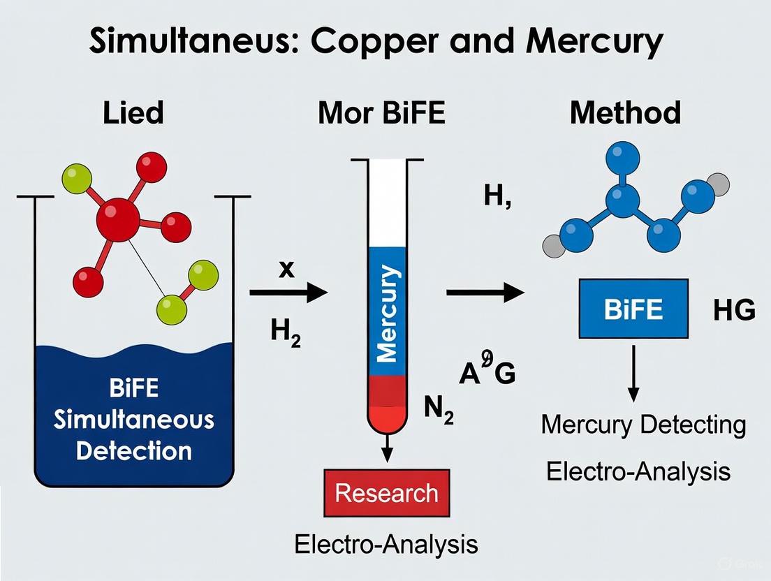
Bismuth-Film Electrodes for Simultaneous Detection of Copper and Mercury: A Researcher's Guide to Methods, Optimization, and Validation
This article provides a comprehensive overview of Bismuth-Film Electrode (BiFE) technology for the simultaneous electrochemical detection of copper (Cu) and mercury (Hg).
Bismuth-Film Electrodes for Simultaneous Detection of Copper and Mercury: A Researcher's Guide to Methods, Optimization, and Validation
Abstract
This article provides a comprehensive overview of Bismuth-Film Electrode (BiFE) technology for the simultaneous electrochemical detection of copper (Cu) and mercury (Hg). Tailored for researchers and drug development professionals, it covers the foundational principles of BiFE as a non-toxic alternative to mercury electrodes, detailed methodological protocols for electrode fabrication and analysis, systematic troubleshooting and optimization strategies using designs of experiments, and rigorous validation techniques against established reference methods. The content aims to serve as a practical guide for developing reliable, sensitive, and applicable sensing methods in biomedical and environmental monitoring contexts.
Why Bismuth-Film Electrodes? Foundations for Replacing Mercury in Heavy Metal Sensing
The Critical Need for Simultaneous Copper and Mercury Detection in Biomedical and Environmental Samples
Copper (Cu) and Mercury (Hg) represent a significant challenge in environmental monitoring and biomedical safety due to their ambiguous yet critical nature. Copper is an essential micronutrient crucial for various enzyme cofactors, proteins, and metabolic functions, playing vital roles in electron transport and regulating neurotransmitters [1]. However, over-accumulation of copper affects the central nervous system and increases the risk of various neurodegenerative diseases including Menken's and Wilson's diseases [1]. In contrast, mercury possesses no beneficial biological role and is highly toxic in all its forms. Excessive intake of Hg²⁺ can cause damage to the nervous system, blood system, kidneys, and reproductive system [2]. The coexistence of these metals in environmental samples poses compounded risks through synergistic toxic effects, making their simultaneous detection a critical analytical challenge [2].
The complexity of detecting this metal pair stems from their contrasting biological roles and the need for highly sensitive techniques capable of distinguishing them in complex matrices. This application note outlines current methodologies and protocols for the simultaneous detection of copper and mercury ions, with particular focus on their integration within Bismuth Film Electrode (BiFE) research contexts.
Current Detection Methodologies and Performance
Comparative Analysis of Simultaneous Detection Methods
Table 1: Performance comparison of simultaneous Cu²⁺ and Hg²⁺ detection methods
| Detection Method | Sensor Platform | Linear Range | Detection Limit | Real Sample Applications |
|---|---|---|---|---|
| Electrochemical (SWASV) | BiVO₄ nanospheres/GCE | 0-110 μM | Cu²⁺: 2.72 μM; Hg²⁺: 1.20 μM | Environmental and industrial samples [3] |
| Colorimetric/Fluorimetric | PYSC chemosensor | Not specified | Cu²⁺: 3 nM; Hg²⁺: 15 nM | Water, food samples, and intracellular imaging [1] |
| Electrochemical | Bi/DL-Ti₃C₂Tₓ/GCE | Not specified | Pb²⁺: 1.73 μg/L; Cd²⁺: 1.06 μg/L | Actual water samples [4] |
| Fluorimetric/Colorimetric | CuNCs@Zr-MOF/NMM | Not specified | Hg²⁺: 0.59 nM (fluorimetric), 36.3 nM (colorimetric) | Real aqueous samples [5] |
Bismuth-Based Electrodes in Heavy Metal Detection
Bismuth-based electrodes have emerged as promising alternatives to traditional mercury electrodes for heavy metal detection due to their low toxicity, excellent electrochemical performance, and insensitivity to dissolved oxygen [4]. The environmentally friendly nature of bismuth electrodes combined with their ability to form multicomponent alloys with heavy metals rather than competing for surface active sites makes them particularly valuable for environmental monitoring applications [4].
The performance of bismuth-based sensors can be significantly enhanced through nanomaterial integration. Studies demonstrate that combining bismuth with delaminated Ti₃C₂Tₓ MXene nanosheets develops sensors with good conductivity and performance for simultaneous detection of heavy metal ions [4]. Similarly, sol-gel synthesized Bismuth Vanadate (BiVO₄) nanospheres integrated onto glassy carbon electrodes have shown exceptional analytical performance for simultaneous detection of Cd²⁺, Pb²⁺, Cu²⁺, and Hg²⁺ ions [3].
Detailed Experimental Protocols
Protocol 1: Pyrene-Based Chemosensor (PYSC) for Dual Detection
Principle and Mechanism
The PYSC chemosensor operates based on aggregation-induced emission properties and complexation-driven fluorescence changes. In organic media, PYSC exhibits violet fluorescence (445 nm), which undergoes a redshift (538 nm) with increasing water content. In a 1:1 DMSO:H₂O mixture, PYSC displays blue fluorescence, while in 99% water, it exhibits orange fluorescence due to aggregation [1]. The presence of Hg²⁺ and Cu²⁺ induces distinct spectral changes enabling their detection.
Reagent Preparation
- PYSC Probe Solution: Dissolve pyrene-based Schiff base in DMSO to prepare 25 μM stock solution
- Metal Ion Standards: Prepare individual 1000 ppm stock solutions of Cu²⁺ and Hg²⁺ in double-distilled water
- Buffer System: Use phosphate buffer (0.01 M, pH 7.4) for aqueous measurements
- Solvent System: Prepare DMSO:water mixtures in varying ratios (1:1 to 1:99 v/v)
Detection Procedure
- Add 2 mL of PYSC probe solution (25 μM) to a quartz cuvette
- Introduce aliquots of sample or standard solutions containing Cu²⁺ and/or Hg²⁺
- Incubate the mixture for 5 minutes at room temperature
- Record absorption spectra from 300-600 nm
- Measure fluorescence emission with excitation at 380 nm
- Generate calibration curves using peak intensities at characteristic wavelengths
Interference Studies
- Test with competing ions including Pb²⁺, Cd²⁺, Zn²⁺, Ni²⁺, Co²⁺, Mn²⁺, Fe²⁺, Cr³⁺, Ag⁺
- Assess selectivity in mixed ion solutions
- Determine detection limits from signal-to-noise ratio (S/N > 3)
Protocol 2: BiVO₄ Nanosphere Modified Electrode for Simultaneous Detection
Synthesis of BiVO₄ Nanospheres
- Prepare Solution A: Dissolve 0.03 M Bi(NO₃)₃·5H₂O in deionized water
- Prepare Solution B: Dissolve 0.03 M NH₄VO₃ in deionized water with heating
- Slowly add Solution B to Solution A under continuous stirring
- Adjust pH to 7-8 using ammonium hydroxide
- Age the resultant suspension for 24 hours at room temperature
- Collect the precipitate by centrifugation and wash repeatedly with deionized water
- Dry at 80°C for 12 hours and calcine at 400°C for 2 hours [3]
Electrode Modification
- Polish glassy carbon electrode (GCE) with alumina slurry
- Clean ultrasonically in ethanol and deionized water
- Prepare BiVO₄ dispersion (1 mg/mL in water) and sonicate for 30 minutes
- Drop-cast 8 μL of dispersion onto GCE surface
- Dry under infrared lamp to form BiVO₄/GCE [3]
Square Wave Anodic Stripping Voltammetry (SWASV) Analysis
- Prepare acetate buffer (0.1 M, pH 4.5) as supporting electrolyte
- Add known concentrations of Cd²⁺, Pb²⁺, Cu²⁺, and Hg²⁺ standards
- Optimize deposition potential and time (-1.2 V for 270 seconds)
- Record SWASV signals from -1.0 V to +0.5 V
- Use peak currents at characteristic potentials for quantification:
- Cd²⁺: ~ -0.8 V
- Pb²⁺: ~ -0.55 V
- Cu²⁺: ~ -0.1 V
- Hg²⁺: ~ +0.3 V [3]
Real Sample Analysis
- Filter water samples through 0.45 μm membrane
- Acidify to pH 2 with nitric acid
- Mix sample with supporting electrolyte in 1:1 ratio
- Apply standard addition method for quantification
- Validate results with ICP-MS reference method
Diagram Title: BiVO₄ Electrode Fabrication and SWASV Analysis Workflow
Advanced Sensing Mechanisms
DNA-Based Recognition Systems
Advanced detection platforms utilize specific DNA interactions for metal ion recognition. T-Hg²⁺-T base pairing provides exceptional specificity for mercury detection, where thymine-rich DNA sequences selectively bind Hg²⁺ ions [5] [6]. Similarly, copper can be detected through its interaction with specific DNAzymes or aptamer sequences.
Field-effect transistor (FET) biosensors based on single-walled carbon nanotubes (SWNTs) functionalized with DNA sequences have achieved ultra-sensitive detection of Hg²⁺ with limits of 5.14 pM [6]. The mechanism involves direct conversion of DNA-Hg²⁺ interactions into electrical signals through changes in source-drain current (ID) when charged biomolecules adsorb to SWNTs [6].
Dual-Mode Sensing Approaches
Dual-mode sensors combining multiple detection principles provide enhanced reliability through self-calibration and anti-interference capabilities [5]. A notable example utilizes nanofluorophores, i.e., fluorescent copper nanoclusters-doped zirconia metal-organic framework (CuNCs@Zr-MOF) nanoconjugate and N-methyl mesoporphyrin IX (NMM) in combination with peroxidase-mimicking G-quadruplex DNAzyme (PMDNAzyme) [5].
This system operates through:
- FRET-based quenching of CuNCs@Zr-MOF fluorescence at 463 nm
- Fluorescence enhancement of NMM at 610 nm upon G-quadruplex formation
- Peroxidase-like activity of G4/hemin DNAzyme for colorimetric detection [5]
Diagram Title: Sensing Mechanisms for Cu²⁺/Hg²⁺ Detection
Research Reagent Solutions Toolkit
Table 2: Essential research reagents for simultaneous Cu²⁺/Hg²⁺ detection
| Reagent/Chemical | Function/Application | Specifications/Notes |
|---|---|---|
| Pyrene-based Schiff base (PYSC) | Dual chemosensing probe for Hg²⁺/Cu²⁺ | Exhibits aggregation-induced emission; Detection limits: Cu²⁺: 3 nM, Hg²⁺: 15 nM [1] |
| Bismuth Vanadate (BiVO₄) nanospheres | Electrode modifier for electrochemical detection | Sol-gel synthesized; enables simultaneous detection of Cd²⁺, Pb²⁺, Cu²⁺, Hg²⁺ [3] |
| Cysteamine-functionalized nanomaterials | Surface functionalization for improved sensing | Free -NH₂ and -SH groups enhance analyte interaction and sensor performance [7] |
| Thymine-rich DNA sequences | Specific recognition element for Hg²⁺ | Forms T-Hg²⁺-T coordination chemistry; used in FET biosensors [6] |
| CuNCs@Zr-MOF | Fluorescent nanomaterial for dual-mode sensing | Blue-emitting nanoconjugate; used in FRET-based Hg²⁺ detection [5] |
| N-methyl mesoporphyrin IX (NMM) | G-quadruplex binding dye | Red-emitting fluorescence; enhanced emission with G4 structure [5] |
| Delaminated Ti₃C₂Tₓ MXene | 2D conductive nanomaterial support | High conductivity, functional groups for material loading [4] |
| Covalent Organic Frameworks (COF) | Porous substrate for probe immobilization | High specific surface area, adjustable pore structure [8] |
The simultaneous detection of copper and mercury ions remains a challenging yet critical analytical task. Current methodologies show promising advances in sensitivity, selectivity, and practical applicability across environmental and biomedical samples. The integration of bismuth-based electrodes with nanomaterials and specific recognition elements provides a robust platform for future developments in this field.
Future research directions should focus on:
- Developing miniaturized portable devices for on-site monitoring
- Creating multi-array sensors for high-throughput screening
- Engineering advanced recognition elements with improved specificity
- Implementing machine learning algorithms for data analysis and pattern recognition
- Validating standardized protocols for regulatory applications
The protocols and methodologies outlined in this application note provide researchers with comprehensive tools for advancing simultaneous copper and mercury detection within the broader context of BiFE research and environmental monitoring.
The detection of toxic heavy metals, such as lead (Pb), cadmium (Cd), copper (Cu), and mercury (Hg), in environmental water samples is a critical concern for public health and ecological safety. For decades, mercury-film electrodes (MFEs) were the cornerstone of electrochemical stripping analysis due to their excellent reproducibility and wide negative potential window [9]. However, the high toxicity of mercury presents significant environmental and safety challenges, driving the search for alternative electrode materials.
Bismuth-film electrodes (BiFEs) have emerged as a highly promising, environmentally friendly replacement for traditional MFEs [9]. Bismuth shares many favorable electrochemical properties with mercury, such as the ability to form fusible alloys with other metals and a wide operational potential window, but with very low toxicity [10] [9]. This application note details the advantages of BiFEs over MFEs, supported by quantitative performance data, and provides a detailed protocol for their application in the simultaneous detection of heavy metals, with a specific focus on copper and mercury within a broader research thesis.
Comparative Advantages of Bismuth over Mercury
The transition from mercury- to bismuth-based electrodes is motivated by both practical performance and environmental, safety, and health (ESG) considerations.
- Environmental and Safety Profile: Bismuth is characterized by its very low toxicity, making it a more sustainable and safer material for routine laboratory use and the development of field-deployable sensors [9]. Mercury, in contrast, is a dangerous heavy metal known for its toxicity and bioaccumulation in many species [9].
- Electrochemical Performance: Bismuth films exhibit excellent stripping performance, characterized by well-defined, sharp peaks, and low background currents [9]. The sensitivity of BiFEs can be remarkably high. For instance, one study reported detection limits of 0.16 µg L⁻¹ for Pb(II) and 0.09 µg L⁻¹ for Cd(II) using an in-situ BiFE, outperforming many historical methods that used mercury [11].
- Practical Applicability: BiFEs can be easily formed in-situ by simply adding a Bi(III) salt to the sample solution, allowing for the use of unmodified carbon electrodes and simplifying the analytical procedure [11].
Table 1: Quantitative Comparison of Bismuth and Mercury Films for Heavy Metal Detection
| Feature | Bismuth-Film Electrode (BiFE) | Mercury-Film Electrode (MFE) |
|---|---|---|
| Toxicity | Very low toxicity [9] | High toxicity and bioaccumulation potential [9] |
| Detection Limit (Example) | Pb(II): 0.16 µg L⁻¹; Cd(II): 0.09 µg L⁻¹ [11] | Varies, but historically the benchmark for sensitivity |
| Sensitivity | High; can be enhanced with complexing agents (e.g., Alizarin Red S) [11] | High, well-established |
| Film Formation | Simple in-situ or ex-situ deposition [11] [9] | Requires careful plating; in-situ or ex-situ deposition [9] |
| Applicability for Cu(II) | Can be determined, though may require optimized conditions [9] | Can be determined [9] |
The Scientist's Toolkit: Key Reagent Solutions
The following table outlines the essential reagents and materials required for preparing and operating an in-situ bismuth film electrode for heavy metal detection.
Table 2: Essential Research Reagents and Materials for In-Situ BiFE Fabrication
| Reagent/Material | Function/Description | Example from Protocol |
|---|---|---|
| Bismuth(III) Salt | Source of Bi³⁺ ions for the simultaneous formation of the bismuth film on the electrode surface during the deposition step. | Bi(III) nitrate salt, 0.75 mg L⁻¹ in solution [11] |
| Supporting Electrolyte | Provides ionic conductivity and fixes the pH of the measurement solution. | 30.0 mmol L⁻¹ Acetic acid buffer (pH ~3.0) [11] |
| Complexing Agent | Enhances analytical sensitivity by forming complexes with the target metals, facilitating their accumulation. | Alizarin Red S (ARS), 40.0 µmol L⁻¹ [11] |
| Electrode Material | The substrate for bismuth film formation. Glassy carbon is commonly used. | Glassy carbon electrode [11] |
| Standard Metal Solutions | Used for calibration and quantification of the target analytes. | 1000 mg L⁻¹ stock solutions of Pb(II), Cd(II), Cu(II), Hg(II) [11] [12] |
Experimental Protocol: Simultaneous Detection of Copper and Mercury Using an In-Situ BiFE
This protocol is adapted from published methodologies for Pb/Cd detection and modified to encompass the simultaneous analysis of copper and mercury, which is the focus of the broader thesis [11].
Materials and Equipment
- Electrochemical Cell: Standard three-electrode system.
- Working Electrode: Glassy carbon electrode (GCE, 3 mm diameter).
- Counter Electrode: Platinum wire.
- Reference Electrode: Ag/AgCl (3 M KCl).
- Potentiostat: Capable of performing Differential Pulse Anodic Stripping Voltammetry (DPASV).
- Reagents:
- Bi(III) stock solution (e.g., from Bi(NO₃)₃, 1000 mg L⁻¹).
- Cu(II), Hg(II), and other target metal standard solutions (1000 mg L⁻¹).
- Alizarin Red S (ARS): 1.0 mmol L⁻¹ aqueous solution.
- Acetic acid (CH₃COOH) and Sodium acetate (CH₃COONa) for 0.1 M acetate buffer, pH 4.5. Note: For Cu/Hg, a different pH may be optimal and requires optimization.
- Potassium hexacyanoferrate(II) (K₄[Fe(CN)₆]): 1.0 mmol L⁻¹ solution.
- High-purity deionized water (18.2 MΩ·cm).
Electrode Preparation
- Glassy Carbon Electrode Polishing: Polish the GCE surface successively with 1.0, 0.3, and 0.05 µm alumina slurry on a microcloth pad.
- Rinsing: Rinse the polished electrode thoroughly with deionized water.
- Sonication: Sonicate the electrode in deionized water and then in ethanol for 1 minute each to remove any adhering alumina particles.
- Drying: Allow the electrode to air dry.
Procedure Workflow
The following diagram illustrates the key stages of the experimental procedure for simultaneous detection of copper and mercury using an in-situ bismuth film electrode.
Detailed Measurement Steps
Solution Preparation: In the electrochemical cell, mix the following to prepare 10 mL of measurement solution:
- Sample or Standard Solution: Containing Cu(II), Hg(II), and other target metals.
- Supporting Electrolyte: 1.0 mL of 0.1 M acetate buffer (pH ~4.5). Note: The optimal pH for simultaneous Cu and Hg detection must be determined empirically, as their redox behavior is pH-dependent.
- Bismuth Source: 75 µL of 100 mg L⁻¹ Bi(III) stock solution (final concentration: 0.75 mg L⁻¹).
- Complexing Agent: 400 µL of 1.0 mmol L⁻¹ ARS solution (final concentration: 40.0 µmol L⁻¹).
- Additive: 500 µL of 1.0 mmol L⁻¹ K₄[Fe(CN)₆] solution (final concentration: 50.0 µmol L⁻¹).
- Dilute to 10 mL with deionized water and degas with nitrogen for 300 s.
Anodic Stripping Voltammetry (ASV):
- Deposition Step: Apply a deposition potential of -1.40 V vs. Ag/AgCl for 60 s while stirring the solution. This step co-deposits Bi and the target metals (Cu, Hg) onto the GCE surface as alloys.
- Equilibration: Stop stirring and allow the solution to equilibrate for 15 s.
- Stripping Step: Record the differential pulse anodic stripping voltammogram by scanning the potential from -1.40 V to 0 V. Use the following DPASV parameters: pulse amplitude 50 mV, pulse width 50 ms, step potential 5 mV, scan rate 20 mV s⁻¹.
Electrode Cleaning: After each measurement, apply a potential of 0 V for 30 s under stirring to remove residual metals and the bismuth film from the electrode surface, ensuring a fresh start for the next analysis.
Data Analysis and Validation
- Calibration: Record stripping voltammograms for a series of standard solutions of Cu(II) and Hg(II) under identical conditions. The peak current (typically in microamperes, µA) is proportional to the metal concentration.
- Peak Identification: Identify the metals based on their characteristic peak potentials (e.g., Pb ~ -0.5 V, Cd ~ -0.8 V, Cu ~ -0.2 V, Hg ~ +0.2 V, vs. Ag/AgCl). The exact position can shift slightly depending on the matrix.
- Quantification: Plot the peak height or area against metal concentration to create a calibration curve. Use this curve to determine the concentration of metals in unknown samples.
- Method Validation: Validate the method by analyzing certified reference materials (CRMs) and spiked real water samples to ensure accuracy and reliability [11].
Bismuth-film electrodes represent a significant advancement in electroanalytical chemistry, successfully replacing toxic mercury films without compromising analytical performance. The provided protocol demonstrates a sensitive and green method for the simultaneous detection of heavy metals, including copper and mercury. The key advantages of BiFEs—low toxicity, high sensitivity, and simple fabrication—make them an ideal platform for routine environmental monitoring and advanced research applications. Future work in this thesis will focus on optimizing the support electrolyte and pH specifically for the Cu/Hg pair and exploring novel nanostructured bismuth surfaces to further enhance sensitivity and selectivity.
Anodic Stripping Voltammetry (ASV) is a highly sensitive electroanalytical technique for determining trace concentrations of metal ions. Its exceptional sensitivity, capable of detecting metals at sub-parts per billion (ppb) levels, stems from a pre-concentration step that accumulates analyte on the electrode surface prior to measurement [13] [14]. This makes ASV particularly valuable for environmental monitoring, pharmaceutical analysis, and food safety, where detecting low levels of toxic metals like lead, cadmium, and mercury is crucial [14]. Within the scope of thesis research focused on the simultaneous detection of copper and mercury using a Bismuth Film Electrode (BiFE), understanding the core principles of ASV is foundational. This document details the fundamental electrochemistry, practical protocols, and key experimental considerations for ASV, providing a framework for method development using environmentally friendly bismuth-based electrodes.
Fundamental Principles of ASV
Anodic Stripping Voltammetry is a two-step technique consisting of an electrodeposition step followed by a stripping step, as illustrated in the workflow below.
The Electrodeposition (Pre-concentration) Step
In the first step, the working electrode is held at a constant potential that is sufficiently negative to reduce the target metal ions (Mⁿ⁺) to their metallic state (M(0)) [14]. The reduced metal is deposited onto the electrode surface. For a traditional mercury electrode, this forms an amalgam; for a bismuth film electrode (BiFE), it forms a fused alloy [15].
[ \text{M}^{n+} + n\text{e}^- \rightarrow \text{M (electrode surface)} ]
The deposition potential must be more negative than the formal reduction potential (E°′) of the target metal. The amount of metal deposited is controlled by the deposition time and mass transport conditions (e.g., stirred solution), effectively pre-concentrating the analyte from the bulk solution onto the electrode surface [13] [14].
The Stripping (Anodic Scan) Step
Following deposition and a brief quiet period, the potential is scanned in an anodic (positive) direction. This oxidizes the deposited metal back into solution, generating a measurable faradaic current.
[ \text{M (electrode surface)} \rightarrow \text{M}^{n+} + n\text{e}^- ]
The resulting plot of current versus applied potential is called a stripping voltammogram. The peak current is proportional to the concentration of the metal in the original solution, while the peak potential is characteristic of the specific metal being oxidized, allowing for identification [14]. The peak shape is often sharp and well-defined, which enhances resolution between different metals and improves the signal-to-noise ratio [13].
Electrode Materials: The Shift to Bismuth
The choice of working electrode is critical for ASV performance. Table 1 compares the properties of common electrode materials.
Table 1: Comparison of Working Electrode Materials for Anodic Stripping Voltammetry
| Electrode Material | Toxicity | Key Characteristics | Ideal for Detection of |
|---|---|---|---|
| Mercury (Hg) [14] | High | Forms homogeneous amalgams; wide cathodic potential window; well-defined, reproducible peaks. | Dozens of metals (e.g., Cd, Pb, Zn); except Hg itself and metals more noble than Hg. |
| Gold (Au) [16] | Low | Excellent for Hg(II) detection; high sensitivity and selectivity. | Mercury (Hg), Lead (Pb) |
| Copper Film (CuFE) [16] | Low (compared to Hg) | Simple in-situ preparation; excellent sensitivity for Hg and Pb; low-cost. | Mercury (Hg), Lead (Pb) |
| Bismuth (BiFE/BiBE) [17] [15] | Low (Environmentally friendly) | Forms "fused alloys" with metals; high hydrogen overpotential; works well in the presence of dissolved oxygen; comparable performance to Hg. | Cadmium (Cd), Lead (Pb), Zinc (Zn) |
The movement towards "green" electroanalysis has driven the adoption of bismuth-based electrodes as a primary replacement for toxic mercury [14] [15]. Bismuth shares key advantageous properties with mercury, including the ability to form alloys with heavy metals and a high overpotential for hydrogen evolution, which allows for the detection of metals like zinc without interference from water reduction [15]. Furthermore, analyses with bismuth electrodes can often be performed without the need for oxygen removal, simplifying the experimental procedure [17] [15].
Key Experimental Parameters and Protocols
Core Research Reagent Solutions
A successful ASV experiment relies on a set of well-prepared reagent solutions. Table 2 lists the essential materials and their functions.
Table 2: Essential Research Reagent Solutions for ASV with a Bismuth Film Electrode
| Reagent / Material | Function / Purpose | Example / Typical Composition |
|---|---|---|
| Supporting Electrolyte | Carries current; fixes ionic strength and pH; can influence metal complexation. | 0.1 M Acetate Buffer (pH ~4.5) [15]; 0.1 M HCl [16]. |
| Bismuth Precursor | Source of Bi(III) ions for the in-situ formation of the bismuth film on the substrate. | 0.02 M Bismuth(III) nitrate pentahydrate (Bi(NO₃)₃·5H₂O) in 1 M HCl [17]. |
| Metal Standard Solutions | Used for calibration curves, standard addition, and method validation. | 1000 mg/L stock solutions of Cd(II), Pb(II), Zn(II), Cu(II), Hg(II) [15]. |
| Substrate Electrode | The conductive base upon which the bismuth film is deposited. | Glassy Carbon [13], Carbon Nanotubes [15], or Brass [17]. |
| pH Buffer | Controls the pH of the measurement solution, which affects metal speciation and stability. | Acetate buffer for pH ~4-5 [17] [15]; Nitric acid (5% HNO₃) [13]. |
Detailed Protocol: Determination of Cd, Pb, and Zn using a Bismuth Bulk Electrode (BiBE)
This protocol, adapted from the work of Hocevar et al., outlines a validated method for the simultaneous detection of trace metals [15].
Experimental Workflow:
Step-by-Step Procedure:
- Electrode Preparation: Fabricate the Bismuth Bulk Electrode (BiBE) by melting bismuth needles into a hand-blown glass casing under a vacuum atmosphere. Polish the freshly exposed electrode surface sequentially with emery paper (200 to 800 grit) and alumina slurries (1.0 μm, 0.3 μm, and 0.05 μm) to a smooth finish [15].
- Solution Preparation: Prepare a 20 mL standard or sample solution in an electrochemical cell. The solution should contain 0.1 M sodium acetate buffer, adjusted to pH 5.0 with acetic acid. Spike with standard solutions of Cd(II), Pb(II), and Zn(II) to achieve concentrations within the desired calibration range (e.g., 10–100 μg L⁻¹) [15].
- Pre-concentration / Electrodeposition: Immerse the working, reference, and counter electrodes in the solution. Hold the potential of the BiBE at -1.4 V (vs. Ag/AgCl) for 180 seconds while stirring the solution at approximately 1200 rpm. This step reduces and accumulates the metal ions as alloys on the bismuth surface [15].
- Stripping Analysis: After the accumulation time, stop the stirring and initiate the stripping step immediately. Use Square-Wave Voltammetry (SWV) with the following parameters to scan anodically:
- Initial E: -1.4 V
- Final E: -0.35 V
- Potential Step: 4 mV
- Pulse Amplitude: 25 mV
- Frequency: 15 Hz
- Quiet Time: 0 s (stirring already stopped) [15].
- Data Collection and Quantification: Record the voltammogram (current vs. potential). Identify the stripping peaks for Pb(II) at approximately -0.50 V, Cd(II) at -0.75 V, and Zn(II) at -1.10 V [15]. Construct a calibration curve by plotting the peak current (or peak area) against metal concentration. For unknown samples, use the standard addition method to account for matrix effects.
- Validation: Validate the method by analyzing a real sample, such as contaminated river water, and comparing the results with those obtained from a standard technique like Inductively Coupled Plasma-Optical Emission Spectroscopy (ICP-OES) [15].
Quantitative Performance Data
Under optimized conditions, ASV offers exceptional sensitivity. Table 3 summarizes the performance metrics achievable with a bismuth bulk electrode for the detection of common heavy metals.
Table 3: Analytical Performance of ASV for Heavy Metal Detection at a Bismuth Bulk Electrode (BiBE) [15]
| Metal Ion | Stripping Peak Potential (V vs. Ag/AgCl) | Linear Range (μg L⁻¹) | Individual Calibration Sensitivity (μA L μg⁻¹) | Limit of Detection (LOD) (ng L⁻¹) |
|---|---|---|---|---|
| Lead (Pb(II)) | -0.50 V | 10 – 100 | 0.125 | 105 |
| Cadmium (Cd(II)) | -0.75 V | 10 – 100 | 0.112 | 54 |
| Zinc (Zn(II)) | -1.10 V | 10 – 100 | 0.187 | 396 |
Critical Considerations for Method Development
Intermetallic Compound Formation
A significant challenge in the simultaneous detection of multiple metals is the formation of intermetallic compounds. These are alloys formed between two different metals on the electrode surface, which can alter their stripping potentials and currents. For instance, the presence of copper can interfere with the detection of other metals [14]. When developing a method for the simultaneous detection of copper and mercury, this potential interaction must be investigated and mitigated, for example, by optimizing the deposition potential and time or by using complexing agents to mask interfering ions [16].
The Influence of Metal Speciation
ASV typically detects the fraction of metal that is electroactive, which includes free hydrated ions and weakly bound (labile) complexes [14]. The speciation of a metal in a sample is highly dependent on pH and the presence of organic or inorganic ligands. This is a critical distinction from techniques like ICP-MS, which typically measure total metal content after acid digestion. Therefore, careful control and reporting of solution pH and composition are essential for obtaining reproducible and meaningful results, especially in complex matrices like environmental waters or biological fluids.
The pursuit of environmentally friendly and highly sensitive electroanalytical methods has established Bismuth Film Electrodes (BiFEs) as a cornerstone technology for the detection of heavy metals. As a non-toxic alternative to mercury electrodes, bismuth offers exceptional performance in stripping voltammetry, characterized by its ability to form alloys with metals, well-defined stripping signals, insensitivity to dissolved oxygen, and a wide operational potential window [18] [19]. This document details the principal configurations of BiFEs—graphite supports, screen-printed electrodes (SPEs), and in-situ modification methods—framed within advanced research for the simultaneous detection of copper and mercury. The protocols and data herein are designed to provide researchers and scientists with reliable methodologies for sensor fabrication and application.
Key BiFE Configurations and Performance
The performance of a Bismuth Film Electrode is profoundly influenced by its support material and the method of bismuth deposition. The table below summarizes the core configurations and their validated performance in heavy metal detection.
Table 1: Key Configurations of Bismuth Film Electrodes for Heavy Metal Detection
| Configuration | Support Material/Modification | Target Analytes | Electrochemical Technique | Limit of Detection (LOD) | Linear Range | Key Findings |
|---|---|---|---|---|---|---|
| Graphite Support | Graphite rod | Hg(II) and Pb(II) | Square Wave Anodic Stripping Voltammetry (SWASV) | Hg(II): 1 ppbPb(II): 10 ppb | Not Specified | Optimal synthesis: 3 mM [Bi(III)], 10 s deposition time. The Bi/graphite electrode is low-cost and suitable for field analysis [20]. |
| Screen-Printed Electrode (SPE) | Boron-Doped Diamond (BDD) | Pb(II) and Hg(II) | Square Wave Voltammetry (SWV) | Pb(II): 6.7 µg/LHg(II): 7.5 µg/L | 31.3 - 2000 µg/L | Method allows direct determination in complex matrices like beer with minimal sample treatment (40 µL) [21]. |
| Screen-Printed Electrode (SPE) | Poly(bromocresol purple) polymer film | Cd(II) and Pb(II) | Differential Pulse Anodic Stripping Voltammetry (DPASV) | Cd(II): 0.036 µg/LPb(II): 0.027 µg/L | 0 - 250 µg/L | The polymer-modified SPCE demonstrated excellent repeatability, reproducibility, and stability in wastewater [22]. |
| In-Situ Modification | Screen-printed carbon electrode (untreated) | Cd(II) and Pb(II) | Anodic Stripping Voltammetry (ASV) | Not explicitly stated | Not explicitly stated | Bismuth is added directly to the sample solution and co-deposited with the target metals during the pre-concentration step [18]. |
| Ex-Situ Modification | Pre-oxidized Screen-printed carbon electrode (Type A/B) | Cd(II) and Pb(II) | Differential Pulse Stripping Voltammetry | Not explicitly stated | Not explicitly stated | Electrode is pre-coated with a bismuth film in a separate step prior to exposure to the sample [18]. |
Experimental Protocols for BiFE Fabrication and Measurement
Protocol 1: Fabrication of a Graphite-Supported BiFE
This protocol is adapted from the synthesis of a graphite-supported bismuth film working electrode for the simultaneous quantification of Hg(II) and Pb(II) [20].
Research Reagent Solutions:
- Bismuth Precursor: 3 mM Bismuth nitrate pentahydrate (Bi(NO₃)₃·5H₂O) in 1 M HNO₃.
- Supporting Electrolyte (for deposition): 1 M Nitric Acid (HNO₃).
- Acetate Buffer (for measurement): 1 M Acetic acid buffer, pH 4.7.
- Standard Solutions: 1000 ppm Hg(NO₃)₂ and Pb(NO₃)₂ for calibration.
Methodology:
- Graphite Electrode Pretreatment:
- Insulate the side of a cylindrical graphite segment with polytetrafluoroethylene (PTFE), exposing only the circular end (4 mm diameter).
- Polish the exposed surface with 2500-grit aluminum oxide sandpaper.
- Sonicate the polished electrode in deionized water for 5 minutes to remove residual particles.
- Electrochemical Activation:
- Assemble a three-electrode system with the graphite electrode as the working electrode, a platinum wire as the counter electrode, and an Ag/AgCl (3 M NaCl) reference electrode.
- Immerse the electrodes in a 1 M HNO₃ solution containing the optimized 3 mM Bi(III) concentration.
- Perform Cyclic Voltammetry (CV) by scanning the potential between -0.5 V and +0.3 V for 5 complete cycles. Use a step potential of 0.004 V and a scan rate of 0.05 V/s.
- Bismuth Film Electrodeposition:
- In the same solution, apply a constant deposition potential (Edep) of -0.5 V vs. Ag/AgCl for a deposition time (tdep) of 10 seconds under constant stirring (6 Hz). This step forms the bismuth film on the graphite surface.
Protocol 2: In-Situ vs. Ex-Situ Modification of Screen-Printed Electrodes
This protocol outlines the strategies for modifying screen-printed carbon electrodes (SPCEs) with bismuth films, highlighting the critical role of bismuth chemistry [18].
Research Reagent Solutions:
- Bismuth Stock Solution: 1000 mg/L Bi(III) in nitric acid.
- Acetate Buffer: 0.1 M, pH 4.4.
- Nafion Solution: 5 wt% in lower aliphatic alcohols/water.
Methodology: A. In-Situ BiFE Modification:
- Preparation: Clean the SPCE by rinsing with ethanol and then water.
- Analysis: Add the sample or standard solution containing the target metals (e.g., Cd(II), Pb(II)) directly to the electrochemical cell.
- Co-deposition: Introduce the bismuth ion precursor (e.g., from a 1000 mg/L stock) directly into the same sample solution to achieve the desired concentration (e.g., 0.1 mM).
- Measurement: Apply the deposition potential (e.g., -1.20 V). The bismuth and target metals are simultaneously reduced and co-deposited onto the SPCE surface as an alloy, after which the stripping scan is performed.
B. Ex-Situ BiFE Modification (with Surface Pre-oxidation):
- SPCE Pre-oxidation (Treatment A):
- Immerse the SPCE in 0.1 M acetate buffer (pH 4.4).
- Apply a potential of +1.50 V for 120 seconds to oxidize the carbon surface and introduce oxygen-containing functional groups.
- Bismuth Film Electrodeposition:
- Transfer the pre-oxidized SPCE to a separate plating solution of 0.1 mM Bi(III) in acetate buffer (pH 4.4).
- Apply a reduction potential of -1.20 V for 30 seconds to electrodeposit the bismuth film.
- Protective Layer Casting (Optional):
- Immediately after bismuth deposition, drop-cast 1 µL of the Nafion solution onto the BiF-modified carbon surface.
- Allow the film to dry in air. The electrode must be used immediately after preparation to minimize bismuth oxidation.
Protocol 3: Simultaneous Detection via SWASV
This is a generalized protocol for the simultaneous detection of multiple heavy metals, such as Cu(II) and Hg(II), using Square Wave Anodic Stripping Voltammetry (SWASV) [20] [19].
Methodology:
- Electrode Preparation: Fabricate the BiFE using one of the protocols above (e.g., graphite-supported or SPCE-modified).
- Pre-concentration/Deposition:
- Place the BiFE into the sample solution containing the target metal ions and the supporting electrolyte (e.g., acetate buffer).
- Apply a deposition potential of -1.0 V to -1.2 V (vs. Ag/AgCl) for 60-120 seconds under constant stirring. This causes the reduction and accumulation of metal ions (and bismuth, if in-situ) onto the electrode surface.
- Equilibration: After deposition, stop stirring and allow the solution to become quiescent for a brief period (e.g., 10-15 seconds).
- Stripping Scan:
- Initiate the SWASV scan from a negative potential towards a more positive potential (e.g., -1.0 V to +0.7 V).
- The applied square wave parameters typically include an amplitude of 25 mV and a frequency of 25 Hz.
- During this scan, the accumulated metals are oxidized (stripped) back into the solution, generating characteristic current peaks at their respective oxidation potentials.
- Electrode Cleaning: After each measurement, apply a positive potential (e.g., +0.7 V) for 30-60 seconds in a clean supporting electrolyte to strip off any residual metals and rejuvenate the electrode surface.
Diagram 1: Generalized workflow for heavy metal detection using a Bismuth Film Electrode (BiFE) with Square Wave Anodic Stripping Voltammetry (SWASV), incorporating both in-situ and ex-situ modification pathways.
The Scientist's Toolkit: Essential Research Reagents
The following table catalogs the key reagents and materials required for the fabrication and application of BiFEs as discussed in the protocols.
Table 2: Essential Research Reagents for BiFE Fabrication and Analysis
| Reagent/Material | Function / Role in BiFE Analysis | Exemplary Application |
|---|---|---|
| Bismuth Nitrate Pentahydrate (Bi(NO₃)₃·5H₂O) | Primary precursor for bismuth film formation. Source of Bi(III) ions for electrodeposition. | Standard source for in-situ and ex-situ bismuth film formation in various supporting electrolytes [20] [18]. |
| Screen-Printed Carbon Electrodes (SPCEs) | Disposable, mass-producible, and portable substrate for the working electrode. Enables decentralized analysis. | Base transducer for bismuth modification; used in polymer-coated, pre-oxidized, and in-situ configurations [18] [22]. |
| Graphite Electrodes / Inks | Support material for bismuth film. Provides high electrical conductivity, low cost, and ease of modification. | Used as a support for bismuth electrodeposition to create a low-cost, sensitive sensor for Hg(II) and Pb(II) [20]. |
| Nafion Perfluorinated Resin | Cation-exchange polymer coating. Used to protect the bismuth film, improve mechanical stability, and alleviate interferences from surfactants or macromolecules. | Cast as a protective layer on ex-situ plated BiFEs to enhance robustness and selectivity [18]. |
| Acetate Buffer (pH ~4.4-4.7) | Common supporting electrolyte. Provides optimal pH for the deposition and stripping of many heavy metal ions and for bismuth film stability. | Used as the medium for analysis in the detection of Cd(II), Pb(II), Hg(II), and Cu(II) [20] [18]. |
| Nitric Acid (HNO₃) | Acidic medium and supporting electrolyte. Used for the electrodeposition of bismuth films, particularly from Bi(III) nitrate solutions. | Serves as the supporting electrolyte (1 M) during the electrodeposition of bismuth onto graphite supports [20]. |
| Dimethylglyoxime (DMG) | Chelating agent for adsorptive stripping voltammetry. Forms complexes with specific metals (e.g., Pt, Pd) for enhanced pre-concentration. | Used as a complexing ligand for the sensitive detection of Platinum Group Metals (PGMs) at BiFEs [23]. |
Step-by-Step Protocol: Fabricating Your BiFE and Detecting Cu(II) and Hg(II)
Within the framework of developing a novel method for the simultaneous detection of copper (Cu) and mercury (Hg) using a Bismuth Film Electrode (BiFE), the selection and meticulous preparation of the underlying electrode substrate is a critical foundational step. The substrate governs the stability, uniformity, and overall analytical performance of the subsequently formed bismuth film. This application note provides detailed protocols for the pre-treatment of three common electrode substrates—graphite, glassy carbon, and screen-printed electrodes—tailored specifically for researchers and scientists engaged in electroanalytical method development for trace metal analysis. Proper electrode preparation ensures the reproducibility, sensitivity, and low detection limits required for environmental monitoring and drug development applications [18] [24].
Electrode Substrate Selection and Comparison
The choice of substrate influences the morphology of the bismuth film, the background current, and the overall signal-to-noise ratio in stripping voltammetry. The following table summarizes the key characteristics of the three substrates in the context of BiFE preparation for heavy metal detection.
Table 1: Comparison of Electrode Substrates for Bismuth Film Electrode Preparation
| Substrate Type | Key Advantages | Key Limitations | Ideal for Cu/Hg Detection? | Typical Pre-treatment Method |
|---|---|---|---|---|
| Graphite (e.g., Exfoliated Graphite) | High surface area, porous structure, cost-effective [25]. | Surface heterogeneity can affect film uniformity. | Yes, high surface area aids pre-concentration [25]. | Mechanical polishing, electrochemical activation. |
| Glassy Carbon (GC) | Dense, impermeable surface, excellent electrochemical stability, wide potential window [26]. | Requires rigorous surface polishing for reproducibility. | Yes, provides a stable, well-defined base [27]. | Sequential mechanical polishing with alumina slurry. |
| Screen-Printed Electrodes (SPEs) | Disposable, mass-producible, portable for field use, small sample volume [18] [24]. | Inks can dissolve in organic solvents; performance batch-dependent. | Yes, excellent for disposable sensors; Au-SPEs are good for Hg [24]. | Often used as-received; oxidative pre-treatment can enhance performance [18]. |
Detailed Experimental Protocols
Protocol 1: Pre-treatment of Graphite Substrates (e.g., Exfoliated Graphite Electrode)
This protocol is adapted from the work on bismuth-modified exfoliated graphite electrodes for the co-detection of heavy metals [25].
Objective: To clean and electrochemically activate the graphite surface to ensure a uniform and adherent bismuth film.
Materials:
- Exfoliated Graphite (EG) electrode
- 0.1 M Acetate buffer (pH 5.0)
- 0.1 M Nitric Acid (HNO₃)
- Bismuth oxide (Bi₂O₃)
- Ultrasonic bath
- Potentiostat
Procedure:
- Mechanical Cleaning (if applicable): For solid graphite electrodes, gently polish the surface with a soft, clean paper to remove any visible debris. Avoid abrasive powders that may clog the porous structure.
- Electrochemical Activation:
- Immerse the EG electrode in 0.1 M Acetate buffer (pH 5.0).
- Using a potentiostat, apply a potential of -1.0 V vs. Ag/AgCl for 300 seconds under stirring. This step helps reduce surface oxides and clean the surface.
- Record a cyclic voltammogram in a 5 mM ferrocene solution to confirm electroactive surface area and cleanliness [25].
- Bismuth Film Formation (Ex-situ):
- Prepare a plating solution of 0.02 M Bi(NO₃)₃ in 1 M HCl with 0.5 M LiBr [26].
- Transfer the pre-treated EG electrode to the plating solution.
- Apply a deposition potential of -0.28 V vs. Ag/AgCl for 20-30 seconds to electrodeposit bismuth nanoparticles onto the EG surface [26] [25].
- Rinse the newly formed Bi/EG electrode thoroughly with ultrapure water before transferring it to the sample solution for Cu and Hg analysis.
Protocol 2: Pre-treatment of Glassy Carbon Electrodes (GCE)
This protocol ensures a mirror-finish, reproducible surface on GCE, which is crucial for forming a uniform bismuth film.
Objective: To achieve a pristine, polished, and oxide-free glassy carbon surface.
Materials:
- Glassy Carbon Electrode (3 mm diameter typical)
- Alumina polishing slurries (1.0, 0.3, and 0.05 μm)
- Polishing cloths
- Deionized water
- Ethanol
- Ultrasonic bath
Procedure:
- Sequential Mechanical Polishing:
- Place the GCE on a clean polishing cloth.
- Apply a slurry of 1.0 μm alumina powder in deionized water and polish the electrode surface in a figure-8 pattern for 60 seconds.
- Repeat this process with successively finer alumina slurries (0.3 μm and finally 0.05 μm).
- Ultrasonic Rinsing:
- After each polishing step, sonicate the GCE in deionized water for 60 seconds to remove embedded alumina particles.
- After the final polish, sonicate sequentially in ethanol and deionized water, each for 60 seconds [27].
- Electrochemical Cleaning (Optional but Recommended):
- In a clean supporting electrolyte (e.g., 0.1 M H₂SO₄ or acetate buffer), perform cyclic voltammetry between -0.5 V and +1.0 V until a stable voltammogram is achieved, indicating a clean surface.
- Bismuth Film Formation (In-situ or Ex-situ):
- The polished GCE is now ready for bismuth film formation. For in-situ deposition, simply add Bi(III) ions (e.g., 3 mM [28]) directly to the sample solution containing your target analytes (Cu and Hg). The bismuth will co-deposit with the target metals during the pre-concentration step at -1.0 V to -1.3 V [28] [27].
Protocol 3: Pre-treatment of Screen-Printed Electrodes (SPEs)
This protocol outlines oxidative pre-treatment methods to enhance the performance of carbon-based SPEs, as their as-received state may be suboptimal for bismuth film formation [18].
Objective: To functionalize the carbon surface of SPEs, increasing the density of oxygen-containing groups that improve the adhesion and uniformity of the bismuth film.
Materials:
- Carbon Screen-Printed Electrodes (C-SPEs)
- 0.1 M Acetate Buffer (pH 4.4)
- Saturated Sodium Carbonate (Na₂CO₃) solution
- 0.1 M HCl
- Nafion perfluorinated resin solution (5 wt%)
- Potentiostat
Procedure:
- Oxidative Pre-treatment (Two Methods):
- Bismuth Film Deposition:
- Immediately after oxidative treatment, transfer the SPE to a deaerated acetate buffer (pH 4.4) containing 0.1 mM Bi(III).
- Apply a reduction potential of -1.20 V for 30 seconds to electrodeposit the bismuth film [18].
- Application of a Protective Layer (Optional):
- To improve stability and alleviate interferences, drop-cast 1 μL of a Nafion solution onto the BiF/SPE surface and allow it to dry in air.
- Note: The electrode should be used immediately after preparation, as the bismuth film can oxidize over time [18].
The workflow for the pre-treatment and modification of these electrodes is summarized in the diagram below.
Critical Parameters for Bismuth Film Formation and Analysis
The analytical performance for the simultaneous detection of Cu and Hg is highly dependent on the parameters used for bismuth deposition and the subsequent stripping analysis. The following table consolidates optimized parameters from recent studies.
Table 2: Key Parameters for Bismuth Film Formation and Anodic Stripping Voltammetry
| Parameter | Typical Range / Optimal Value | Impact on Analysis |
|---|---|---|
| Bismuth Concentration ([Bi]) | 0.1 mM – 3 mM [28] [18] | Higher concentrations can lead to thicker, less porous films; 3 mM was optimal for Hg/Pb detection [28]. |
| Deposition Potential (E_dep) | -0.28 V to -1.3 V vs. Ag/AgCl [28] [27] [26] | Must be negative enough to reduce Bi(III) and target metals; too negative can cause H₂ evolution. -1.0 V to -1.3 V is common for multiple metals [28] [27]. |
| Deposition Time (t_dep) | 10 s – 300 s [28] [25] | Controls the amount of metal pre-concentrated; longer times increase sensitivity but reduce throughput. 10 s was sufficient for ppb-level Hg [28]. |
| Supporting Electrolyte | Acetate buffer (pH 4.4-5.0), HNO₃, HCl [28] [18] [26] | Affects deposition efficiency, film morphology, and peak resolution. Acetate buffer is common for multiple metal detections [28] [18]. |
| Stripping Technique | Square Wave Anodic Stripping Voltammetry (SWASV) [28] [25] | Provides high sensitivity and speed, effective for simultaneous multi-metal detection. |
The Scientist's Toolkit: Essential Research Reagent Solutions
Table 3: Key Reagents and Materials for BiFE Preparation and Analysis
| Reagent / Material | Function / Role | Example & Notes |
|---|---|---|
| Bismuth Salt | Source of Bi(III) ions for film electrodeposition. | Bismuth nitrate pentahydrate (Bi(NO₃)₃·5H₂O) dissolved in dilute HNO₃ is a common stock solution [18]. |
| Supporting Electrolyte | Provides ionic conductivity and controls pH. | 0.1 M Acetate Buffer (pH 4.4-5.0) is versatile for many metals. 1 M HNO₃ or HCl can also be used [28] [18]. |
| Polishing Abrasives | To create a smooth, reproducible electrode surface. | Alumina (Al₂O₃) powders, 1.0, 0.3, and 0.05 μm for sequential polishing of GC [27]. |
| Ion-Exchange Polymer | Protective membrane to improve film stability and selectivity. | Nafion solution, drop-cast onto the BiFE to form a cation-exchange layer [18]. |
| Metal Standard Solutions | For calibration and quantitative analysis. | AA standard solutions of Cu, Hg, Bi (1000 mg/L). Dilute prior to use [18] [27]. |
| Oxygen Scavenger | To remove dissolved O₂, though BiFE is less sensitive [18]. | High-purity Nitrogen or Argon gas for deaeration of solutions. |
Common Issues:
- Poorly Defined Stripping Peaks: Often due to an improperly polished substrate (GC), contaminated solutions, or non-optimized deposition potential.
- High Background Current: Can indicate a dirty electrode surface or an unstable bismuth film. Re-polish the substrate and ensure fresh plating solutions.
- Low Reproducibility: Standardize pre-treatment times and potentials precisely. For SPEs, use electrodes from the same batch and consider a Nafion coating to stabilize the film [18].
In summary, the successful deployment of a BiFE for the simultaneous detection of copper and mercury hinges on a disciplined approach to electrode selection and pre-treatment. By adhering to these detailed protocols for graphite, glassy carbon, and screen-printed surfaces, researchers can establish a robust and reliable foundation for their electroanalytical methods, ensuring high-quality data for both environmental and pharmaceutical applications.
Bismuth Film Electrodes (BiFEs) have emerged as a robust, environmentally friendly alternative to mercury-based electrodes for the anodic stripping voltammetry (ASV) of heavy metals. Their low toxicity, insensitivity to dissolved oxygen, and ability to form alloys with various metal ions make them particularly suitable for environmental monitoring [29]. The electroanalytical performance of a BiFE is profoundly influenced by its fabrication method, primarily categorized into in-situ and ex-situ electrodeposition techniques. This application note details these two fundamental fabrication protocols, providing a structured comparison and detailed experimental procedures tailored for research on the simultaneous detection of copper and mercury.
Comparative Analysis: In-Situ vs. Ex-Situ Electrodeposition
The choice between in-situ and ex-situ BiFE fabrication significantly impacts the electrode's sensitivity, stability, and applicability. The table below summarizes the core characteristics of each method.
Table 1: Comparative analysis of in-situ and ex-situ bismuth film electrodeposition techniques.
| Feature | In-Situ BiFE Deposition | Ex-Situ BiFE Deposition |
|---|---|---|
| Core Principle | Bismuth ions and target analytes are co-deposited from the sample solution onto the substrate during the pre-concentration step [29]. | The bismuth film is pre-plated onto the substrate electrode from a separate, optimized plating solution before exposure to the sample [29]. |
| Typical Bi(III) Concentration | ~3 mM in the sample solution [28]. | ~1 mM in a separate plating solution [30]. |
| Deposition Potential/Current | -1.0 V (vs. Ag/AgCl) in the sample solution [28]. | Multi-pulse galvanostatic protocol or constant potential (-0.5 V to -1.0 V) in plating solution [29] [28]. |
| Deposition Time | 10 seconds to 5 minutes [28] [27]. | 10 seconds to 2 minutes [28] [29]. |
| Advantages | Simplified procedure; fresh, reproducible film for each measurement; ideal for centralized analysis [29]. | Superior mechanical and functional stability; suitable for multiple measurements; essential for flow analysis systems and field applications [29] [31]. |
| Disadvantages/Limitations | Not suitable for samples where Bi(III) addition is prohibited (e.g., natural waters, in-vivo); film stability can be lower [29]. | Requires an extra plating step; optimization of plating solution is critical [29]. |
| Ideal Application Context | Laboratory analysis of samples where reagent addition is permissible; single-use, high-sensitivity detection [28]. | On-site monitoring, flow-injection systems, and analysis of samples where Bi(III) addition is not possible [31] [29]. |
Experimental Protocols
Protocol for In-Situ BiFE Fabrication and ASV
This protocol is adapted for the simultaneous detection of Hg(II) and Pb(II) [28], and can be optimized for Cu(II) and Hg(II) detection.
1. Reagents and Solutions:
- Supporting Electrolyte: 1 M Acetic acid/Acetate buffer, pH ~4.5.
- Bismuth Stock Solution: 1000 mg/L Bi(III) in nitric acid.
- Analyte Standards: 1000 mg/L stock solutions of Cu(II), Hg(II), etc., in dilute nitric acid.
- Working Solutions: Prepared daily by diluting stock solutions in the supporting electrolyte. The final measurement solution should contain 3 mM Bi(III) [28].
2. Electrode System and Pretreatment:
- Working Electrode: Glassy Carbon Electrode (GCE).
- Reference Electrode: Ag/AgCl (3 M KCl).
- Counter Electrode: Platinum wire.
- GCE Pretreatment: Polish the GCE surface sequentially with 1.0 µm and 0.3 µm alumina slurry on a microcloth pad. Sonicate in deionized water and ethanol for 1-2 minutes each to remove adsorbed alumina particles [27].
3. In-Situ Deposition and Stripping Voltammetry:
- Transfer 20 mL of the sample/standard solution containing the target metals and 3 mM Bi(III) into the electrochemical cell.
- Deposition Step: Apply a deposition potential of -1.0 V under stirring for a defined period (e.g., 10 s to 5 min) to co-deposit Bi and the target metals as an alloy onto the GCE [28] [27].
- Equilibration: After deposition, stop stirring and allow the solution to become quiescent for 10 seconds.
- Stripping Step: Initiate the square-wave anodic stripping voltammetry (SWASV) scan from a negative to a positive potential. A typical scan may run from -1.0 V to +0.3 V [32]. The bismuth film and accumulated metals are stripped off, generating characteristic current peaks.
Protocol for Ex-Situ BiFE Fabrication and ASV
This protocol, based on the multi-pulse galvanostatic method, produces a nanostructured BiFE (nsBiFE) with enhanced performance [29].
1. Plating Solution:
- 0.1 M Acetate buffer (pH 4.5) containing 1 mM Bi(III) and 0.1 M NaBr. The Br⁻ ions act as an auxiliary ligand, promoting the formation of a finer-grained nanostructured film [29].
2. Electrode Pretreatment:
- Identical to the in-situ protocol (Section 3.1, Step 2).
3. Ex-Situ Multi-Pulse Galvanostatic Deposition:
- Transfer the plating solution to the electrochemical cell.
- Use a multi-pulse galvanostatic protocol to deposit the bismuth film. A typical sequence involves applying a pulse current of -0.8 mA for 0.5 s, followed by a relaxation period of 0.5 s at 0 mA. This cycle is repeated for a total deposition time of 60-120 s [29].
- After deposition, rinse the modified electrode (nsBiFE) carefully with deionized water to remove residual plating solution.
4. Anodic Stripping Voltammetry with Ex-Situ BiFE:
- Transfer the sample/standard solution (without added Bi(III)) to the cell.
- Perform the Deposition Step at a potential of -1.3 V for 300 s under stirring to pre-concentrate the target metals.
- After equilibration, perform the Stripping Step using SWASV. The pre-plated nsBiFE can be used for multiple measurements, demonstrating good repeatability (RSD < 5.1%) [29].
The Scientist's Toolkit: Essential Research Reagents
Table 2: Key reagents and materials for BiFE fabrication and heavy metal detection.
| Reagent/Material | Typical Specification | Function in Protocol |
|---|---|---|
| Bismuth Standard Solution | 1000 mg/L Bi(III) in 2-3% HNO₃ [29] | Source of bismuth for film formation, both in-situ and ex-situ. |
| Acetate Buffer | 0.1 M, pH 4.5 [29] | Supporting electrolyte; provides a consistent pH environment for deposition and stripping. |
| Sodium Bromide (NaBr) | Analytical Grade [29] | Auxiliary ligand in ex-situ plating; promotes formation of a nanostructured bismuth film. |
| Metal Standard Solutions | 1000 mg/L Cu(II), Hg(II), Pb(II), etc., in HNO₃ [28] | For preparation of calibration standards and spiked samples. |
| Glassy Carbon Electrode (GCE) | 3 mm diameter, mirror-like polished surface [27] | Common substrate for BiFE formation due to its good electrical conductivity and smooth surface. |
| Alumina Slurry | 1.0 µm, 0.3 µm, and 0.05 µm particle sizes [27] | For mechanical polishing and rejuvenation of the GCE surface before film deposition. |
Workflow and Signaling Pathways
The following diagram illustrates the procedural workflow for the two BiFE fabrication methods, highlighting their parallel paths and key differences.
Diagram 1: Comparative workflow for in-situ and ex-situ BiFE fabrication and analysis.
Within the broader scope of developing a method for the simultaneous detection of copper (Cu) and mercury (Hg) using a Bismuth Film Electrode (BiFE), the optimization of the supporting electrolyte, deposition potential, and deposition time is paramount. These parameters directly control the sensitivity, selectivity, and reproducibility of the anodic stripping voltammetry (ASV) technique. As mercury electrodes face increasing regulatory pressure due to toxicity concerns, bismuth film electrodes have emerged as a promising, environmentally friendly alternative with comparable analytical performance [33]. This protocol details the optimized procedures for the simultaneous electrochemical detection of Cu and Hg, leveraging the advantageous properties of BiFEs.
Experimental Protocols
Reagents and Solutions
- Bismuth Stock Solution (1000 mg/L): Prepare by dissolving an appropriate amount of bismuth(III) nitrate pentahydrate (Bi(NO₃)₃·5H₂O) in 1% (v/v) nitric acid.
- Metal Ion Standard Solutions (1000 mg/L): Prepare stock solutions of Cu(II) and Hg(II) from their certified nitrate or chloride salts. Dilute daily to required working concentrations using the supporting electrolyte.
- Supporting Electrolytes:
- Acetate Buffer (0.1 M, pH 4.35): Mix appropriate volumes of 0.1 M acetic acid and 0.1 M sodium acetate to achieve the desired pH. This is a common and effective medium for bismuth film formation and metal deposition [34].
- Other electrolytes such as ammonia buffer or hydrochloric acid can be evaluated for optimal response.
- All solutions should be prepared using high-purity deionized water (resistivity ≥ 18 MΩ·cm) and analytical grade reagents.
Apparatus and Equipment
- Electrochemical Workstation: A potentiostat capable of performing Square-Wave Anodic Stripping Voltammetry (SWASV) or Differential Pulse Anodic Stripping Voltammetry (DPASV).
- Electrochemical Cell: A standard three-electrode system is used.
- Working Electrode: The bismuth film electrode (BiFE), prepared as detailed in Section 2.3.
- Counter Electrode: A platinum wire or foil.
- Reference Electrode: Ag/AgCl (with KCl filling solution) or Saturated Calomel Electrode (SCE).
- pH Meter: For accurate adjustment of buffer solutions.
- Ultrasonic Bath: For cleaning electrodes.
Bismuth Film Electrode (BiFE) Preparation
The bismuth film can be formed via in-situ or ex-situ plating. The in-situ method, where Bi³⁺ is added directly to the sample solution, is often preferred for its simplicity.
- Substrate Preparation: If using a solid substrate like Glassy Carbon Electrode (GCE), polish the surface sequentially with 1.0, 0.3, and 0.05 μm alumina slurry on a microcloth. Rinse thoroughly with deionized water and ethanol between each polishing step, and dry [4].
- In-situ BiFE Preparation: Add a known concentration of Bi(III) (e.g., 200-400 μg/L) directly to the sample solution containing the target analytes (Cu(II) and Hg(II)) and the supporting electrolyte [4] [22]. The bismuth film and target metals are then simultaneously deposited onto the substrate during the deposition step.
- Ex-situ BiFE Preparation: Immerse the pre-cleaned substrate into a separate plating solution containing 0.02 M Bi(III) in 1 M HCl or a 0.1 M acetate buffer (pH ~4.5). Apply a deposition potential of -0.15 V to -1.2 V (vs. Ag/AgCl) for 300 seconds under stirred conditions [34]. Remove the electrode, rinse gently with deionized water, and transfer it to the measurement cell.
Anodic Stripping Voltammetry Procedure
- Solution Deaeration (Optional): Bubble high-purity nitrogen or argon through the solution for 300-600 seconds to remove dissolved oxygen. Note that BiFEs are often reported to be less sensitive to oxygen, allowing for analysis without deaeration in many cases [4] [33].
- Preconcentration/Deposition: Immerse the working electrode into the sample solution under stirred conditions. Apply the optimized deposition potential (Edep) for a specific deposition time (tdep) to electroreduce and codeposit Bi, Cu, and Hg onto the electrode surface as an alloy.
- Equilibrium Period: After deposition, stop stirring and allow the solution to become quiescent for a brief period (e.g., 15 seconds) [34].
- Stripping Scan: Initiate the voltammetric scan (e.g., SWASV or DPASV) from a negative potential towards positive potentials. This step oxidizes (strips) the deposited metals back into the solution, generating characteristic current peaks for each metal.
- Electrode Cleaning: After each measurement, apply a conditioning potential (e.g., +0.3 V to +0.5 V) for 30-60 seconds under stirred conditions to ensure complete removal of residual metals and renew the electrode surface for the next analysis.
Optimization of Key Parameters
The following parameters are critical and must be optimized for the simultaneous detection of Cu and Hg. The table below summarizes the typical ranges and optimized values based on literature for heavy metal detection using BiFEs.
Table 1: Optimization Ranges and Values for Key Analytical Parameters
| Parameter | Investigation Range | Optimized Value for Cu/Hg (General Guidance) | Impact on Signal |
|---|---|---|---|
| Supporting Electrolyte | Acetate buffer (pH 3.5-5.5), HCl, Nitric acid, Ammonia buffer | Acetate buffer, pH ~4.35 [34] | Affects complexation, deposition efficiency, and peak resolution. |
| Deposition Potential (Edep) | -0.9 V to -1.4 V vs. Ag/AgCl | -1.2 V vs. Ag/AgCl [4] [34] | Must be sufficiently negative to reduce all target ions; overly negative values can cause hydrogen evolution. |
| Deposition Time (tdep) | 60 - 600 seconds | 120 - 300 seconds [4] [34] | Longer times increase sensitivity but can lead to saturated films and longer analysis time. |
| Bi(III) Concentration | 200 - 1000 μg/L | 200 - 400 μg/L [4] | Critical for forming a sensitive and uniform bismuth film. |
| Solution pH | 3.5 - 6.5 | 4.0 - 5.0 | Influences metal hydrolysis, stability of the bismuth film, and stripping peak current. |
Signaling Pathway and Workflow
The following diagram illustrates the core electrochemical process and the experimental workflow for the simultaneous detection of Cu and Hg using a BiFE.
The Scientist's Toolkit: Essential Research Reagents and Materials
A successful experiment requires careful preparation and the use of specific, high-purity materials. The following table lists the key reagents and their functions in the protocol for simultaneous Cu and Hg detection.
Table 2: Essential Research Reagents and Materials
| Item | Function/Description | Example/Note |
|---|---|---|
| Bismuth(III) Nitrate Pentahydrate | Source of Bi³⁺ ions for forming the sensitive bismuth film on the electrode surface. | Use high-purity grade (>99.99%) to minimize interference [22]. |
| Metal Standard Solutions | Certified reference materials for calibration and quantification of Cu(II) and Hg(II). | 1000 mg/L stock solutions in dilute acid, e.g., from NIST. |
| Acetate Buffer | Supporting electrolyte; maintains optimal pH (~4.35) for deposition and stripping. | 0.1 M concentration is typical; prepare with CH₃COOH and CH₃COONa [34]. |
| Glassy Carbon Electrode (GCE) | Common substrate electrode for depositing the bismuth film. | Requires meticulous polishing before each film deposition [4]. |
| Screen-Printed Carbon Electrode (SPCE) | Disposable, planar substrate ideal for portable and field-deployable sensors. | Enables mass production and single-use applications [22]. |
| Polishing Alumina Slurry | For renewing and cleaning the surface of solid substrate electrodes (e.g., GCE). | Use different particle sizes (1.0, 0.3, 0.05 μm) sequentially [4]. |
Anticipated Results and Data Interpretation
Under optimized conditions, the SWASV stripping voltammogram for a solution containing both Cu(II) and Hg(II) should show two well-defined, sharp, and resolved anodic peaks. The peak potential (Ep) is characteristic of each metal (e.g., Hg at a more positive potential than Cu), while the peak current (Ip) is proportional to the concentration of the metal in the solution.
- Calibration: Plotting peak current against metal concentration should yield a linear calibration curve over a defined range. This allows for the quantification of unknown samples.
- Detection Limit: The limits of detection (LOD) for Cu and Hg using BiFE-SPCE can be very low. For analogous systems detecting Pb and Cd, LODs below 1 μg/L have been reported, suggesting potential for similar high sensitivity with Cu and Hg [4] [22].
- Interference: The presence of other cations (e.g., Zn²⁺, Cd²⁺, Pb²⁺) may cause interference if their stripping peaks overlap. The choice of supporting electrolyte and waveform parameters can help mitigate these effects. The use of a three-in-one combined electrode system for medium exchange has also been shown to effectively eliminate some interferences [35].
Troubleshooting Guide
- Broad or Poorly Resolved Peaks: Can result from slow scan rates, an overly thick bismuth film, or an inappropriate supporting electrolyte. Re-optimize deposition time and Bi(III) concentration, and ensure the electrolyte pH is correct.
- Low Stripping Current: Caused by insufficient deposition time, a non-optimal (insufficiently negative) deposition potential, or a degraded electrode surface. Check parameters and renew the bismuth film.
- High Background Current: May indicate a contaminated electrode or solution. Thoroughly clean the electrode substrate and use high-purity reagents and water.
- Poor Reproducibility: Often due to inconsistent electrode surface renewal. Ensure the polishing procedure is consistent and the bismuth film is deposited under identical conditions for each measurement. The use of disposable screen-printed electrodes can greatly enhance reproducibility [22].
Square Wave Anodic Stripping Voltammetry (SWASV) Parameters for Simultaneous Detection
This application note details the optimized methodology for the simultaneous electrochemical detection of copper (Cu) and mercury (Hg) using a Bismuth Film Electrode (BiFE). Square Wave Anodic Stripping Voltammetry (SWASV) is a highly sensitive technique for trace metal analysis, combining an effective pre-concentration step with an advanced electrochemical measurement of the accumulated analytes [27]. The bismuth film electrode serves as an environmentally friendly alternative to traditional mercury electrodes, offering comparable analytical performance with low toxicity and ease of handling [36] [15] [37]. The protocols herein are framed within a broader thesis research context, providing a reliable foundation for drug development professionals and researchers requiring precise heavy metal quantification in complex matrices.
Current Research and Performance Data
Recent advancements in sensor modifiers have demonstrated significant improvements in the simultaneous detection of heavy metals. The following table summarizes quantitative performance data from contemporary studies for the detection of Hg and Cu, alongside other commonly co-detected metals.
Table 1: Recent Performance Data for Simultaneous Heavy Metal Detection
| Sensor Modifier | Target Metals (LOD) | Linear Range | Supporting Electrolyte | Reference |
|---|---|---|---|---|
| Bi/graphite electrode | Hg(II): 1 ppbPb(II): 10 ppb | Hg(II): N/APb(II): N/A | 1 M Acetic Acid buffer | [28] |
| MXene-NH₂@CeFe-MOF-NH₂ | Cd²⁺: 0.69 nMPb²⁺: 0.95 nMHg²⁺: 0.33 nM | Not Specified | 0.1 M Acetate Buffer (pH 5.0) | [38] |
| MIL-101(Cr)-(COOH)₂@MWCNTs | Pb(II): 0.08 μM (16.5 ppb)Cu(II): 0.09 μM (5.7 ppb)Hg(II): 0.04 μM (8.0 ppb) | Pb(II): 0.11–20.1 μMCu(II): 0.11–20.1 μMHg(II): 0.06–20.1 μM | 0.1 M Acetate Buffer | [39] |
| Gold Interdigitated Microband | Pb: N/ACu: 5-100 ppbHg: 1-75 ppb | Pb: 10-100 ppbCu: 5-100 ppbHg: 1-75 ppb | In-situ pH control | [40] |
Optimization of key parameters is critical for achieving maximum sensor performance. The table below consolidates optimized SWASV parameters from recent studies for the sensitive detection of heavy metals, including copper and mercury.
Table 2: Optimized SWASV Parameters for Heavy Metal Detection
| Parameter | Optimized Condition for Hg/Cu Detection | Impact on Analytical Signal |
|---|---|---|
| Deposition Potential (Edep) | -1.0 V to -1.4 V (vs. Ag/AgCl) [28] [27] | Governes the efficiency of metal reduction and amalgamation. Must be sufficiently negative to reduce all target metals. |
| Deposition Time (tdep) | 180 - 300 seconds [15] [4] | Directly influences pre-concentration; longer times increase sensitivity but can reduce throughput. |
| Bismuth Concentration | 3 mM (in-situ) [28] | Critical for forming a uniform and electroactive Bi film that facilitates amalgam formation. |
| Supporting Electrolyte | 0.1 M Acetate Buffer, pH ~4.5-5.0 [36] [27] | Provides ionic conductivity and controls the pH, which affects metal hydrolysis and deposition efficiency. |
| Square Wave Frequency | 15 - 25 Hz [36] [27] | Affects scan rate and current response; higher frequencies can enhance sensitivity but may broaden peaks. |
| Step Potential | 4 - 8 mV [4] [27] | Defines the resolution of the potential scan. |
Experimental Protocols
Sensor Preparation and Modification
Protocol 1: In-situ Bismuth Film Electrode (BiFE) Preparation
This protocol describes the formation of a bismuth film directly on a glassy carbon electrode (GCE) substrate simultaneously with the target metals during the pre-concentration step [36] [4].
- Electrode Pretreatment: Polish the bare GCE successively with 1.0 μm, 0.3 μm, and 0.05 μm alumina slurry on a microcloth polishing pad. Rinse thoroughly with deionized water between each polish. Sonicate the electrode in deionized water and then ethanol for 1 minute each to remove any adsorbed alumina particles. Dry under a gentle stream of nitrogen gas [15] [27].
- Electrochemical Activation (Optional): Immerse the polished GCE in a 0.1 M acetate buffer solution (pH 4.7). Perform cyclic voltammetry scans between -0.5 V and +0.5 V (vs. Ag/AgCl) at a scan rate of 50 mV/s until a stable voltammogram is obtained.
- In-situ BiFE Preparation: Transfer the pretreated GCE into the electrochemical cell containing the sample or standard solution with 3 mM Bi³⁺ in 0.1 M acetate buffer (pH 4.7) [28]. Apply the optimized deposition potential (e.g., -1.2 V vs. Ag/AgCl) for the selected deposition time (e.g., 270 s) with solution stirring. The bismuth film and target metals (Cu, Hg) are co-deposited onto the electrode surface during this step [4].
Protocol 2: Preparation of a Nanocomposite-Modified BiFE
For enhanced sensitivity, a sensor platform can be developed using advanced nanomaterials.
- Substrate Modification: Prepare a delaminated Ti₃C₂Tₓ (DL-Ti₃C₂Tₓ) suspension (1 mg/mL) in ultrapure water and sonicate for 2 hours. Dispense 8 μL of this suspension onto the pre-polished surface of a GCE and allow it to dry under an infrared lamp to form the DL-Ti₃C₂Tₓ/GCE [4].
- Co-deposition of Bismuth and Metals: Use the in-situ plating method described in Protocol 1, using the DL-Ti₃C₂Tₓ/GCE as the substrate instead of the bare GCE. The nanocomposite layer provides a larger active surface area and enhances the enrichment of target metals [38] [4].
SWASV Measurement Procedure
Protocol 3: Simultaneous Detection of Copper and Mercury
This protocol outlines the core SWASV measurement following the co-deposition of metals and bismuth.
- Solution Preparation: Prepare the standard or sample solution in an electrochemical cell. The solution should contain the target analytes (Cu²⁺ and Hg²⁺) and 3 mM Bi³⁺ ion in a 0.1 M acetate buffer (pH 4.7) as the supporting electrolyte [36] [28].
- Pre-concentration / Deposition: Immerse the working electrode, along with the reference (e.g., Ag/AgCl) and counter (e.g., Pt wire) electrodes, into the solution. While stirring the solution at a constant rate (e.g., 1200 rpm), apply a deposition potential of -1.2 V (vs. Ag/AgCl) for a period of 180-300 seconds. This step reduces the metal ions and co-deposits them with bismuth onto the electrode surface [15] [4].
- Equilibration: After the deposition time elapses, stop the stirring and allow the solution to become quiescent for a brief period (e.g., 10 seconds) before initiating the stripping scan.
- Stripping Scan: Initiate the square-wave anodic stripping voltammetry scan from a negative initial potential (e.g., -1.0 V) to a more positive final potential (e.g., +0.2 V) that covers the oxidation potentials of all target metals. The following are typical SWASV parameters:
- Frequency: 15-25 Hz
- Step Potential: 4-8 mV
- Amplitude: 25 mV [15]
- The stripping step oxidizes the amalgamated metals, generating characteristic anodic peak currents for each metal.
- Electrode Renewal: Between measurements, renew the electrode surface by applying a conditioning potential of +0.3 V for 30 seconds in a fresh portion of the supporting electrolyte (without Bi³⁺ or analytes) to ensure complete stripping of any residual metals and the bismuth film.
Data Analysis and Quantification
- Peak Identification: Identify the anodic peaks for copper and mercury based on their characteristic peak potentials. Under the conditions described, Hg typically strips at around -0.1 V to 0.0 V, while Cu strips at approximately -0.2 V to -0.1 V (vs. Ag/AgCl) [28] [40]. Exact potentials should be confirmed by standard additions.
- Calibration: Construct a calibration curve by plotting the peak current (μA) against the concentration (μg L⁻¹ or nM) of standard solutions for each metal. The relationship should be linear within the determined working range.
- Sample Quantification: Use the standard addition method for complex sample matrices like herbal decoctions or environmental waters to account for matrix effects [36] [37]. Spike the sample with known concentrations of the target metals, measure the increase in peak current, and calculate the original concentration in the sample.
The Scientist's Toolkit: Research Reagent Solutions
Table 3: Essential Reagents and Materials
| Reagent/Material | Function in Experiment | Example Specification |
|---|---|---|
| Bismuth Standard Solution | Source of Bi³⁺ for in-situ bismuth film formation. | 1000 mg L⁻¹ Bi(III) in 1-5% HNO₃ [36]. |
| Acetate Buffer | Supporting electrolyte; maintains pH and ionic strength. | 0.1 M, pH 4.5-5.0 (prepared from sodium acetate and acetic acid) [36] [27]. |
| Metal Standard Solutions | For calibration and standard addition; primary analytical standards. | 1000 mg L⁻¹ Cu(II), Hg(II) in 1-5% HNO₃ [36] [28]. |
| High-Purity Water | Preparation of all solutions to minimize background contamination. | Resistivity of 18.2 MΩ·cm at 25°C [36]. |
| Nitric Acid | For cleaning glassware and sample digestion/preservation. | Trace metal grade, purified by sub-boiling distillation. |
| Glassy Carbon Electrode (GCE) | Common substrate for BiFE and nanocomposite modifications. | 3.0 mm diameter, mirror-finish surface [27]. |
Experimental Workflow Visualization
The following diagram illustrates the logical sequence and key steps for the simultaneous detection of copper and mercury using a BiFE and SWASV.
The simultaneous detection of copper (Cu) and mercury (Hg) in complex matrices such as biological fluids and pharmaceutical samples presents significant challenges due to matrix effects and low concentration requirements. Table 1 summarizes the key analytical performance metrics of contemporary techniques applicable to these samples.
Table 1: Performance Metrics of Techniques for Simultaneous Cu(II) and Hg(II) Detection
| Analytical Technique | Sensor/Method Details | Linear Range | Limit of Detection (LOD) | Key Advantages for Complex Matrices |
|---|---|---|---|---|
| Electrochemical (DPASV) | Bismuth/Poly(BCP) modified SPCE [22] | 0 – 250 μgL⁻¹ | Cu: N/A, Hg: N/A | Eco-friendly (Bi replaces Hg), good repeatability & reproducibility in real samples [22] |
| Electrochemical (DPV) | AuNP-modified carbon thread electrode [12] | 1–100 μM | Cu: 1.38 μM, Hg: 0.72 μM | Effective in acidic conditions; suitable for multiplexed detection with IoT integration [12] |
| Fluorescence Sensing | DNA-Ag Nanoclusters (DNA-Ag NCs) [41] | N/A | Cu: 10 nM, Hg: 5 nM | High sensitivity, renewable with EDTA addition; minimal sample volume required [41] |
| Electrochemical (DPASV) | MIL-101(Cr)-(COOH)₂@MWCNTs/GCE [39] | Cu: 0.11–20.1 μM, Hg: 0.06–20.1 μM | Cu: 0.09 μM, Hg: 0.04 μM | Exceptional sensitivity & selectivity; successful in real water sample analysis [39] |
| Electrochemical Stripping | In-situ pH control with gold interdigitated electrode [40] | Cu: 5-100 ppb, Hg: 1-75 ppb | Highly sensitive (ppb range) | Analysis in neutral pH possible; minimal sample pretreatment for complex matrices [40] |
Detailed Experimental Protocols
Protocol: Simultaneous Detection using a Bismuth/Poly(Bromocresol Purple) Modified Screen-Printed Carbon Electrode (Bi/poly(BCP)/SPCE)
This protocol is adapted for analyzing biological fluids such as urine or serum [22].
2.1.1. Reagents and Materials
- Screen-printed carbon electrodes (SPCEs)
- Bromocresol purple (BCP) monomer
- Bismuth precursor solution (e.g., bismuth nitrate in acetate buffer, pH 4.5)
- Standard solutions of Cu(II) and Hg(II)
- Acetate buffer (0.1 M, pH 4.5) as supporting electrolyte
- Biological fluid samples (e.g., urine, serum)
2.1.2. Sensor Preparation (Electropolymerization and Bismuth Coating)
- Electropolymerization of BCP: Place the SPCE in a solution containing 0.5 mM bromocresol purple in acetate buffer. Perform cyclic voltammetry (CV) between 0.0 V and +1.5 V (vs. Ag/AgCl reference) for 15 cycles at a scan rate of 50 mV/s to form a poly(bromocresol purple) film on the electrode surface [22].
- Bismuth Film Formation: Transfer the poly(BCP)/SPCE to a deaerated solution containing 300 μgL⁻¹ bismuth precursor in acetate buffer. Using differential pulse anodic stripping voltammetry (DPASV), simultaneously deposit bismuth and target metals by applying a deposition potential of -1.4 V for 120 seconds with stirring [22].
2.1.3. Sample Preparation
- Urine/Serum Sample: Dilute the biological sample 1:1 with acetate buffer (0.1 M, pH 4.5). Centrifuge at 10,000 rpm for 10 minutes to remove particulate matter. Filter the supernatant through a 0.45 μm membrane filter [22] [42].
- Standard Addition: Use the standard addition method for quantification. Spike the prepared sample with known concentrations of Cu(II) and Hg(II) standards.
2.1.4. Anodic Stripping Voltammetry Measurement
- Pre-concentration/Deposition: Immerse the Bi/poly(BCP)/SPCE in the prepared sample solution. Apply a deposition potential of -1.4 V for 120 seconds with continuous stirring.
- Stripping Scan: After a 10-second equilibration period, perform a DPASV scan from -1.2 V to +0.4 V using a pulse amplitude of 50 mV, pulse time of 50 ms, and step potential of 10 mV [22].
- Peak Identification: Identify Cu(II) and Hg(II) based on their characteristic peak potentials (approximately -0.20 V for Cu and +0.20 V for Hg under these conditions) [12].
- Regeneration: Between measurements, apply a potential of +0.5 V for 30 seconds in clean supporting electrolyte to remove residual metals.
Protocol: Fluorescent Detection using DNA-Ag Nanoclusters for Biological Samples
This protocol is suitable for detecting Cu(II) and Hg(II) in biological fluids with high sensitivity [41].
2.2.1. Reagents and Materials
- DNA oligonucleotide (sequence: 5'-ACC CGA ACC TGG GCT ACC ACC CTT AAT CCC C-3')
- Silver nitrate (AgNO₃)
- Sodium borohydride (NaBH₄)
- Sodium phosphate buffer (PBS, 20 mM, pH 6.6)
- Ethylenediaminetetraacetic acid (EDTA) solution for regeneration
- Standard solutions of Cu(II) and Hg(II)
- Biological samples (serum, plasma)
2.2.2. Synthesis of DNA-Ag Nanoclusters
- Preparation: Combine 6 µL of DNA (250 µM) with 9 µL of AgNO₃ (1 mM) in sodium phosphate buffer (PBS, 20 mM, pH 6.6).
- Incubation: Incubate the mixture at room temperature in the dark for 20 minutes.
- Reduction: Add 90 µM of freshly prepared NaBH₄ solution to the reaction mixture with vigorous shaking for 30 seconds.
- Maturation: Allow the reaction to proceed in the dark at room temperature for 4 hours before use. The resulting DNA-Ag NCs should exhibit intense red fluorescence under UV light (365 nm) with emission maximum at approximately 615 nm when excited at 535 nm [41].
2.2.3. Sample Preparation and Measurement
- Serum/Plasma Processing: Dilute biological fluid samples 1:10 with PBS buffer. Deproteinize by centrifugation using a 3 kDa molecular weight cut-off filter at 14,000 × g for 20 minutes.
- Detection Procedure: Mix 100 µL of as-synthesized DNA-Ag NCs with 100 µL of processed sample in a quartz cuvette.
- Incubation: Allow the mixture to incubate for 5 minutes at room temperature.
- Fluorescence Measurement: Record fluorescence emission spectrum from 550 nm to 700 nm with excitation at 535 nm. Observe fluorescence quenching in the presence of Cu(II) and Hg(II).
- Regeneration: Add 10 µL of 0.1 M EDTA to the sample-NC mixture and incubate for 5 minutes to chelate and remove metals, renewing the sensor for subsequent measurements [41].
The Scientist's Toolkit: Essential Research Reagent Solutions
Table 2: Key Reagents and Materials for Simultaneous Cu/Hg Detection in Complex Matrices
| Reagent/Material | Function/Application | Specific Examples/Notes |
|---|---|---|
| Bismuth Precursor | Eco-friendly alternative to mercury for electrode modification | Bismuth nitrate in acetate buffer (pH 4.5); co-deposited with target metals during analysis [22] |
| Functional Oligonucleotides | Template for fluorescent nanocluster synthesis | 31-nucleotide sequence (5'-ACC CGA ACC TGG GCT ACC ACC CTT AAT CCC C-3') for DNA-Ag NCs formation [41] |
| Metal-Organic Frameworks (MOFs) | Electrode modifier for enhanced sensitivity | MIL-101(Cr)-(COOH)₂@MWCNTs composite increases active surface area and electrocatalytic response [39] |
| Chemical Modifiers for Electrodes | Improve selectivity and antifouling properties | Poly(bromocresol purple) electropolymerized on SPCE surface; prevents fouling in complex samples [22] |
| Ionic Strength & pH Buffers | Control analytical conditions in variable matrices | Acetate buffer (pH 4.5) for electrochemical systems; HCl-KCl buffer (pH 2.0) for AuNP-based sensors [22] [12] |
| Anti-fouling Agents | Protect electrode surface from biomacromolecule adsorption | Dilution of samples with appropriate buffer; use of Nafion membranes or surfactant additives [22] [41] |
| Standard Reference Materials | Method validation and quality control | Certified reference materials (CRMs) matched to sample matrices (e.g., polyethylene, soil, wastewater) [42] |
Signaling Pathways and Experimental Workflows
Detection Mechanism of DNA-Ag Nanoclusters for Cu(II) and Hg(II)
Experimental Workflow for Electrochemical Detection in Biological Fluids
Maximizing Performance: A Systematic Approach to Troubleshooting and Optimizing BiFE Sensors
Identifying and Overcoming Common Interferences Between Copper and Mercury Peaks
The simultaneous electrochemical detection of copper (Cu) and mercury (Hg) using bismuth-film electrodes (BiFEs) presents significant analytical challenges due to the overlapping stripping signals and competitive deposition behaviors of these metals. While BiFEs have emerged as an environmentally friendly alternative to traditional mercury electrodes for trace metal analysis, the close proximity of copper and bismuth stripping potentials, combined with the unique electrochemical behavior of mercury, creates substantial interference problems that compromise analytical accuracy [43] [20]. These interferences are particularly problematic in environmental monitoring and biological sample analysis where copper and mercury frequently coexist as contaminants [21] [44].
This application note systematically addresses these interference challenges by presenting optimized methodologies that enable reliable simultaneous quantification of copper and mercury at BiFEs. We provide detailed protocols incorporating chemical modifiers and operational parameters that resolve signal overlap while maintaining the sensitivity and reproducibility required for trace-level analysis in complex matrices.
The Interference Problem: Fundamental Challenges
Signal Overlap and Competitive Deposition
The primary interference mechanism between copper and mercury at BiFEs stems from two interrelated phenomena: overlapping stripping peaks and competition for active electrode sites during the deposition phase. Copper typically exhibits a stripping potential very close to that of bismuth itself, causing severe peak overlapping that obscures the copper signal when using conventional electrolytes [43]. This overlap is exacerbated by the fact that copper and bismuth compete for deposition sites on the underlying electrode substrate (typically glassy carbon), leading to unpredictable film formation and anomalous stripping behavior [43].
Meanwhile, mercury presents unique challenges due to its ability to form amalgams with bismuth, potentially altering the electrode morphology and electrochemical characteristics during successive measurement cycles [20]. When both copper and mercury are present simultaneously, these effects combine to create complex interference patterns that manifest as peak suppression, broadening, or shifting, ultimately compromising quantification accuracy.
Documented Interference Effects
Recent studies have quantified these interference effects under various electrochemical conditions. One investigation using graphite-supported BiFEs reported that uncontrolled interferences between mercury and copper, among other heavy metals, limit the applicability of stripping voltammetry in real water monitoring due to selectivity problems [20]. The sensitivity of bismuth electrodes has been observed to decrease when the first heavy metal is stripped during analysis involving metal mixtures, particularly affecting subsequent peaks in the stripping sequence [20].
Table 1: Documented Interference Effects Between Copper and Other Metals at BiFEs
| Interfering Pair | Observed Interference | Impact on Quantification | Citation |
|---|---|---|---|
| Cu(II) & Bi(III) | Severe peak overlap at similar stripping potentials | Precludes direct Cu determination at BiFE | [43] |
| Cu(II) & Hg(II) | Mutual interference in simultaneous determination | Compromised accuracy for both metals | [45] |
| Cu(II) & Tl(I) | Peak proximity in complexing media | Requires separation or masking agents | [46] |
Overcoming Interferences: Strategic Approaches
Hydrogen Peroxide Modification Strategy
A particularly effective approach for resolving copper-bismuth interferences involves the addition of hydrogen peroxide (H₂O₂) to the electrochemical cell. This method promotes complete resolution between the re-dissolution peaks of bismuth and copper by shifting the copper stripping peak to more positive potentials while simultaneously eliminating competition between copper and bismuth for glassy carbon substrate sites [43].
The mechanism is believed to involve peroxide-mediated alteration of copper deposition and stripping kinetics, possibly through formation of different copper species or surface interactions. This approach enables sensitive determination of copper without compromising the bismuth film characteristics, allowing for reproducible BiFE formation regardless of copper concentration in the sample [43].
Table 2: Hydrogen Peroxide Method Optimization Parameters
| Parameter | Optimal Condition | Effect | Considerations |
|---|---|---|---|
| H₂O₂ Concentration | 0.1-0.5% v/v | Shifts Cu peak to +212 mV (vs. Bi at -180 mV) | Higher concentrations may affect other metal signals |
| Supporting Electrolyte | Acetate buffer | Maintains optimal BiFE formation | pH-dependent response requires control |
| Bi(III) Concentration | Adjusted for target metals | Prevents competitive substrate occupation | Typically 100-500 µg/L for trace analysis |
| Deposition Potential | -1.2 V to -1.4 V | Simultaneous deposition of Bi, Cu, and Hg | Optimization required for specific matrices |
Electrode Optimization Through Experimental Design
Response Surface Methodology (RSM) with Box-Behnken designs has proven effective for optimizing BiFE synthesis parameters to minimize interference effects. Systematic optimization of bismuth concentration and deposition time can significantly reduce mutual interference between mercury and lead, with similar principles applying to copper-mercury systems [20].
This statistical approach allows researchers to identify the "sweet spot" where sensitivity for both target metals is maximized while interference is minimized. For graphite-supported BiFEs, optimal conditions were identified at bismuth concentrations of 3 mM with deposition times of 10 seconds at -0.5 V, substantially improving peak resolution for heavy metal mixtures [20].
Experimental Protocols
Reagents and Materials
Table 3: Essential Research Reagents and Their Functions
| Reagent/Material | Function | Application Notes |
|---|---|---|
| Bismuth Nitrate Pentahydrate | Bismuth film source | ≥99% purity; prepare fresh solutions in 1% HNO₃ |
| Hydrogen Peroxide (30%) | Copper peak shifting agent | Dilute to working concentration daily |
| Acetate Buffer (1.0 M, pH 4.7) | Supporting electrolyte | Optimal for BiFE stability and metal deposition |
| Nitric Acid (Ultrapure) | Electrode activation | Trace metal grade to prevent contamination |
| Mercury and Copper Standards | Calibration | 1000 mg/L stock solutions; serial dilution |
| Boron-Doped Diamond Electrode | Alternative substrate | For screen-printed configurations [21] |
Hydrogen Peroxide-Assisted SWASV Protocol
Equipment Setup:
- Potentiostat with square wave anodic stripping voltammetry (SWASV) capability
- Three-electrode system: BiFE working electrode, Ag/AgCl reference electrode, platinum counter electrode
- Magnetic stirrer with constant rotation speed (e.g., 600 rpm)
- Oxygen purge system (nitrogen or argon gas)
Step-by-Step Procedure:
Electrode Pretreatment:
- Polish graphite electrode with 2500-grit aluminum oxide sandpaper
- Sonicate in deionized water for 5 minutes
- Rinse with ultrapure water and dry under infrared lamp
Bismuth Film Formation (in-situ):
- Prepare deposition solution containing 0.5-5 mg/L Bi(III) in acetate buffer (0.1 M, pH 4.5)
- Transfer 10 mL to electrochemical cell
- Apply deposition potential of -1.2 V for 60-300 seconds with stirring
- Note: Bismuth can be deposited simultaneously with target metals
Sample Modification:
- Add hydrogen peroxide to sample solution to achieve 0.1-0.3% final concentration
- Allow 2 minutes for equilibration before measurement
Metal Deposition:
- Transfer 10 mL of sample solution (with H₂O₂ modifier) to electrochemical cell
- Purge with nitrogen for 5 minutes to remove dissolved oxygen
- Apply deposition potential of -1.2 V for 60-300 seconds with constant stirring
Stripping Analysis:
- Initiate square-wave anodic scan from -1.2 V to +0.3 V
- Use optimal parameters: frequency 25 Hz, amplitude 25 mV, step potential 5 mV
- Record voltammogram showing resolved peaks for Cu (approximately +0.21 V) and Hg (approximately +0.25 V) vs. Bi (-0.18 V)
Electrode Regeneration:
- Apply cleaning potential of +0.5 V for 30 seconds between measurements
- Verify electrode performance with standard solutions after every 5-10 measurements
Interference Check and Quality Control
Verification of Method Performance:
- Analyze calibration standards without H₂O₂ to confirm peak overlap
- Demonstrate peak separation after H₂O₂ addition
- Conduct recovery tests with spiked samples (recommended: 90-110%)
- Evaluate repeatability through consecutive measurements (RSD < 5%)
- Test specificity with potential interfering ions (e.g., Pb, Cd, Zn)
Troubleshooting Common Issues:
- Poor peak resolution: Optimize H₂O₂ concentration and deposition time
- Signal degradation: Renew bismuth film and check reference electrode
- High background current: Extend purging time and check electrolyte purity
- Irreproducible peaks: Standardize stirring rate and electrode alignment
Applications in Environmental and Biological Analysis
The hydrogen peroxide-modified BiFE method has been successfully applied to complex sample matrices including alcoholic beverages, with results showing excellent correlation with reference methods like graphite furnace atomic absorption spectrometry [43]. Similarly, optimized BiFE approaches have demonstrated practical utility for beer analysis, enabling direct determination of lead and mercury without sample pretreatment [21].
For environmental applications, the method shows particular promise in water monitoring, where it achieved detection limits complying with WHO guidelines for mercury (2 ppb) and lead (10 ppb) in drinking water [20]. The portability of BiFE-based systems combined with minimal sample requirements (as low as 40 μL) enables field-deployable analysis for rapid contamination screening [21].
The strategic incorporation of hydrogen peroxide provides a robust solution to the challenging interference problems between copper and mercury peaks at bismuth-film electrodes. This approach, coupled with systematic electrode optimization, enables researchers to achieve the sensitive, simultaneous determination of these toxic metals while maintaining the environmental benefits of mercury-free electrodes. The protocols detailed in this application note provide a reliable framework for implementing this methodology across diverse analytical scenarios, from environmental monitoring to quality control in food and beverage production.
Using Design of Experiments (DoE) and Response Surface Methodology for Multi-Parameter Optimization
The simultaneous detection of heavy metals, such as copper (Cu) and mercury (Hg), is a critical challenge in environmental monitoring and toxicological research. The bismuth film electrode (BiFE) has emerged as a promising, environmentally friendly alternative to traditional mercury electrodes for the electrochemical detection of trace metals [28] [47]. However, optimizing the analytical procedures for detecting multiple metals presents a complex multi-parameter problem. Key factors such as bismuth concentration, deposition time, and deposition potential interact in ways that significantly impact the sensitivity, selectivity, and detection limits of the method [28].
This application note details the integration of Design of Experiments (DoE) and Response Surface Methodology (RSM) to systematically optimize these parameters for the simultaneous detection of copper and mercury at a BiFE. By employing a structured statistical approach, researchers can efficiently navigate the experimental space, model complex variable interactions, and identify optimal conditions that maximize multiple performance responses concurrently [48] [49] [50].
Key Principles of DoE and RSM
Response Surface Methodology is a collection of statistical and mathematical techniques for developing, improving, and optimizing processes [51]. When combined with a strategically designed DoE, RSM allows an experimenter to:
- Model the relationship between multiple input variables (factors) and one or more output variables (responses).
- Optimize response variables simultaneously, often using a desirability function approach [50].
- Visualize the relationships through contour plots and 3D response surfaces to identify robust optimum conditions [49].
For the simultaneous detection of multiple analytes, multiple response optimization is essential, as the ideal conditions for one metal may not be optimal for another. The desirability function is a key tool in these scenarios, as it transforms individual responses into a unified composite metric that can be maximized [48] [50].
Experimental Design and Optimization Workflow
The following diagram illustrates the systematic workflow for applying DoE and RSM to optimize an electrochemical method.
Case Study: Simultaneous Quantification of Hg(II) and Cu(II)
Background and Objective
This case study is based on recent research optimizing a graphite-supported BiFE for the simultaneous quantification of Hg(II) and Pb(II) at parts-per-billion (ppb) levels using square wave anodic stripping voltammetry (SWASV) [28]. While the original study focused on Hg and Pb, the principles are directly transferable and highly relevant for methods targeting Hg and Cu. The objective was to optimize bismuth concentration ([Bi]) and deposition time (t_dep) to achieve the highest sensitivity and selectivity for the target metals.
Optimized Experimental Protocol
Title: Simultaneous Detection of Copper and Mercury using an In-Situ Bismuth Film Electrode (BiFE) with SWASV.
1. Reagents and Materials Table 1: Key Research Reagent Solutions
| Reagent/Solution | Specification | Function in the Protocol |
|---|---|---|
| Bismuth Stock Solution | 1000 mg L⁻¹ in 0.5 M HNO₃ | Source of Bi(III) for in-situ bismuth film formation [47]. |
| Metal Ion Standards | 1000 mg L⁻¹ Cu(II) & Hg(II) in 0.5 M HNO₃ | Primary analytes for calibration and quantification [28]. |
| Supporting Electrolyte | 1 M Acetic Acid/Acetate Buffer | Provides consistent ionic strength and pH for electrodeposition [28]. |
| Nitric Acid (HNO₃) | 0.5 M & 1 M | Used for dilution of stock solutions and as a supporting electrolyte for Bi electrodeposition [28] [47]. |
| Gallium Solution (Optional) | 1000 mg L⁻¹ Ga(III) | Added to improve resolution and reproducibility of the copper signal [47]. |
2. Equipment
- Potentiostat/Galvanostat with SWASV capability.
- Three-electrode cell: Graphite working electrode, Ag/AgCl reference electrode, Platinum wire counter electrode [28].
- pH meter.
- Analytical balance.
3. Step-by-Step Procedure 1. Electrode Preparation: Clean the graphite working electrode surface according to the manufacturer's protocol. 2. Solution Preparation: Prepare the measurement solution in a voltammetric cell containing: - Supporting electrolyte (1 M acetic acid/acetate buffer). - Target analytes (Cu(II) and Hg(II)). - Bismuth ions at the optimized concentration of 3.0 mM from the Bi stock solution [28]. - (Optional) For enhanced copper signal, add gallium at a 4:1 Ga:Cu mole ratio [47]. 3. In-Situ Bismuth Film Formation & Analyte Pre-concentration: - Purge the solution with nitrogen or argon for 300 seconds to remove dissolved oxygen. - While stirring, apply a deposition potential of -1.0 V for an optimized deposition time of 10 seconds [28]. This step simultaneously deposits Bi and the target metals onto the electrode surface. 4. Stripping Analysis: - After the deposition step, stop stirring and allow a 15-second equilibration period. - Initiate the square-wave anodic stripping voltammogram by scanning the potential from -1.0 V to +0.3 V (or a suitable range to encompass all metal peaks). - Use the following typical SWASV parameters: amplitude: 25 mV; frequency: 25 Hz; step potential: 5 mV. 5. Data Analysis: - Measure the peak currents for Cu and Hg. - Construct a calibration curve by repeating the procedure with standard additions of the target analytes.
Data Analysis and Optimization Output
The application of RSM to the experimental data from the design yields a quantitative model and optimal parameter settings.
Table 2: Summary of Optimized Parameters and Performance from RSM Analysis
| Factor / Response | Original Value / Range | Optimized Value | Model R² |
|---|---|---|---|
| Bismuth Concentration ([Bi]) | 1 - 5 mM | 3.0 mM | - |
| Deposition Time (t_dep) | 5 - 15 s | 10 s | - |
| Deposition Potential (E_dep) | - | -1.0 V | - |
| Hg(II) Detection Limit | - | 1 ppb | 0.988 [28] |
| Pb(II) Detection Limit | - | 10 ppb | 0.982 [28] |
| Cu(II) Detection Limit (with Ga) | - | 1.4 μg L⁻¹ (ppb) [47] | >0.994 [47] |
The relationship between the factors and the response can be visualized using a contour plot, which is instrumental in identifying the optimum region.
Troubleshooting and Technical Notes
- Overlapping Peaks: If the stripping peaks for Cu and Bi overlap, the addition of gallium (III) to the solution is recommended. A 4:1 Ga:Cu mole ratio has been shown to provide excellent resolution and reproducibility [47].
- Model Adequacy: Always check the statistical adequacy of the fitted RSM model using Analysis of Variance (ANOVA), lack-of-fit tests, and R-squared values. A low p-value (<0.05) for the model and a non-significant lack-of-fit are desirable [49] [52].
- Multiple Response Optimization: When responses for Cu and Hg suggest different optimal factor settings, use the desirability function to find a compromise that delivers satisfactory performance for both analytes simultaneously [48] [50].
The integration of Design of Experiments and Response Surface Methodology provides a powerful, systematic framework for optimizing complex multi-parameter analytical procedures. The detailed protocol and case study presented herein demonstrate how researchers can efficiently develop a highly sensitive and robust method for the simultaneous detection of copper and mercury using a bismuth film electrode. This approach minimizes the number of required experiments while maximizing the information gained, leading to reliable and optimized analytical methods.
Within the development of an electrochemical method for the simultaneous detection of copper (Cu) and mercury (Hg) using a bismuth film electrode (BiFE), signal degradation presents a significant challenge to analytical reliability. Electrode stability and measurement reproducibility are paramount for transforming laboratory findings into a validated analytical method suitable for environmental monitoring or drug development quality control. This application note details the primary sources of signal degradation in BiFE-based systems and provides standardized protocols to mitigate these issues, ensuring robust analytical performance for the simultaneous quantification of Cu and Hg.
Understanding and Mitigating Signal Degradation in BiFE Systems
The principal challenge in simultaneous copper and mercury analysis at the BiFE is the overlapping stripping signals of copper and bismuth, which leads to poor resolution, inconsistent bismuth signals, and a subsequent decline in data quality [47]. The co-deposition of multiple metals can create intermetallic compounds or compete for limited active sites on the electrode surface, directly causing signal fading and poor reproducibility [53] [47].
A proven strategy to overcome the Cu/Bi interference is the introduction of gallium (III) into the sample matrix [47]. Gallium acts as a resolution-enhancing agent, forming a complex with copper or modifying the bismuth film morphology. This results in well-separated, reproducible stripping peaks. Investigations indicate a 4:1 gallium-to-copper mole ratio is optimal for this purpose [47].
Additional critical factors influencing signal stability include:
- Film Deposition Parameters: The morphology and stability of the bismuth film are highly dependent on the deposition potential and time. A compact, uniform film is essential for reproducible results [36].
- Supporting Electrolyte: The composition and pH of the electrolyte significantly impact both the film plating efficiency and the stripping behavior of the target analytes [53] [21].
- Electrode Substrate and Pre-treatment: A consistently clean and well-polished electrode substrate is crucial for the uniform adhesion of the bismuth film [53].
Table 1: Key Parameters and Their Impact on Signal Stability
| Parameter | Effect on Signal Stability | Optimization Strategy |
|---|---|---|
| Cu/Bi Signal Overlap | Poor peak resolution, irreproducible Bi signal | Add Gallium (III) (4:1 Ga:Cu mole ratio) [47] |
| Deposition Potential | Influences Bi film morphology & analyte deposition | Optimize for compact film formation (e.g., -1.2 V) [53] [36] |
| Deposition Time | Affects film thickness & analyte pre-concentration | Balance sensitivity with analysis time (e.g., 150-300 s) [53] [47] |
| Solution pH | Impacts hydrolysis of Bi³⁺ and analyte stability | Use acidic acetate buffer (pH ~4.5) for film formation [53] |
| Electrode Substrate | Affects film adhesion and uniformity | Implement rigorous pre-polishing and cleaning protocol [53] |
Optimized Protocols for Stable BiFE Performance
The following protocols are adapted from established electrochemical methods and optimized for the context of simultaneous copper and mercury detection.
Protocol A: Preparation of a Stable Nanostructured Bismuth Film Electrode (nsBiFE)
This ex-situ protocol generates a stable, nanostructured bismuth film, minimizing variability introduced by in-situ plating.
Table 2: Reagent Solutions for nsBiFE Preparation
| Research Reagent | Function in the Protocol |
|---|---|
| Bismuth(III) Stock Solution (e.g., 1000 mg L⁻¹ in HNO₃) | Source of bismuth for film formation on the electrode surface [54]. |
| Acetate Buffer (0.1 M, pH 4.5) | Optimized electrolyte for the ex-situ deposition of a uniform bismuth film [54]. |
| High-Purity Water (≥18 MΩ·cm) | Prevents contamination from interfering ions in all solution preparations [47]. |
| Nitric Acid (0.5 M, trace metal grade) | Used for diluting stock solutions and cleaning procedures to minimize contamination [47]. |
| Glassy Carbon Electrode (GCE) | A common, well-defined substrate for the formation of the bismuth film [53] [54]. |
Procedure:
- Electrode Pre-treatment: Polish the glassy carbon working electrode with alumina slurry (e.g., 0.3 μm) on a suede polishing cloth, using a figure-8 motion. Rinse thoroughly with high-purity water followed by ethanol and water again. Dry under a gentle stream of inert gas or an infrared lamp [53].
- Electrochemical Cleaning: Perform cyclic voltammetry (CV) in a clean supporting electrolyte (e.g., 0.1 M acetate buffer) until a stable and characteristic CV profile is achieved, indicating a clean surface [53].
- Nanostructured Film Deposition: Transfer the pre-treated GCE to a modification cell containing 5 mg L⁻¹ Bi(III) in 0.1 M acetate buffer (pH 4.5). Using a multi-pulse galvanostatic protocol, deposit the film by applying 50 consecutive cycles of the following [54]:
- Deposition Pulse: -100 μA for 5 seconds.
- Relaxation Pulse: +10 μA for 2 seconds.
- Post-modification: Rinse the modified electrode (now nsBiFE) thoroughly with high-purity water to remove loosely adsorbed ions. The electrode is now ready for use.
Protocol B: Anodic Stripping Voltammetry with Gallium Enhancement for Cu and Hg
This method details the analysis step, incorporating gallium to ensure stable and resolved signals for copper and mercury.
Procedure:
- Sample/Standard Preparation: Prepare standards and samples in an appropriate supporting electrolyte (e.g., acetate buffer). Add Bi(III) at a low concentration (e.g., 160 μg L⁻¹) if an in-situ BiFE is preferred. Critical Step: Introduce gallium(III) to the solution to achieve a final Ga:Cu mole ratio of 4:1 [47].
- Pre-concentration/Deposition: Transfer the solution to the electrochemical cell. Under stirred conditions, apply a deposition potential of -1.2 V for 180 seconds to simultaneously deposit bismuth, copper, and mercury onto the electrode surface [53] [21].
- Equilibration: Stop stirring and allow the solution to become quiescent for a brief period (e.g., 15 seconds) [54].
- Stripping Scan: Initiate a square-wave anodic stripping voltammetry (SWASV) scan from a negative potential towards positive potentials.
- Suggested SWASV parameters: Frequency: 25 Hz; Amplitude: 40 mV; Step Potential: 8 mV [54].
- Electrode Renewal: Between measurements, apply a conditioning potential (e.g., +0.3 V) for 30 seconds under stirring to fully strip any residual metals and refresh the electrode surface.
Protocol C: Electrode Performance Validation and Stability Check
- Reproducibility Test: Perform 10 consecutive measurements of a standard solution containing Cu and Hg at a medium concentration (e.g., 100 μg L⁻¹). The relative standard deviation (RSD) of the peak currents should be <5% for a stable system [47].
- Calibration: Run a calibration curve with at least 5 different concentrations. A correlation coefficient (R²) of >0.995 indicates a linear and stable response [36].
- Signal Stability Monitoring: The bismuth stripping peak itself should be monitored over multiple runs; a reproducible peak height and shape indicates a stable film [47].
Expected Outcomes and Performance Metrics
Implementing the above protocols should yield a significant improvement in analytical performance for the simultaneous detection of Cu and Hg.
Table 3: Typical Performance Metrics Achievable with an Optimized BiFE Method
| Analytical Metric | Target Performance for Cu & Hg | Supporting Reference |
|---|---|---|
| Linear Range | Up to 2000 μg L⁻¹ | [21] |
| Limit of Detection (LOD) | < 10 μg L⁻¹ | [21] [47] |
| Reproducibility (RSD) | < 5% (n=10) | [47] |
| Analysis Time per Sample | < 5 minutes (incl. deposition) | [36] |
Schematic Workflow for a Stable BiFE Analysis
The following diagram visualizes the logical sequence of steps and key decision points for ensuring electrode stability and reproducibility, from initial preparation to troubleshooting.
Diagram 1: Stable BiFE analysis workflow.
Signal degradation in BiFE-based simultaneous detection of copper and mercury is a manageable challenge. By understanding the root causes, such as signal overlap and unstable film formation, and implementing the detailed protocols for electrode preparation and gallium-enhanced analysis provided in this document, researchers can achieve the high levels of stability and reproducibility required for rigorous scientific and regulatory applications. This paves the way for the development of a reliable and robust analytical method.
Optimizing Bismuth Ion Concentration and Deposition Time for Enhanced Sensitivity and Selectivity
This application note provides a detailed experimental framework for optimizing bismuth film electrodes (BiFEs) for the simultaneous detection of heavy metals, with specific emphasis on copper and mercury. Bismuth-based electrodes have emerged as environmentally friendly alternatives to traditional mercury electrodes, offering comparable analytical performance with significantly lower toxicity [55]. The sensitivity and selectivity of BiFEs in anodic stripping voltammetry are critically dependent on two key parameters: the concentration of bismuth ions ([Bi(III)]) in the plating solution and the electrodeposition time (t_dep). This document provides optimized protocols based on statistical design of experiments and response surface methodology to systematically enhance sensor performance for trace metal detection.
Experimental Optimization and Data Presentation
Quantitative Optimization Parameters for BiFE Development
Table 1: Key optimization parameters for bismuth film electrodes in heavy metal detection
| Target Analyte | Optimal [Bi(III)] | Optimal t_dep | Supporting Electrolyte | Deposition Potential | Limit of Detection |
|---|---|---|---|---|---|
| Hg(II) & Pb(II) | 3 mM | 10 s | 1 M HNO₃ | -0.5 V | Hg(II): 1 ppb; Pb(II): 10 ppb [20] |
| Pb(II) & Cd(II) | 400 µg/L | 270 s | Acetate buffer (pH 4.5) | -1.2 V | Pb(II): 1.73 µg/L; Cd(II): 1.06 µg/L [4] |
| Pb(II) | N/A | 240 s | Acetate buffer (pH 4.5) | -1.2 V | 0.1 µg/L [56] |
| General BiFE | 0.1 mM | 30 s | Acetate buffer (pH 4.5) | -1.2 V | Method-dependent [55] |
Table 2: Impact of bismuth concentration and deposition time on stripping signal response
| [Bi(III)] | t_dep | Pb(II) Signal Response | Cd(II) Signal Response | Hg(II) Signal Response | Optimal Application |
|---|---|---|---|---|---|
| Low (0.1-1 mM) | Short (10-30 s) | Moderate | Moderate | High | Hg(II) detection [20] |
| Medium (1-3 mM) | Medium (30-120 s) | High | High | Moderate | Simultaneous Pb(II)/Cd(II) detection [4] |
| High (>3 mM) | Long (>120 s) | Declining | Declining | Declining | Not recommended (signal suppression) |
Research Reagent Solutions
Table 3: Essential reagents and materials for BiFE preparation and optimization
| Reagent/Material | Specification | Function | Example Sources |
|---|---|---|---|
| Bismuth Nitrate Pentahydrate | Bi(NO₃)₃·5H₂O, ≥99% | Bismuth ion source for film formation | Sigma-Aldrich, Merck Millipore [20] [55] |
| Supporting Electrolyte | HNO₃ (1 M) or acetate buffer (pH 4.5-4.7) | Provides conductive medium; controls pH | Fluka (TraceSelect) [20] [55] |
| Metal Standard Solutions | Hg(II), Pb(II), Cd(II), Cu(II) (1000 mg/L) | Calibration and method validation | Sigma-Aldrich, Merck Millipore [56] [20] |
| Electrode Support Material | Graphite, glassy carbon, screen-printed carbon | Substrate for bismuth film deposition | Various manufacturers [20] [23] |
| pH Adjustment Solutions | Sodium acetate, acetic acid, NaOH | Buffer preparation and pH control | Sigma-Aldrich, Fluka [20] [55] |
Detailed Experimental Protocols
Protocol 1: Response Surface Methodology Optimization for Hg(II) and Cu(II) Detection
Principle: This protocol employs a Box-Behnken experimental design to simultaneously optimize bismuth ion concentration and deposition time for sensitive detection of mercury and copper [20].
Materials and Equipment:
- Potentiostat/Galvanostat (e.g., μStat 400, PalmSens)
- Three-electrode system: graphite working electrode (4 mm diameter), platinum wire counter electrode, Ag/AgCl reference electrode
- Bismuth nitrate pentahydrate (Bi(NO₃)₃·5H₂O)
- Nitric acid (65%, TraceSelect grade)
- Mercury and copper standard solutions (1000 mg/L)
- Acetate buffer: 0.1 M acetic acid/sodium acetate (pH 4.7)
Procedure:
- Electrode Pretreatment:
- Polish graphite electrode surface with 2500-grit aluminum oxide sandpaper
- Sonicate in deionized water for 5 minutes
- Dry under nitrogen stream or infrared lamp
Experimental Design Implementation:
- Prepare 15 solutions according to Box-Behnken design with [Bi(III)] ranging from 0.1-3.6 mM and t_dep from 10-300 s
- Perform all experiments in triplicate at constant stirring speed (6 Hz)
- Use deposition potential of -0.5 V vs. Ag/AgCl
Bismuth Film Electrodeposition:
- Activate electrode by cyclic voltammetry from -0.5 V to +0.3 V (5 cycles) in 1 M HNO₃ containing Bi(III)
- Perform bismuth electrodeposition at -0.5 V for predetermined time
- Rinse electrode gently with deionized water
Stripping Analysis:
- Transfer electrode to acetate buffer (pH 4.7) containing target metals
- Apply deposition potential of -1.0 V for 120 s with stirring
- Record square wave anodic stripping voltammograms from -1.0 V to +0.7 V
- Use step potential of 0.06 V, amplitude of 0.04 V, and frequency of 25 Hz
Data Analysis:
- Measure peak current densities for Hg(II) and Cu(II)
- Generate response surfaces using statistical software (e.g., Minitab)
- Identify optimal region where both metals show maximum response
Validation:
- Verify optimal conditions ([Bi(III)] = 3 mM, t_dep = 10 s) using standard addition method
- Confirm linear range (1-50 μg/L) and detection limits (<1 μg/L) for both metals
- Test interference effects from common cations (Zn²⁺, Cd²⁺, Pb²⁺)
Protocol 2: In-situ Bismuth Film Formation for Simultaneous Metal Detection
Principle: This protocol describes the in-situ formation of bismuth films during the analyte deposition step, simplifying the electrode preparation process while maintaining high sensitivity for copper and mercury detection [4].
Materials and Equipment:
- Screen-printed carbon electrodes or glassy carbon electrodes
- Bismuth nitrate solution (400 μg/L in acetate buffer)
- Acetate buffer (0.1 M, pH 4.5)
- Nitrogen gas for deaeration
Procedure:
- Electrode Preparation:
- Clean screen-printed electrodes by immersion in 0.01 M HCl for 5 minutes
- Rinse thoroughly with deionized water
- Optional: Apply oxidative pretreatment at +1.50 V in acetate buffer for 120 s
In-situ Bismuth Film and Analyte Deposition:
- Prepare measurement solution containing acetate buffer, 400 μg/L Bi(III), and target metals
- Decorate with nitrogen for 5 minutes to remove dissolved oxygen
- Apply deposition potential of -1.2 V for 270 s with constant stirring
- Note: Simultaneous deposition of bismuth film and target metals occurs
Stripping Analysis:
- After deposition, quiet time of 10 s
- Record differential pulse anodic stripping voltammogram from -1.2 V to +0.2 V
- Use pulse amplitude of 50 mV, pulse width of 50 ms, and scan rate of 20 mV/s
Electrode Renewal:
- For repeated measurements, clean electrode at +0.3 V for 30 s in fresh acetate buffer
- Verify surface renewal by comparing consecutive measurements
Optimization Notes:
- The bismuth to target metal concentration ratio should exceed 5:1 for optimal film formation
- Maximum sensitivity achieved after 3-5 deposition/stripping cycles
- Electrode stability maintained for up to 15 measurements with RSD <5%
Protocol 3: Ex-situ Bismuth Film Preparation with Controlled Morphology
Principle: This protocol focuses on ex-situ bismuth film formation allowing better control over film morphology and composition, particularly beneficial for complex sample matrices [55].
Materials and Equipment:
- Polyvinyl chloride (PVC) coating solution for electrode protection
- Dimethylglyoxime (DMG) as complexing agent for enhanced metal adsorption
- Controlled atmosphere chamber for reproducible film formation
Procedure:
- Electrode Surface Modification:
- Pre-oxidize screen-printed carbon electrode at +1.20 V in saturated sodium carbonate for 240 s
- Rinse with deionized water and dry at room temperature
Ex-situ Bismuth Film Formation:
- Immerse electrode in acetate buffer (pH 4.4) containing 0.1 mM Bi(III)
- Apply reduction potential of -1.20 V for 30 s without hydrogen evolution
- Rinse gently with deionized water to remove loosely adsorbed bismuth
Surface Protection:
- Coat electrode with Nafion membrane (5 wt% in aliphatic alcohols)
- Allow to dry for 30 minutes at room temperature
- Alternative: Use Methocel 90HG or poly(sodium-4-styrene sulfonate)
Analytical Measurement:
- Transfer modified electrode to sample solution containing target metals
- Apply deposition potential of -1.2 V for 180 s
- Record stripping voltammogram using parameters specific to target metals
Quality Control:
- Verify film uniformity by scanning electron microscopy
- Test reproducibility across multiple electrode batches (RSD <8%)
- Validate using certified reference materials
Workflow Visualization
Figure 1: Experimental workflow for systematic optimization of bismuth film electrodes, integrating electrode preparation, experimental design, film formation, and analytical validation.
The optimization of bismuth ion concentration and deposition time represents a critical step in developing high-performance bismuth film electrodes for simultaneous detection of copper and mercury. The protocols outlined herein enable researchers to systematically explore the parameter space and identify conditions that maximize sensitivity while maintaining selectivity. The response surface methodology approach provides a statistically rigorous framework for this optimization, particularly valuable when dealing with multiple target metals that may have competing optimal conditions.
For application in simultaneous copper and mercury detection, specific considerations include the relatively positive stripping potential of mercury, which may require adjusted deposition potentials, and potential intermetallic compound formation between target metals. The recommended approach involves initial optimization using single-metal solutions followed by verification with metal mixtures to identify and address potential interference effects.
The optimized BiFE systems demonstrate sufficient sensitivity for environmental monitoring of copper and mercury at levels below regulatory limits, with the additional advantage of portability for field-based analysis. Further development could focus on extending these principles to more complex sample matrices and multiplexed detection systems.
Proving Efficacy: Validation Protocols and Comparative Analysis of BiFE Performance
In the development and validation of any analytical method, the establishment of key performance parameters, known as Analytical Figures of Merit (AFOM), is paramount to ensure the method is "fit for purpose" [57]. These figures characterize a methodology's prediction ability and detection capability, with the most critical being the Limit of Detection (LOD), Limit of Quantification (LOQ), linear range, and sensitivity [57]. For research focusing on the simultaneous detection of copper and mercury using bismuth film electrodes (BiFE), a rigorous approach to determining these parameters is essential to demonstrate the method's reliability for detecting trace heavy metals in environmental samples such as river water [4] [40]. This document provides detailed application notes and protocols for establishing these critical figures of merit within the context of BiFE-based electrochemical stripping techniques.
Theoretical Foundations
Definitions and Significance
- Limit of Detection (LOD): The lowest concentration of an analyte that can be reliably distinguished from the background noise or a blank sample, but not necessarily quantified as an exact value [58] [59]. It represents the threshold at which detection is feasible. For a sensor detecting heavy metals, this defines the lowest concentration at which the presence of lead or copper can be confidently confirmed.
- Limit of Quantification (LOQ): The lowest concentration of an analyte that can be quantified with acceptable levels of precision and accuracy under stated experimental conditions [58] [59]. It is the minimum level at which the analyte can be used for reliable quantitative measurements.
- Linear Range: The concentration interval over which the analytical response is directly proportional to the analyte concentration, as determined by a linear calibration curve [60]. This range is bounded at the lower end by the LOQ and at the upper end by a departure from linearity.
- Sensitivity: In the context of calibration, sensitivity refers to the slope of the calibration curve, indicating how much the instrumental response changes for a unit change in analyte concentration [57] [60]. A steeper slope signifies a more sensitive method.
The relationship between these parameters is foundational to method validation. The following workflow outlines the strategic process for their determination.
Mathematical Models for Calculation
Several mathematical approaches are accepted for calculating LOD and LOQ, each with its own requirements and applications [57] [58]. The most common methods are summarized in the table below.
Table 1: Common Methods for Calculating LOD and LOQ
| Method | Basis | LOD Formula | LOQ Formula | Key Considerations |
|---|---|---|---|---|
| Signal-to-Noise (S/N) [58] [60] | Instrumental noise | S/N ≥ 3 | S/N ≥ 10 | Quick and practical; often used for initial estimates in chromatographic and electrochemical methods. |
| Standard Deviation of the Blank [57] [59] | Response of analyte-free matrix | MeanBlank + 1.645(SDBlank) [For LC] 3.3(SD)/S | 10(SD)/S | Requires a true, analyte-free blank sample. Can be challenging for complex matrices [57]. |
| Calibration Curve (ICH Q2(R1)) [60] | Standard error of regression | LOD = 3.3σ / S | LOQ = 10σ / S | σ = standard deviation of the response (e.g., standard error of the y-intercept or regression); S = slope of the calibration curve. Considered robust and scientifically satisfying [60]. |
| CLSI EP17 Protocol [59] | Statistical distinction from blank | LoB + 1.645(SD_Low Concentration Sample) | Lowest concentration meeting predefined bias/imprecision goals | A more rigorous, multi-step protocol that empirically verifies the limits. LoB = Limit of Blank. |
For the BiFE research, the calibration curve method is highly recommended for its statistical robustness, while the S/N method can serve as a quick verification tool [4] [60].
Experimental Protocols
Reagents and Materials
Table 2: Research Reagent Solutions for BiFE-based Heavy Metal Detection
| Reagent/Material | Function/Role in Experiment |
|---|---|
| Bismuth Precursor (e.g., Bi(NO₃)₃) | Source of Bi³⁺ ions for the in-situ or ex-situ formation of the bismuth film on the electrode surface, which facilitates the formation of alloys with target metals [4]. |
| Metal Standard Solutions | Certified reference materials of Cu, Hg, Pb, etc., used to prepare calibration standards and fortify samples for recovery studies [4] [40]. |
| Supporting Electrolyte (e.g., Acetate Buffer) | Provides a consistent ionic strength and pH medium, crucial for controlling the deposition efficiency and stripping peak shape in voltammetric analysis [40]. |
| Delaminated Ti₃C₂Tₓ (DL-Ti₃C₂Tₓ) MXene | A two-dimensional conductive nanomaterial used to modify the electrode surface, enhancing conductivity, providing active sites, and improving the sensitivity for metal detection [4]. |
| Ultrapure Water | Used for preparing all solutions to minimize contamination and background signals from trace metals. |
| River Water Samples | A complex natural matrix used to validate the method's performance in real-world conditions and assess matrix effects [4] [40]. |
Step-by-Step Protocol for AFOM Determination
This protocol outlines the procedure for determining LOD, LOQ, linear range, and sensitivity using square wave anodic stripping voltammetry (SWASV) with a Bi/DL-Ti₃C₂Tₓ modified electrode, adaptable for the simultaneous detection of copper and mercury [4].
1. Electrode Preparation and Modification
- Polish the glassy carbon electrode (GCE) with alumina slurry on a suede cloth and clean ultrasonically in water and ethanol [4].
- Deposit a suspension of DL-Ti₃C₂Tₓ MXene (e.g., 8 µL of 1 mg/mL) onto the GCE surface and allow it to dry, creating the DL-Ti₃C₂Tₓ/GCE [4].
2. Preparation of Calibration Standards and Blanks
- Prepare a blank solution containing the supporting electrolyte (e.g., 0.1 M acetate buffer, pH 4.5) and the bismuth precursor (e.g., 300 µg/L Bi³⁺) [4].
- Prepare a series of at least 5 standard solutions by spiking the blank solution with known, increasing concentrations of the target analytes (copper and mercury). The concentration range should be expected to bracket the LOD/LOQ at the low end and exceed the anticipated linear range at the high end. For example: 0, 1, 5, 10, 25, 50, 100 µg/L.
3. SWASV Measurement and Data Collection
- For each standard solution (including the blank), perform the SWASV analysis using optimized parameters (e.g., deposition potential: -1.2 V vs Ag/AgCl, deposition time: 270 s, amplitude: 25 mV, frequency: 15 Hz) [4].
- Record the stripping peak current for each analyte (e.g., Pb ~ -0.5 V, Cd ~ -0.7 V, Cu ~ -0.2 V, Hg ~ +0.3 V) as the analytical response (y). Measure each standard in replicate (n ≥ 3).
4. Data Analysis and Calculation of Figures of Merit
- Calibration Curve: Plot the mean peak current (y) against the analyte concentration (x) for each metal. Perform linear regression analysis (y = Sx + a) to obtain the slope (S, which is the sensitivity) and the y-intercept (a).
- Linear Range: Assess the correlation coefficient (R²) and the residuals to identify the concentration range over which the response is linear.
- LOD and LOQ via Calibration Curve: From the regression output, obtain the standard error of the regression (σ, also known as s_y/x). Calculate [60]:
- LOD = 3.3 × σ / S
- LOQ = 10 × σ / S
- LOD and LOQ via Standard Deviation of the Blank: If a true blank is available, measure its response multiple times (n ≥ 10). Calculate the standard deviation (SD) of these responses. Then [58] [59]:
- LOD = 3.3 × SD / S
- LOQ = 10 × SD / S
5. Experimental Verification
- It is mandatory to experimentally verify the calculated LOD and LOQ [60]. Prepare and analyze independent samples (n=6) at the calculated LOD and LOQ concentrations.
- For LOD: At least 5 out of 6 samples should produce a detectable peak (e.g., S/N > 3) [59].
- For LOQ: The measured concentration should have an acceptable precision (e.g., RSD ≤ 15%) and accuracy (e.g., bias ±15%) [60].
Application to BiFE Research: A Practical Example
In a study on a Bi/DL-Ti₃C₂Tₓ/GCE sensor for detecting Pb and Cd, the calibration data was used to compute the LOD and LOQ [4]. Applying the ICH Q2(R1) method, the LODs were found to be 1.73 µg/L for Pb and 1.06 µg/L for Cd, demonstrating the high sensitivity achievable with this platform. This methodology is directly transferable to the detection of copper and mercury.
When applying this protocol to the simultaneous detection of copper and mercury in a complex matrix like river water, special consideration must be given to the sample matrix. The use of in-situ pH control with a protonator electrode has been shown to effectively adjust the local pH, enabling the deposition of metals without bulk acidification of the sample, thus simplifying the analysis and improving sensor performance [40]. Furthermore, the linear range for each metal must be established individually, as they may differ. A well-optimized method might demonstrate linearity for copper from 5-100 µg/L and for mercury from 1-75 µg/L, as an example [40].
Table 3: Exemplary AFOM Data for a BiFE-based Sensor
| Analyte | Sensitivity (nA/µg/L) | Linear Range (µg/L) | R² | LOD (µg/L) | LOQ (µg/L) |
|---|---|---|---|---|---|
| Lead (Pb) | 250 | 5 - 100 | 0.997 | 1.73 | 5.2 |
| Cadmium (Cd) | 380 | 5 - 100 | 0.999 | 1.06 | 3.2 |
| Copper (Cu) | Data from experiment | e.g., 5 - 100 [40] | >0.995 | Calculated Value | Calculated Value |
| Mercury (Hg) | Data from experiment | e.g., 1 - 75 [40] | >0.995 | Calculated Value | Calculated Value |
The rigorous determination of LOD, LOQ, linear range, and sensitivity is a non-negotiable component of analytical method validation. For researchers developing BiFE-based sensors for the simultaneous detection of copper and mercury, adhering to the protocols outlined herein—particularly the calibration curve method coupled with experimental verification—will ensure the reported figures of merit are reliable, defensible, and meaningful for assessing the method's capability to monitor trace heavy metals in environmental waters.
The development of any new analytical method, such as the simultaneous detection of copper (Cu) and mercury (Hg) using a Bismuth Film Electrode (BiFE), requires rigorous validation against established reference methods to confirm its accuracy, precision, and reliability. Within drug development and environmental monitoring, Inductively Coupled Plasma Mass Spectrometry (ICP-MS) and Atomic Absorption Spectrometry (AAS) represent two gold-standard techniques for elemental analysis [61] [62]. This application note provides a structured framework for validating a novel BiFE method against these reference techniques, complete with experimental protocols and data interpretation guidelines. The successful validation ensures that the electroanalytical method generates data of comparable quality, offering a potential complementary tool that is simpler, more cost-effective, and less resource-intensive.
Performance Comparison of Reference Techniques
Selecting appropriate reference methods is the first critical step in validation. ICP-MS and AAS, including its variants, offer different performance characteristics, costs, and operational complexities. The choice depends on the required sensitivity, the number of elements to be analyzed, and available resources.
Table 1: Comparison of ICP-MS and AAS Techniques for Metal Analysis
| Feature | ICP-MS | Flame AAS | Graphite Furnace AAS | CVG-AAS |
|---|---|---|---|---|
| Detection Limit | Very low (μg L⁻¹ to ng L⁻¹) [61] | Moderate (μg L⁻¹) [62] | Very low (ng L⁻¹) [62] | Very low for specific elements [63] |
| Multielement Capability | Excellent [61] [62] | Single element [62] | Single element [62] | Sequential multielement possible [63] |
| Sample Throughput | High for multiple elements [62] | Fast for single element [62] | Slow [62] | Moderate |
| Operational Cost | High [62] | Low [62] | Moderate [62] | Moderate |
| Interferences | Spectral, matrix [61] | Few chemical interferences | More interferences than flame | Spectral from NOx, O₂ [63] |
| Best For | Ultratrace multielement analysis | Routine, low-cost single-element analysis | Trace single-element analysis | Specific volatile elements (Hg, As, Se, etc.) [63] [64] |
Table 2: Analytical Figures of Merit for Hg and Cu Determination by Different Techniques
| Analyte | Technique | Limit of Detection (LOD) | Linear Range | Key Application Notes |
|---|---|---|---|---|
| Mercury (Hg) | ICP-MS [64] | 1.9 μg kg⁻¹ (in sediment) | 0.050 to 5.0 μg L⁻¹ [64] | Requires sample digestion; high sensitivity [64]. |
| TDA AAS [64] | 0.35 μg kg⁻¹ (in sediment) | 0.1 to 10.0 ng (absolute) [64] | Direct solid sampling; no pretreatment needed [64]. | |
| CVG-HR-CS QTAAS [63] | 0.031 mg kg⁻¹ | Not specified | High sensitivity after microwave digestion [63]. | |
| Magnetic Field-SAGD [65] | Improved vs. standard SAGD | Not specified | Emerging, miniaturized technique [65]. | |
| Copper (Cu) | ICP-MS [61] | Very low (trace/ultratrace) | Wide | Ideal for multielement panels including Cu [61]. |
| Flame AAS [62] | Moderate (μg L⁻¹ range) | Wide | Fast and cost-effective for single-element analysis [62]. | |
| GF AAS [62] | Low (ng L⁻¹ range) | Wide | Excellent sensitivity for trace Cu [62]. |
Experimental Design and Protocols
A robust validation study involves analyzing a statistically significant number of real-world samples and certified reference materials (CRMs) using both the novel BiFE method and the reference techniques to ensure the data's reliability.
Sample Preparation Workflow
The following diagram outlines the core experimental workflow for parallel method validation:
Protocol 1: Sample Preparation and Digestion
This protocol is critical for ICP-MS and AAS analysis to transfer metals into a soluble form for accurate measurement [66] [61].
- Reagents: Ultrapure water (Type I), high-purity nitric acid (HNO₃, 63-69%), hydrogen peroxide (H₂O₂, 30%), hydrochloric acid (HCl, 37%) [64].
- Equipment: Microwave-assisted digestion system, calibrated analytical balance, polypropylene digestion vessels, and pipettes.
- Procedure:
- Weighing: Accurately weigh 0.2 - 0.5 g of homogenized solid sample (e.g., sediment, plant material, or certified reference material) into a clean digestion vessel.
- Acid Addition: Add 6 mL of concentrated HNO₃ and 2 mL of H₂O₂ to the vessel [64].
- Digestion: Place the vessels in the microwave digester and run the appropriate program. A typical program involves ramping to 180°C over 15 minutes and holding for 20 minutes.
- Cooling and Dilution: After cooling, carefully release pressure and open vessels. Transfer the digestate quantitatively to a 50 mL volumetric flask using ultrapure water and dilute to the mark.
- Filtration: Filter the solution through a 0.45 μm membrane filter to remove any particulate matter before analysis.
Protocol 2: Analysis by ICP-MS
This protocol is for the determination of total Cu and Hg, as well as other elements, with high sensitivity [61].
- Instrumentation: ICP-MS spectrometer with a collision/reaction cell.
- Key Parameters: RF power (1300-1600 W), plasma gas flow (15-18 L/min Ar), nebulizer gas flow, and sampler/skimmer cone material (Ni) [64].
- Procedure:
- Tuning: Tune the instrument for optimal sensitivity (Ce, Co, Li, Tl) and low oxide levels (CeO/Ce < 2.5%) using a manufacturer-recommended tuning solution.
- Calibration: Prepare a blank and a series of calibration standards (e.g., 0.05, 0.5, 1.0, 5.0 μg L⁻¹) in a 2% HNO₃ matrix. Include an internal standard (e.g., Rh, In, or Ir) online to correct for signal drift and matrix suppression [64].
- Analysis: Introduce samples, quality control (QC) standards, and CRMs. Monitor isotopes: ⁶³Cu and ²⁰²Hg.
- Quality Control: Ensure that the recovery of the QC standard is within 85-115% and that the CRM results are within the certified uncertainty range.
Protocol 3: Analysis by AAS
This protocol outlines the determination of Cu and Hg, with specific considerations for the latter.
- For Copper by GF AAS:
- Instrumentation: Graphite furnace AAS with background correction.
- Procedure: Inject an aliquot (e.g., 20 μL) of the digested sample into the graphite tube. Use a temperature program: drying (~100°C), pyrolysis (~600°C), atomization (~2300°C), and clean-out. Construct a calibration curve using matrix-matched standards [62].
- For Mercury by TDA AAS:
- Instrumentation: Direct Mercury Analyzer (e.g., DMA-80).
- Procedure: This technique requires no liquid digestion. Weigh ~0.1 g of solid sample directly into a sample boat. The instrument performs thermal decomposition, catalytic reduction, amalgamation, and AAS detection in an automated cycle [64]. Calibration is performed using aqueous Hg standards or certified solid materials.
Protocol 4: Analysis by BiFE for Cu and Hg
This is a generalized protocol for the simultaneous detection of Cu and Hg using a BiFE, which must be optimized by the researcher.
- Reagents: Bismuth nitrate, acetate buffer solution (0.1 mol L⁻¹, pH 4.0), standard solutions of Cu(II) and Hg(II) [67].
- Instrumentation: Potentiostat/Galvanostat, three-electrode system: BiFE working electrode, Ag/AgCl reference electrode, and platinum wire counter electrode.
- Procedure:
- BiFE Preparation: Prepare the BiFE by electrodepositing a bismuth film in-situ (by adding Bi(III) to the measurement solution) or ex-situ onto a suitable substrate (e.g., glassy carbon) from a solution of Bi(NO₃)₃ in acetate buffer [67].
- Stripping Voltammetry:
- Deposition Step: Apply a negative deposition potential (e.g., -1.2 V vs. Ag/AgCl) to the electrode for a fixed time (e.g., 60-300 s) while stirring the solution. This reduces and pre-concentrates Cu(II) and Hg(II) onto the BiFE.
- Stripping Step: Record the square-wave anodic stripping voltammogram by scanning the potential positively (e.g., from -1.0 V to 0.5 V). The oxidation (stripping) of each metal occurs at a characteristic potential [67].
- Calibration: Construct calibration curves by plotting the peak current versus the concentration of Cu and Hg in standard solutions.
Validation Parameters and Statistical Analysis
After data acquisition, the results from the BiFE method must be systematically compared against those from the reference methods using standard validation parameters [68].
Table 3: Key Validation Parameters and Acceptance Criteria
| Parameter | Definition | Recommended Acceptance Criteria |
|---|---|---|
| Selectivity/Specificity | Ability to measure analyte in presence of interferences [68]. | No significant interference from sample matrix. |
| Linearity | Ability to produce results proportional to analyte concentration [68]. | Correlation coefficient (r) > 0.995. |
| Range | Interval between upper and lower concentration levels [68]. | Must cover expected sample concentrations. |
| Limit of Detection (LOD) | Lowest analyte concentration detectable [68]. | Typically 3× signal-to-noise ratio. |
| Limit of Quantification (LOQ) | Lowest analyte concentration quantifiable [68]. | Typically 10× signal-to-noise ratio. |
| Accuracy | Closeness of results to true value [68]. | Recovery of 85-115% from CRMs/spiked samples. |
| Precision | Closeness of results to each other (Repeatability & Reproducibility) [68]. | Relative Standard Deviation (RSD) < 15% (at LOQ, < 20%). |
The following diagram illustrates the logical sequence for assessing the success of the validation process:
The Scientist's Toolkit: Essential Research Reagent Solutions
Table 4: Key Reagents and Materials for Method Validation
| Reagent/Material | Function | Application Notes |
|---|---|---|
| Certified Reference Materials (CRMs) | To validate accuracy and traceability of results. | Choose CRMs with certified values for Cu and Hg in a matrix similar to your samples. |
| High-Purity Acids (HNO₃, HCl) | For sample digestion and preparation of standards [64]. | Use trace metal grade to minimize background contamination. |
| Multi-Element Standard Solutions | For calibration of ICP-MS and AAS [64] [62]. | Commercially available, certified solutions. |
| Bismuth Nitrate | Source of Bi(III) for the formation of the bismuth film electrode [67]. | Enables sensitive anodic stripping voltammetry for heavy metals. |
| Supporting Electrolyte (Acetate Buffer) | Provides a conductive medium and controls pH for BiFE analysis [67]. | Optimizes deposition and stripping efficiency. |
| Internal Standards (e.g., Rh, In) | Corrects for signal drift and matrix effects in ICP-MS [64]. | Should not be present in samples and not suffer from interferences. |
The comprehensive validation of a novel BiFE method for simultaneous Cu and Hg detection against established techniques like ICP-MS and AAS is paramount for demonstrating its analytical credibility. By adhering to the structured protocols and validation parameters outlined in this document, researchers can robustly characterize the performance of their method. A successfully validated BiFE method presents a significant advantage, offering a simpler, more cost-effective, and potentially portable alternative for routine analysis, thereby expanding the toolkit available to scientists in drug development and environmental monitoring.
Within the broader scope of developing a novel method for the simultaneous detection of copper (Cu) and mercury (Hg) using BiFE (Bio-inspired Functionalized Electrodes) research, the demonstration of accuracy and precision is paramount. Recovery studies in spiked real-world samples form the cornerstone of this validation, providing evidence that the method produces reliable quantitative results in complex matrices. This document outlines detailed application notes and protocols for conducting these essential recovery studies, ensuring the method's robustness for researchers, scientists, and drug development professionals who require stringent quality control in environmental and pharmaceutical analysis.
Analytical Performance of Representative Methods
To contextualize the expected performance for the BiFE method, the table below summarizes the accuracy and precision (as demonstrated by recovery rates and detection limits) of several established and novel techniques for detecting Cu and Hg in real samples. These methods serve as a benchmark.
Table 1: Analytical Performance of Various Methods for Copper and Mercury Detection
| Detection Method | Target Analyte | Real Sample Matrix | Limit of Detection (LOD) | Reported Recovery Range/Notes | Key Reference |
|---|---|---|---|---|---|
| DNA-Ag Nanoclusters (Fluorescence) | Hg2+ and Cu2+ | Domestic Water | Hg2+: 5 nM; Cu2+: 10 nM | Technique was renewably employed; successfully applied to real samples. | [69] |
| Graphite Furnace AAS (GFAAS) | Cu2+ | Seawater | 0.07 – 0.4 µg/L | High precision and accuracy reported. | [70] |
| Flame AAS (FAAS) | Cu2+ | Wastewater | 4 ppb (µg/L) | Study demonstrated reliable detection in refinery wastewater. | [70] |
| FAAS with Liquid-Liquid Microextraction | Cu2+ | Water Solution | 0.60 μg/L | Pre-concentration technique enhanced LOD. | [70] |
| Functionalized Microwave Sensor | Cu2+ | Mining-Impacted Water | Not Explicitly Stated | Strong linear correlation (R2 = 0.99) for concentration quantification. | [71] |
Experimental Protocol: Recovery Studies for BiFE-based Simultaneous Detection
This protocol details the procedure for assessing the accuracy and precision of the simultaneous Cu and Hg detection method using spiked real samples.
The Scientist's Toolkit: Essential Research Reagent Solutions
Table 2: Key Reagents and Materials for Recovery Studies
| Item | Function/Description | Example & Notes |
|---|---|---|
| BiFE Sensors | The core sensing element. Bio-inspired functionalized electrodes tailored for simultaneous binding of Cu and Hg ions. | Electrodes functionalized with biomimetic ligands like L-cysteine, which has high affinity for heavy metals [71]. |
| Standard Solutions | Primary standards for spiking. Used to introduce a known quantity of the analyte into the sample. | 1000 mg/L certified atomic absorption standards for Cu2+ and Hg2+ in high-purity nitric acid. |
| Real Sample Matrices | The complex environment in which method accuracy is tested. | Samples include tap water, river water, seawater, and synthetic wastewater to mimic various application scenarios [69] [70] [71]. |
| Supporting Electrolyte/Buffer | Provides a consistent ionic strength and pH for the electrochemical measurement. | 0.1 M acetate buffer (pH 4.5) or 0.1 M phosphate buffer saline (PBS). pH is critical for metal-ligand binding stability. |
| Complexing Agent (for Regeneration) | Used to strip bound metals from the sensor for re-use, demonstrating reusability. | Ethylenediaminetetraacetic acid (EDTA) is an effective chelator for renewing sensors [69]. |
| Calibration Standards | A series of solutions with known analyte concentrations used to generate the calibration curve. | Prepared in the same supporting electrolyte from serial dilution of primary standards. Cover a range from below to above the expected sample concentrations. |
Detailed Procedure
Step 1: Sample Collection and Pre-treatment
- Collect real-world water samples (e.g., tap water, river water) in clean, acid-washed polypropylene containers.
- Filter samples immediately after collection using a 0.45 µm membrane filter to remove suspended particulates.
- If the sample contains residual chlorine (e.g., tap water), add a small, stoichiometric amount of sodium thiosulfate to quench it.
- Adjust the pH of all samples to the optimal value for the BiFE sensor (e.g., pH 4.5 using acetate buffer) to ensure consistent analyte speciation and sensor response.
Step 2: Preparation of Spiked Samples
- Divide the pre-treated real sample into four aliquots.
- Aliquot 1 (Unspiked): Analyze directly to determine the endogenous concentration of Cu and Hg, if any.
- Aliquot 2 (Low Spike): Spike with a known volume of standard solution to achieve a concentration equivalent to 1-2 times the estimated Limit of Quantification (LOQ) of the method.
- Aliquot 3 (Medium Spike): Spike to achieve a concentration near the mid-point of the calibration curve.
- Aliquot 4 (High Spike): Spike to achieve a concentration near the upper limit of the calibration curve.
- Perform each spike level in triplicate (n=3) to assess precision.
Step 3: Analysis Using the BiFE Sensor
- Calibration: First, run the series of calibration standards with the BiFE sensor using the optimized electrochemical technique (e.g., Square Wave Anodic Stripping Voltammetry). Plot the signal (e.g., peak current) versus concentration to generate a linear calibration curve.
- Sample Measurement: Immerse the BiFE sensor in each sample aliquot (unspiked and spiked) and perform the measurement under identical conditions. Record the analytical signal for each replicate.
Step 4: Data Calculation and Analysis
- Calculate the concentration of Cu and Hg in each sample from the calibration curve.
- For the spiked aliquots, calculate the percentage recovery using the formula:
- Recovery (%) = (Measured Concentration - Endogenous Concentration) / Spiked Concentration × 100%
- Assess accuracy by how close the mean recovery value is to 100%.
- Assess precision by calculating the Relative Standard Deviation (RSD%) of the recovery for the triplicate measurements at each spike level.
- Precision (RSD%) = (Standard Deviation of Recovery / Mean Recovery) × 100%
Experimental Workflow
The following diagram illustrates the logical workflow for the recovery study protocol, from sample preparation to data interpretation.
Troubleshooting and Data Interpretation
- Low Recovery (<90%): Suggests analyte loss, incomplete extraction from the matrix, or matrix interference suppressing the signal. Re-evaluate sample pre-treatment and consider using the method of standard additions for quantification.
- High Recovery (>110%): Indicates potential contamination during sample handling or a signal enhancement effect from the sample matrix. Verify cleanliness of glassware and reagents.
- High RSD (>10-15%): Points to poor precision, often due to inconsistent sensor surface regeneration, pipetting errors, or inhomogeneous samples. Ensure rigorous adherence to the protocol and check instrument stability.
A well-validated method for simultaneous Cu and Hg detection should demonstrate mean recovery rates between 90-110% with an RSD of less than 10% across all tested spike levels and sample matrices, proving its accuracy and precision for application in real-world scenarios.
Comparative Analysis of BiFE Performance with Other Electrode Materials and Modifications
The accurate and simultaneous detection of heavy metal ions, such as copper (Cu²⁺) and mercury (Hg²⁺), is a critical challenge in environmental monitoring, food safety, and clinical toxicology. Electrochemical sensors, particularly those utilizing bismuth-based electrodes, have emerged as a leading solution due to their favorable electrochemical properties and low toxicity. This application note provides a comparative analysis of Bismuth Film Electrode (BiFE) performance against other prominent electrode materials and modifications, delivering structured experimental protocols to facilitate method development for researchers and scientists engaged in drug development and environmental analysis. The data presented herein supports a broader thesis on developing a robust method for the simultaneous detection of copper and mercury using BiFE technology.
Performance Data Comparison
The following tables summarize key performance metrics for various electrode materials used in heavy metal ion detection, providing a quantitative basis for comparative analysis.
Table 1: Comparative Sensor Performance for Copper (Cu²⁺) Ion Detection
| Electrode Material | Modification/Composite | Detection Technique | Linear Range (µM) | Detection Limit | Reference |
|---|---|---|---|---|---|
| Glassy Carbon Electrode | Bi-Metal Organic Framework (Bi-MOF) | Cyclic Voltammetry | Not Specified | 10 µM | [72] |
| Carbon Paste Electrode | Metallic Copper (Cu) | Cyclic Voltammetry & Square Wave Voltammetry | Not Specified | Simultaneous detection of Cd²⁺, Pb²⁺, Fe²⁺ demonstrated | [73] |
| Glassy Carbon Electrode | Bismuth Nanoparticles / Delaminated Ti₃C₂Tₓ MXene (Bi/DL-Ti₃C₂Tₓ) | Square Wave Anodic Stripping Voltammetry (SWASV) | Optimized for Pb²⁺ and Cd²⁺ | Method applicable for Cu²⁺ and Hg²⁺ | [4] |
Table 2: Comparative Sensor Performance for Lead (Pb²⁺) and Cadmium (Cd²⁺) Ion Detection (Reference Metrics)
| Electrode Material | Modification/Composite | Detection Technique | Linear Range (µM) | Detection Limit | Reference |
|---|---|---|---|---|---|
| Laser-Induced Graphene | Boron and Nitrogen co-doping (LIGBN) | Square Wave Voltammetry (SWV) | 8.0 to 80 (for both Pb²⁺ and Cd²⁺) | Pb²⁺: 0.21 µM; Cd²⁺: 0.25 µM | [74] |
| Carbon Paste Electrode | Green AgNPs / Polyaniline (PANI) | Square Wave Voltammetry (SWV) | Not Specified | Pb²⁺: 0.09 µg/L; Cd²⁺: 0.05 µg/L | [75] |
| Glassy Carbon Electrode | Bismuth Nanoparticles / Delaminated Ti₃C₂Tₓ MXene (Bi/DL-Ti₃C₂Tₓ) | Square Wave Anodic Stripping Voltammetry (SWASV) | Not Specified | Pb²⁺: 1.73 µg/L; Cd²⁺: 1.06 µg/L | [4] |
Experimental Protocols
Protocol 1: Fabrication of a Bi-MOF Modified Glassy Carbon Electrode for Copper Detection
This protocol is adapted from the synthesis and application of a bismuth-metal organic framework (Bi-MOF) for electrochemical sensing of copper ions [72].
- Principle: A solvothermally synthesized bismuth-based Metal-Organic Framework (MOF) provides a porous nanostructure with high surface area and strong interaction with metal ions, enhancing electrochemical sensitivity and selectivity for copper detection in aqueous solutions.
- Reagents:
- Bismuth Nitrate Pentahydrate (Bi(NO₃)₃·5H₂O)
- 1,3,5-Benzenetricarboxylic Acid (H₃BTC)
- Dimethylformamide (DMF), Ethanol, Chloroform
- Deionized water
- Supporting electrolyte (e.g., acetate buffer)
- Equipment:
- Stainless steel autoclave with Teflon cuvette
- Glazy Carbon Electrode (GCE)
- Transmission Electron Microscope (TEM), Scanning Electron Microscope (SEM), X-ray Diffractometer (XRD)
- Potentiostat with standard three-electrode cell
- Step-by-Step Procedure:
- Synthesis of Bi-MOF: Combine 0.25 g of Bi(NO₃)₃·5H₂O and 0.25 g of H₃BTC in a mixture of DMF, ethanol, and chloroform. Transfer the solution to a Teflon-lined autoclave and react at 150°C for 24 hours. Cool the product to room temperature, then wash and dry the resulting precipitate [72].
- Electrode Modification: Prepare a suspension of the synthesized Bi-MOF in water. Drop-cast a precise volume of this suspension onto the pre-polished and cleaned surface of a Glassy Carbon Electrode (GCE). Allow the solvent to evaporate, forming a stable Bi-MOF/GCE modified electrode [72].
- Electrochemical Detection: Perform Cyclic Voltammetry (CV) in an electrolyte solution containing the target analyte (e.g., Cu²⁺ ions). Conduct measurements across a scan rate range of 10–100 mV s⁻¹. The anodic and cathodic peak currents for Cu²⁺ are enhanced linearly within this range, enabling quantitative detection with a reported lowest sensing limit of 10 µM [72].
Protocol 2: In-Situ Preparation of a Bismuth-Modified MXene Electrode for Simultaneous Heavy Metal Detection
This protocol details the construction of a highly sensitive sensor for the simultaneous detection of heavy metals, such as lead and cadmium, using a bismuth nanoparticle and MXene composite [4].
- Principle: Delaminated Ti₃C₂Tₓ MXene nanosheets provide a highly conductive and hydrophilic substrate with abundant functional groups. Bismuth nanoparticles are co-deposited in-situ with target metal ions, forming alloys that facilitate sensitive and selective detection via Square Wave Anodic Stripping Voltammetry (SWASV).
- Reagents:
- Ti₃AlC₂ (MAX phase precursor)
- Lithium Fluoride (LiF), Hydrochloric Acid (HCl)
- Bismuth Nitrate (Bi(NO₃)₃)
- Acetate buffer solution
- Standard solutions of target metal ions (e.g., Pb²⁺, Cd²⁺, Cu²⁺, Hg²⁺)
- Equipment:
- Ultrasonic bath, Centrifuge, Freeze-dryer
- CO₂ Laser cutter (for alternative LIG fabrication)
- Potentiostat with three-electrode cell
- Step-by-Step Procedure:
- Synthesis of Delaminated Ti₃C₂Tₓ (DL-Ti₃C₂Tₓ) MXene: Etch 2 g of Ti₃AlC₂ powder in a mixture of 3.2 g LiF and 50 mL of 9 M HCl at 45°C for 48 hours under continuous stirring. Wash the resulting suspension repeatedly with deionized water until a near-neutral pH is achieved. Sonicate the washed sediment for 3 hours and centrifuge to collect the delaminated nanosheets. Finally, freeze-dry the product to obtain DL-Ti₃C₂Tₓ powder [4].
- Electrode Modification: Prepare a 1 mg/mL aqueous dispersion of DL-Ti₃C₂Tₓ. Drop-cast 8 µL of this suspension onto a pre-polished GCE and allow it to dry under an infrared lamp, resulting in a DL-Ti₃C₂Tₓ/GCE [4].
- In-Situ Bismuth Film Deposition and SWASV Analysis: Prepare an analyte solution containing the target heavy metal ions (e.g., Pb²⁺, Cd²⁺) and Bi³⁺ ions in a suitable supporting electrolyte like acetate buffer (pH ~4.5). Using the DL-Ti₃C₂Tₓ/GCE as the working electrode, apply a deposition potential of -1.2 V for 270 seconds with stirring. This step co-deposits Bi and the target metals onto the electrode surface. Following deposition, record the Square Wave Anodic Stripping Voltammetry (SWASV) signal by scanning the potential in a positive direction. The distinct, well-separated stripping peaks for each metal allow for simultaneous quantification [4].
Experimental Workflow Visualization
The following diagram illustrates the logical workflow for the modification of an electrode and the subsequent electrochemical detection of heavy metal ions, integrating principles from the cited protocols.
The Scientist's Toolkit: Essential Research Reagents and Materials
This section lists critical materials and their functions for developing and working with bismuth-based electrochemical sensors.
Table 3: Key Reagent Solutions for BiFE-based Heavy Metal Detection
| Reagent/Material | Function/Application | Examples from Literature |
|---|---|---|
| Bismuth Salts (e.g., Bi(NO₃)₃) | Source of Bi³⁺ ions for in-situ or ex-situ bismuth film formation on electrode surfaces. The bismuth film facilitates the formation of alloys with target metals, enhancing stripping signals. | Used in Bi/DL-Ti₃C₂Tₓ/GCE preparation [4]. |
| Conductive 2D Materials (e.g., MXene, MoS₂) | Serve as a high-surface-area, highly conductive substrate to support bismuth and enhance electron transfer rates. | Delaminated Ti₃C₂Tₓ MXene [4]; MoS₂ in BiFeO₃/MoS₂/MWCNT composites [76]. |
| Carbon Nanomaterials (e.g., MWCNTs, Graphene) | Improve electrical conductivity, structural stability, and provide a porous network for efficient ion diffusion and charge transport. | MWCNTs in BiFeO₃/MoS₂/MWCNT composites [76]; Boron-doped LIG for EMI shielding [77]. |
| Metal-Organic Frameworks (MOFs) | Provide porous nanostructures with high surface area and tunable functionality for selective analyte preconcentration and interaction. | Bi-MOF for copper ion sensing [72]. |
| Dopants for Graphene (e.g., Boron, Nitrogen) | Introduce defects and active sites, modify the electronic structure, and improve the electrocatalytic properties of carbon-based electrodes. | B and N co-doped LIG for Pb²⁺ and Cd²⁺ detection [74]; B and F co-doped LIG for supercapacitors [78]. |
| Supporting Electrolytes (e.g., Acetate Buffer) | Provide a consistent ionic strength and pH medium for electrochemical measurements, optimizing the stripping response and metal-ion stability. | Acetate buffer (pH 4.5) used in AgNPs/PANI-CPE sensing [75] and Bi/DL-Ti₃C₂Tₓ/GCE optimization [4]. |
This comparative analysis underscores the significant versatility and performance enhancements achieved through the modification of electrode surfaces. While materials like doped graphene and conductive polymers offer excellent properties, the integration of bismuth—particularly with advanced nanomaterials like MXenes and MOFs—provides a powerful pathway for developing sensitive, selective, and robust sensors. The detailed protocols and structured data provided herein offer a practical foundation for researchers aiming to refine and implement BiFE-based methodologies for the simultaneous detection of copper and mercury, contributing valuable tools for environmental and pharmaceutical analysis.
Evaluating Repeatability, Reproducibility, and Long-Term Stability of the Sensor
The bismuth film electrode (BiFE) has emerged as a highly effective, environmentally friendly platform for the electrochemical detection of heavy metal ions, serving as a superior alternative to traditional mercury-based electrodes [79] [80]. This application note provides a detailed experimental framework for validating the analytical performance of a BiFE-based sensor configured for the simultaneous detection of copper (Cu²⁺) and mercury (Hg²⁺). The protocols herein are designed to systematically evaluate the critical performance parameters of repeatability (intra-assay precision), reproducibility (inter-assay and inter-electrode precision), and long-term stability under defined storage conditions. Ensuring these parameters is paramount for transforming a laboratory sensor into a reliable tool for environmental monitoring, drug development, and industrial quality control, where the accurate quantification of toxic metals in complex matrices is essential [81] [82].
Performance Evaluation Protocols
The following section outlines the core experimental workflows and methodologies for the systematic assessment of sensor performance.
Experimental Workflow for Sensor Validation
The comprehensive evaluation of the sensor follows a logical sequence, from initial electrode preparation through to final data analysis, as illustrated below.
Detailed Experimental Methodologies
Sensor Preparation: Fabrication of the Bismuth Film Electrode (BiFE)
The foundation of a reliable sensor is a consistent and optimized preparation protocol.
- Working Electrode: A screen-printed carbon electrode (SPCE) is recommended as the substrate due to its cost-effectiveness, disposability, and suitability for mass production [79] [81]. The surface can be further modified with nanomaterials like porous graphene or graphene quantum dots (GQDs) to enhance the active surface area and electrical conductivity [79] [81].
- Bismuth Film Deposition: The bismuth film is typically formed via in-situ or ex-situ electrodeposition. For in-situ deposition, a solution containing 200–400 µg L⁻¹ Bi³⁺ is added to the sample solution containing the target analytes (Cu²⁺ and Hg²⁺). A potentiostatic electrodeposition is then performed, often at an optimal potential of -1.20 V (vs. pseudo-Ag/AgCl) for a duration of 200 seconds [79]. This process results in the formation of bismuth nanostructures (e.g., nanoneedles) on the electrode surface, which facilitate the amalgamation and detection of metals [79].
- Electrochemical Measurement: The analysis is performed using Square-Wave Anodic Stripping Voltammetry (SWASV), a highly sensitive technique [79] [80]. The protocol involves:
- Pre-concentration/Deposition: Applying a negative potential to reduce and deposit Cu²⁺ and Hg²⁺ onto the BiFE surface, forming amalgams.
- Stripping: Scanning the potential in a positive direction, which oxidizes the deposited metals back into solution, generating characteristic current peaks. The peak potential is identity-specific, and the peak current is proportional to concentration [80].
Protocol for Evaluating Repeatability (Intra-Assay Precision)
This test assesses the sensor's precision under the same operating conditions within a short period.
- Objective: To determine the variation in sensor response for repeated measurements of the same sample using a single electrode.
- Procedure:
- Prepare a standard solution containing Cu²⁺ and Hg²⁺ at a mid-range concentration within the sensor's linear detection range (e.g., 10 µg L⁻¹ for each ion).
- Using a single, freshly prepared BiFE, perform SWASV measurements (n ≥ 5) consecutively from the same solution.
- Between each measurement, an electrochemical cleaning step should be performed by applying a positive potential for a set time to strip off any residual metals.
- Data Analysis: Calculate the mean peak current, standard deviation (SD), and relative standard deviation (RSD) for the stripping peaks of Cu²⁺ and Hg²⁺ across all replicates. An RSD of less than 5% is typically indicative of excellent repeatability [79].
Protocol for Evaluating Reproducibility (Inter-Assay and Inter-Electrode Precision)
This test evaluates the sensor's performance across different batches and operators.
- Objective: To determine the variation in sensor response when the experiment is replicated using different electrodes, on different days, or by different analysts.
- Procedure:
- Inter-Electrode Reproducibility: Fabricate multiple BiFE sensors (e.g., n = 5) following the same preparation protocol. Measure the standard solution (from 2.2.2) with each electrode and record the SWASV response.
- Inter-Assay Reproducibility: Using a single preparation protocol, have a second analyst fabricate electrodes and perform the measurement on a different day.
- Data Analysis: Compute the RSD for the peak currents obtained from the multiple electrodes and analysts. An RSD of less than 10% is generally considered acceptable for sensor reproducibility [79].
Protocol for Evaluating Long-Term Stability
This test determines the sensor's shelf life and operational robustness over time.
- Objective: To monitor the change in sensor response when the electrode is stored under controlled conditions and tested periodically.
- Procedure:
- Fabricate a batch of electrodes and store them under specified conditions (e.g., dried and stored in a vacuum desiccator at 4°C).
- At regular intervals (e.g., day 1, 3, 7, 14, 30), retrieve one electrode and measure the SWASV response for the standard solution.
- Compare the peak current obtained at each time point to the initial response (day 1).
- Data Analysis: The stability is expressed as the percentage of initial response retained over time. A sensor that retains >90% of its initial signal after 2-4 weeks demonstrates excellent long-term stability [81].
The quantitative outcomes from the validation experiments should be consolidated for clear interpretation and comparison.
Table 1: Key Performance Metrics for a BiFE Sensor for Cu²⁺ and Hg²⁺ Detection
| Performance Parameter | Target Value | Experimental Result (Example) | Assessment Method |
|---|---|---|---|
| Repeatability (Intra-assay RSD) | < 5% | ~3.5% (for n=5) | Consecutive measurements with one electrode [79] |
| Reproducibility (Inter-electrode RSD) | < 10% | ~6.5% (for n=5 electrodes) | Measurements across multiple electrode batches [79] |
| Long-Term Stability | >90% signal retention after 4 weeks | ~92% of initial response after 30 days | Periodic testing under defined storage [81] |
| Linear Detection Range | - | Cu²⁺: 0.01 - 50 µg mL⁻¹ (example) | Calibration curve with standard solutions [79] [81] |
| Limit of Detection (LOD) | - | Hg²⁺: < 0.1 µg L⁻¹ (example from literature) [83] | 3σ of blank signal / slope of calibration curve [79] |
Table 2: The Scientist's Toolkit: Essential Research Reagent Solutions
| Reagent/Material | Function / Rationale | Example Specification / Note |
|---|---|---|
| Screen-Printed Carbon Electrode (SPCE) | Disposable substrate; provides a stable and reproducible carbon surface for bismuth modification [79] [81]. | Pre-fabricated three-electrode systems (WE: carbon, CE: carbon, RE: Ag/AgCl) are commercially available. |
| Bismuth Nitrate (Bi(NO₃)₃) | Source of Bi³⁺ ions for the electrodeposition of the sensing bismuth film [79]. | "Mercury-free" and non-toxic, forming the core of the BiFE platform [79]. |
| Graphene Quantum Dots (GQDs) | Nanomaterial modifier; increases electrode surface area and enhances electron transfer kinetics, improving sensitivity [81]. | Can form supramolecular complexes (e.g., with porphyrins) to enhance selectivity [81]. |
| Acetate Buffer (0.1 M, pH 4.5) | Common supporting electrolyte; provides optimal pH and ionic strength for the deposition and stripping of Cu and Hg [80]. | Ensures consistent electrochemical conditions and supports the stability of the Bi³⁺-EDTA complex [79]. |
| Metal Standard Solutions | Used for calibration curves and validation tests (e.g., Cu(NO₃)₂, Hg(NO₃)₂) [81]. | Traceable certified reference materials are recommended for accurate quantification. |
Troubleshooting and Technical Notes
A successful validation requires attention to potential pitfalls. The following diagram outlines a logical troubleshooting pathway for addressing common issues identified during performance evaluation.
- Low or Drifting Signal: Ensure the Bi³⁺ solution is fresh, as Bi³⁺ ions can be unstable in aqueous solutions over long periods [79]. Verify the deposition potential and time are optimized for the specific electrode geometry.
- High Background Noise: Check for oxygen dissolution in the solution; degassing with an inert gas (e.g., nitrogen or argon) for 5-10 minutes prior to measurement is recommended. Ensure all connections in the electrochemical cell are secure.
- Poor Peak Resolution between Cu and Hg: Optimize the SWASV parameters, including the square-wave amplitude, frequency, and step potential. A slight adjustment in the supporting electrolyte's pH can also improve peak separation.
The rigorous evaluation of repeatability, reproducibility, and long-term stability is a critical milestone in the development lifecycle of any electrochemical sensor. The standardized protocols detailed in this application note provide a clear roadmap for researchers to validate BiFE-based sensors for the simultaneous detection of copper and mercury. By adhering to these methodologies and achieving the target performance metrics, scientists can ensure their sensor generates reliable, high-fidelity data, thereby strengthening the foundations for its application in environmental monitoring, pharmaceutical quality control, and broader public health safety initiatives [79] [82].
Conclusion
The development of Bismuth-Film Electrodes for the simultaneous detection of copper and mercury presents a robust, eco-friendly, and highly sensitive alternative to traditional methods. By integrating foundational electrochemistry with systematic optimization and rigorous validation, researchers can create reliable sensors suitable for complex applications in drug development and clinical diagnostics. Future directions should focus on creating disposable, point-of-care sensors for rapid biomarker screening, developing advanced nanostructured bismuth composites for ultra-trace detection, and validating these methods in a wider range of clinical samples to fully realize their potential in preventing heavy metal toxicity and improving public health outcomes.
References
- 1. Instant detection of environment pollutants (Hg 2+ /Cu ... [https://www.sciencedirect.com/science/article/abs/pii/S0167732224028460]
- 2. High-reliability simultaneous detection of trace heavy metal ... [https://www.sciencedirect.com/science/article/abs/pii/S0308814625023453]
- 3. Simultaneous electrochemical detection of heavy metal ions ... [https://pubs.rsc.org/en/content/articlehtml/2025/na/d5na00102a]
- 4. Simultaneously Selective Detection of Trace Lead and ... [https://pmc.ncbi.nlm.nih.gov/articles/PMC12194994/]
- 5. Detection of Hg2+ Using a Dual-Mode Biosensing Probe ... [https://pmc.ncbi.nlm.nih.gov/articles/PMC11650877/]
- 6. Ultra-Sensitive Detection of Mercury by Using Field-Effect ... [https://www.mdpi.com/2079-6374/15/12/779]
- 7. A Review on Biomedical, Biomolecular, and Environmental ... [https://www.mdpi.com/2072-666X/16/10/1144]
- 8. The trace detection of Hg 2+ by flexible electrochemical ... [https://www.sciencedirect.com/science/article/abs/pii/S0925400524009511]
- 9. Paper-Based Working Electrodes Coated with Mercury or ... [https://www.mdpi.com/2079-6374/10/5/52]
- 10. A review article on: Voltammetric detection of lead, mercury ... [https://www.sciencedirect.com/science/article/pii/S1388248125001353]
- 11. Determination of Pb(II) and Cd(II) via anodic stripping ... [https://www.sciencedirect.com/science/article/abs/pii/S0026265X20310754]
- 12. IoT integrated and deep learning assisted electrochemical ... [https://www.nature.com/articles/s41545-025-00441-x]
- 13. c) Anodic Stripping Voltammetry [https://chem.libretexts.org/Bookshelves/Analytical_Chemistry/Supplemental_Modules_(Analytical_Chemistry)/Analytical_Sciences_Digital_Library/Courseware/Analytical_Electrochemistry%3A_The_Basic_Concepts/04_Voltammetric_Methods/A._Basics_of_Voltammetry/02_Potential_Sweep_Methods/c)_Anodic_Stripping_Voltammetry]
- 14. Addressing the practicalities of anodic stripping voltammetry ... [https://pubs.rsc.org/en/content/articlehtml/2019/an/c9an01437c]
- 15. Individual and simultaneous determination of lead ... [https://pmc.ncbi.nlm.nih.gov/articles/PMC2898743/]
- 16. Insights into the anodic stripping voltammetric behaviour of ... [https://www.sciencedirect.com/science/article/abs/pii/S0026265X18316382]
- 17. Electrochemical Detection of Cadmium Using a Bismuth ... [https://www.mdpi.com/1424-8220/25/1/159]
- 18. Exploiting Chemistry to Improve Performance of Screen ... [https://pmc.ncbi.nlm.nih.gov/articles/PMC5039657/]
- 19. Screen-Printed Electrodes Modified with “Green” Metals for ... [https://pmc.ncbi.nlm.nih.gov/articles/PMC5948781/]
- 20. Simultaneous quantification of Hg(II) and Pb(II) by square wave anodic... [https://pmc.ncbi.nlm.nih.gov/articles/PMC11734074/]
- 21. A simple and reliable electroanalytical method employing a ... [https://www.sciencedirect.com/science/article/pii/S088915752200182X]
- 22. Simultaneous quantification of Cd(II) and Pb(II) by bismuth ... [https://www.sciencedirect.com/science/article/pii/S1452398123198853]
- 23. Screen-printed carbon electrodes modified with a bismuth ... [https://www.sciencedirect.com/science/article/abs/pii/S0013468613022548]
- 24. Screen-Printed Electrodes Modified with “Green” Metals for ... [https://www.mdpi.com/1424-8220/18/4/1032]
- 25. Electrochemical co-detection of As(III), Hg(II) and Pb(II) on ... [https://www.sciencedirect.com/science/article/abs/pii/S0039914016301308]
- 26. Bismuth film electrode for adsorptive stripping voltammetry [https://www.sciencedirect.com/science/article/abs/pii/S1388248103003059]
- 27. Simultaneous Stripping Detection of Pb(II), Cd(II) and Zn ... [https://pmc.ncbi.nlm.nih.gov/articles/PMC3108327/]
- 28. Simultaneous quantification of Hg(II) and Pb(II) by square ... [https://www.sciencedirect.com/science/article/pii/S2405844024106871]
- 29. Multi-pulse galvanostatic preparation of nanostructured ... [https://www.sciencedirect.com/science/article/abs/pii/S0925400517301697]
- 30. Bismuth-film-plated carbon paste electrodes [https://www.sciencedirect.com/science/article/abs/pii/S1388248101003010]
- 31. Development of a flow analysis system based on an ... [https://www.sciencedirect.com/science/article/pii/S2213343725020160]
- 32. An In Situ AFM Study of Electrochemical Bismuth Film ... [https://www.mdpi.com/2673-4591/31/1/27]
- 33. Disposable electrochemical flow cells for catalytic ... [https://link.springer.com/article/10.1007/s00216-008-1948-5]
- 34. Electrochemical Detection of Cadmium Using a Bismuth ... [https://pmc.ncbi.nlm.nih.gov/articles/PMC11722831/]
- 35. Determination of indium by adsorptive stripping ... [https://www.sciencedirect.com/science/article/abs/pii/S0039914024000596]
- 36. A BiFEs-based SWASV method for fast screening of multi- ... [https://www.sciencedirect.com/science/article/abs/pii/S0026265X18305447]
- 37. Quantification of lead in cooking utensils and vegetables ... [https://pmc.ncbi.nlm.nih.gov/articles/PMC5857516/]
- 38. Simultaneous detection of heavy metal ions in food ... [https://www.sciencedirect.com/science/article/abs/pii/S0308814625006132]
- 39. Enhanced simultaneous voltammetric detection of lead ... [https://ui.adsabs.harvard.edu/abs/2025AcAC.133843600R/abstract]
- 40. Simultaneous Detection of Copper, Lead and Mercury in ... [https://chemrxiv.org/engage/chemrxiv/article-details/60c74e5af96a008566287ad6]
- 41. Highly Sensitive Simultaneous Detection of Mercury and ... [https://pmc.ncbi.nlm.nih.gov/articles/PMC4019454/]
- 42. determination in various environmental, food and material... Mercury [https://pubs.rsc.org/en/content/articlehtml/2023/ay/d3ay01468a]
- 43. Use of hydrogen peroxide to achieve interference-free ... [https://www.sciencedirect.com/science/article/abs/pii/S0003267008013299]
- 44. Recent updates on analytical methods for mercury ... [https://www.sciencedirect.com/org/science/article/pii/S0428029625000770]
- 45. Mutual interference problems in the simultaneous ... [https://pubs.rsc.org/doi/c0ay00310g]
- 46. OPTIMIZING THE DIFFERENTIAL PULSE ANODIC STRIPPING ... [https://www.scielo.org.mx/article_plus.php?pid=S0188-49992019000200481&tlng=en&lng=en]
- 47. Utilising gallium for enhanced electrochemical copper ... [https://www.sciencedirect.com/science/article/abs/pii/S0003267007011762]
- 48. 5. Multiple Response Surface Design and Analysis [https://www.theopeneducator.com/doe/Response-Surface-Methodology/Design-and-Analyze-Multiple-Response-Surface]
- 49. Response Surface Methods for Optimization [https://help.reliasoft.com/reference/experiment_design_and_analysis/doe/response_surface_methods_for_optimization.html]
- 50. Experimental design and multiple response optimization. ... [https://pubmed.ncbi.nlm.nih.gov/24767454/]
- 51. Response Surface Methodology (RSM) in Design of ... [https://www.6sigma.us/six-sigma-in-focus/response-surface-methodology-rsm/]
- 52. Response Surface Methodology for Copper Flotation ... [https://www.mdpi.com/2075-163X/12/9/1131]
- 53. Simultaneously Selective Detection of Trace Lead and ... [https://www.mdpi.com/1996-1944/18/12/2828]
- 54. Nanostructured Bismuth Film Electrode for Detection of ... [https://www.mdpi.com/1424-8220/18/12/4233]
- 55. Exploiting Chemistry to Improve Performance of Screen- ... [https://www.mdpi.com/2079-6374/6/3/38]
- 56. A novel bismuth imidazolate-based sensor for detection of ... [https://www.sciencedirect.com/science/article/abs/pii/S0925400519306392]
- 57. A tutorial for computing limits of detection and ... [https://www.sciencedirect.com/science/article/abs/pii/S0003267021011685]
- 58. What Is LOD and LOQ Determination in Analytical ... [https://altabrisagroup.com/lod-and-loq-determination/]
- 59. Limit of Blank, Limit of Detection and Limit of Quantitation [https://pmc.ncbi.nlm.nih.gov/articles/PMC2556583/]
- 60. Determining LOD and LOQ Based on the Calibration Curve [https://www.sepscience.com/hplc-solutions-126-chromatographic-measurements-part-5-determining-lod-and-loq-based-on-the-calibration-curve-6959]
- 61. Determination of Essential and Toxic Elements by ICP–MS ... [https://pmc.ncbi.nlm.nih.gov/articles/PMC12593022/]
- 62. Comparison Between ICP and Atomic Absorption as ... [https://prime.erpnext.com/blog/application-knowledge/comparison-between-icp-and-atomic-absorption-as-analytical-techniques]
- 63. High-resolution continuum source quartz tube atomic ... [https://pubs.rsc.org/en/content/articlehtml/2025/ja/d4ja00468j]
- 64. Comparison of the Performance of ICP-MS, CV-ICP-OES, and ... [https://pmc.ncbi.nlm.nih.gov/articles/PMC11656208/]
- 65. Sensitivity improvement for determination of Zn, Pb, Hg, Cd ... [https://www.sciencedirect.com/science/article/abs/pii/S0026265X25020818]
- 66. Comparison of AAS, EDXRF, ICP-MS and INAA ... [https://www.sciencedirect.com/science/article/abs/pii/S0026265X16300200]
- 67. Application to cladoceran ecotoxicity studies [https://www.sciencedirect.com/science/article/abs/pii/S0026265X18303461]
- 68. Bioanalytical method validation: An updated review - PMC [https://pmc.ncbi.nlm.nih.gov/articles/PMC3658022/]
- 69. (PDF) Highly sensitive simultaneous of detection and... mercury [https://www.academia.edu/143900646/Highly_sensitive_simultaneous_detection_of_mercury_and_copper_ions_by_ultrasmall_fluorescent_DNA_Ag_nanoclusters]
- 70. Recent trends of copper in water detection | Bulletin of the... samples [https://bnrc.springeropen.com/articles/10.1186/s42269-021-00677-w]
- 71. Functionalised microwave sensors for real -time monitoring of copper ... [https://link.springer.com/article/10.1007/s13762-019-02588-w]
- 72. Electrochemical sensing of copper (II) ion in water using bi ... [https://www.sciencedirect.com/science/article/abs/pii/S0278691522005117]
- 73. Simultaneous electro-chemical detection of Pb and Fe by ... [https://www.sciencedirect.com/science/article/abs/pii/S1387700324010888]
- 74. Boron and Nitrogen Co-Doped Porous Graphene ... [https://pmc.ncbi.nlm.nih.gov/articles/PMC11085509/]
- 75. Simultaneous electrochemical detection of Cd 2+ and Pb 2+ ... [https://pubs.rsc.org/en/content/articlehtml/2025/ra/d5ra03135d]
- 76. MWCNT-integrates BiFeO 3 /MoS 2 composite for high- ... [https://www.sciencedirect.com/science/article/abs/pii/S0925963525009574]
- 77. Boron Doping Activated 3D Porous Graphene for High- ... [https://www.sciencedirect.com/science/article/abs/pii/S0008622325009972]
- 78. Boron and fluorine Co-doped laser-induced graphene ... [https://www.sciencedirect.com/science/article/pii/S0008622323003469]
- 79. Single-drop electrodeposition of nanoneedle-like bismuth ... [https://www.sciencedirect.com/science/article/abs/pii/S0039914024005587]
- 80. From Synthesis to Sensing: The Insight into the Properties ... [https://pmc.ncbi.nlm.nih.gov/articles/PMC12472846/]
- 81. Electrochemical determination of heavy metals by a ... [https://www.sciencedirect.com/science/article/abs/pii/S0379677924002868]
- 82. Electrochemical and Colorimetric Nanosensors for ... [https://www.mdpi.com/1424-8220/23/22/9080]
- 83. Recent Advances (2019-2025) in Mercury Ion Detection [https://pubmed.ncbi.nlm.nih.gov/40448700/]