Advanced Strategies for Improving Signal-to-Noise Ratio in Voltammetry: A Comprehensive Guide for Biomedical Research and Drug Development
This article provides a systematic framework for enhancing signal-to-noise ratio (SNR) in voltammetric analyses, addressing critical needs in biomedical research and pharmaceutical development.
Advanced Strategies for Improving Signal-to-Noise Ratio in Voltammetry: A Comprehensive Guide for Biomedical Research and Drug Development
Abstract
This article provides a systematic framework for enhancing signal-to-noise ratio (SNR) in voltammetric analyses, addressing critical needs in biomedical research and pharmaceutical development. Covering foundational principles to advanced applications, we explore electrode engineering strategies, methodological optimizations using response surface methodology, and practical troubleshooting for common instrumentation issues. The content validates electrochemical methods against established techniques like HPLC and demonstrates their efficacy in complex matrices including biological and environmental samples. This comprehensive guide equips researchers with practical knowledge to achieve superior analytical sensitivity, reliability, and detection limits in diverse research applications.
Understanding Signal-to-Noise Ratio Fundamentals in Electrochemical Systems
What is Signal-to-Noise Ratio (SNR) in Voltammetry and Why is it Critical for Research?
Signal-to-Noise Ratio (SNR) is a fundamental metric that quantifies how clearly a voltammetric sensor can detect an analyte against the background "noise" of the system. It is defined as the ratio of the faradaic current (signal produced by the redox reaction of the target analyte) to the random fluctuations in the background current (noise). An SNR ≥ 3 is generally accepted as the threshold for reliable detection [1].
High SNR is essential for reliable and precise detection, especially at low analyte concentrations where differences are subtle. It directly impacts a sensor's Limit of Detection (LOD)—the lowest analyte concentration that can be reliably distinguished from background noise. A higher SNR allows for a lower LOD, which is crucial in applications like drug development and diagnostic sensing where detecting trace amounts is paramount [1].
Key Parameters Affecting SNR in Voltammetry
The SNR in voltammetric measurements is influenced by several interdependent parameters. Understanding and optimizing these is key to improving data quality.
Table 1: Key Parameters Impacting SNR in Voltammetry
| Parameter | Effect on Signal & Noise | Impact on SNR | Typical Optimization Strategy |
|---|---|---|---|
| Electroactive Surface Area (ESA) | A larger ESA increases the faradaic signal by enabling higher loading of biorecognition elements and increasing analyte interaction [1]. | Increases | Use microstructured or porous electrode materials to maximize the true reactive area [1]. |
| Scan Rate | Faster scan rates increase capacitive (charging) current, which can dominate and increase background noise. Slower scan rates allow the capacitive current to decay more than the faradaic current [2] [3]. | Variable | Use slower scan rates to minimize capacitive background, or use pulse techniques that discriminate against charging current [2] [3] [4]. |
| Square-Wave Frequency & Amplitude | Frequency and amplitude strongly influence the measured current from the redox reporter. Optimal pairing maximizes the binding-induced change in signal (gain) [5]. | Can significantly increase | Simultaneously optimize frequency and amplitude for the specific sensor architecture and redox reporter; this can more than double signal gain [5]. |
| Electrode Kinetics & Redox Reporter | The intrinsic electron transfer rate of the redox reporter (e.g., Methylene Blue vs. Ferrocene) dictates the optimal voltammetric parameters for maximum signal [5]. | Determines optimal parameters | Match the voltammetric technique and its parameters (e.g., square-wave frequency) to the electron transfer kinetics of the reporter [5]. |
| Uncompensated Solution Resistance | High resistance can lead to distorted voltammograms, voltage compliance errors, and resistive heating, which contributes noise [6] [3]. | Decreases | Ensure proper electrode connection, use supporting electrolyte at sufficient concentration, and consider instrumental positive feedback compensation (iR compensation) [3]. |
Troubleshooting Common SNR Problems: An FAQ Guide
FAQ 1: My voltammogram has a large, hysteretic background and no visible peaks. How can I reduce the capacitive background?
- Problem: A large, reproducible hysteresis in the baseline is primarily due to charging currents at the electrode-solution interface, which acts like a capacitor [3].
- Solution:
- Switch Technique: Employ pulse voltammetric techniques like Normal Pulse Voltammetry (NPV) or Square-Wave Voltammetry (SWV). These techniques apply potential pulses and sample the current after the capacitive current has decayed exponentially, leaving mostly the faradaic current [2] [4].
- Adjust Parameters: Decrease the scan rate. The charging current is proportional to the scan rate, while the faradaic current is proportional to its square root. Slower scans reduce the capacitive contribution [3].
- Use a Smaller Electrode: Reduce the surface area of the working electrode, as the capacitive current is directly proportional to it [3].
FAQ 2: The potentiostat reports a "voltage compliance error" and the signal is noisy or absent. What should I check?
- Problem: The potentiostat cannot maintain the desired potential between the working and reference electrodes, often due to high circuit resistance [3].
- Solution:
- Check Electrode Connections: Ensure all cables (Working, Counter, Reference) are securely connected to the potentiostat and the corresponding electrodes in the cell [3].
- Verify Electrode Placement: Confirm that all three electrodes are properly submerged in the electrolyte solution. A disconnected reference electrode is a common cause [3].
- Inspect the Reference Electrode: Check that the frit (porous tip) of the reference electrode is not blocked. A blocked frit creates a high resistance connection [3].
- Check for Shorts: Ensure the working and counter electrodes are not touching, as this creates a short circuit [3].
FAQ 3: My baseline is not flat and has an unexpected slope or shape. What could be the cause?
- Problem: A non-straight baseline can originate from problems with the working electrode or unknown processes at the electrodes [3].
- Solution:
- Clean the Working Electrode: Polish the working electrode with a fine alumina slurry (e.g., 0.05 μm) to remove adsorbed species that can cause residual currents. For Pt electrodes, electrochemical cleaning in 1 M H2SO4 by cycling between the potentials for H2 and O2 evolution can be effective [3].
- Check for Impurities: Run a background measurement in the pure electrolyte (without analyte) to identify if the slope comes from impurities in the solvent, electrolyte, or from a degraded component [3].
- Inspect Electrode Integrity: Internal faults in the working electrode, such as poor electrical contacts or compromised seals, can lead to high resistivity and sloping baselines [3].
Step-by-Step Experimental Protocol for SNR Optimization
This protocol outlines a systematic approach to optimizing Square-Wave Voltammetry (SWV) parameters for maximum SNR, based on research into electrochemical DNA sensors [5].
Objective: To find the optimal combination of square-wave frequency and amplitude that maximizes the signal gain (and thus SNR) for a specific sensor and redox reporter.
Materials:
- Potentiostat capable of SWV.
- Fabricated Sensor on electrode.
- Electrolyte solution (with and without target analyte).
Procedure:
- Initial Setup: Place the sensor in the electrolyte solution (without target) and connect it to the potentiostat.
- Parameter Mapping: Program the potentiostat to perform SWV scans over a wide range of frequencies (e.g., 5 Hz to 5 kHz) and amplitudes (e.g., 1 mV to 100 mV). This can often be automated using a script.
- Data Collection (Background): For each frequency/amplitude pair, record the square-wave voltammogram and note the peak current.
- Data Collection (Signal): Introduce a saturating concentration of the target analyte into the cell. Repeat Step 3 to collect voltammograms for all parameter pairs in the presence of the target.
- Data Analysis:
- For each (frequency, amplitude) pair, calculate the signal gain:
Gain = [(I_signal - I_background) / I_background] * 100%. - Create a 2D contour map or numerical table plotting the signal gain as a function of both frequency and amplitude.
- For each (frequency, amplitude) pair, calculate the signal gain:
- Identification of Optima: Identify the frequency/amplitude pairing that yields the highest positive (or most negative, for "signal-off" sensors) signal gain. This is the optimal condition for your sensor.
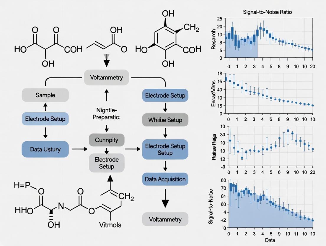
Visual Guide to the Optimization Workflow:
Figure 1: A workflow diagram for the systematic optimization of Square-Wave Voltammetry parameters to achieve maximum signal gain and SNR.
The Scientist's Toolkit: Essential Reagents & Materials
Table 2: Key Research Reagent Solutions for Voltammetric Experiments
| Item | Function / Rationale | Example / Note |
|---|---|---|
| Supporting Electrolyte | Minimizes uncompensated solution resistance (iR drop) by carrying the majority of the ionic current. This prevents distorted voltammograms and improves SNR [3]. | Inert salts at high concentration (e.g., 0.1-1.0 M KCl, LiTFSI, phosphate buffer) [7]. |
| Redox Reporter | A molecule that undergoes reversible electron transfer, providing the measurable faradaic signal. Its intrinsic electron transfer kinetics dictate optimal instrument parameters [5]. | Methylene Blue, Ferrocene, Anthraquinone. Choice affects optimal square-wave frequency [5]. |
| Electrode Polishing Kit | A clean, reproducible electrode surface is critical for a stable baseline and low noise. Polishing removes adsorbed contaminants [3]. | Alumina or diamond slurries (e.g., 0.05 μm alumina) on a microcloth pad [3]. |
| Validated Reference Electrode | Provides a stable, known potential for the working electrode. A blocked or faulty reference is a common source of noise and error [3]. | Ag/AgCl (3M KCl) or Saturated Calomel Electrode (SCE). Always check the frit is not clogged [3]. |
| High-Surface-Area Electrode Material | Increases the electroactive surface area (ESA), leading to higher signal for the same geometric area, thereby improving SNR and lowering the LOD [1]. | Porous carbon materials (e.g., activated carbon), nanostructured gold, or screen-printed electrodes [1] [7]. |
In voltammetry research, the signal-to-noise ratio (SNR) is a critical determinant of data quality, impacting the sensitivity, limit of detection, and overall reliability of analytical measurements. Electrochemical signals are susceptible to a variety of noise sources that can obscure faradaic currents, which are the primary signals of interest in analytical sensing. These noises can be fundamentally categorized as thermal (Johnson-Nyquist) noise, flicker (1/f) noise, and interference (environmental) noise. Effectively troubleshooting these issues is paramount for researchers and scientists developing robust electrochemical sensors for applications in drug development and clinical diagnostics. This guide provides a structured approach to identifying and mitigating these noise sources to improve SNR in voltammetric experiments.
Fundamental Noise Types: Characteristics and Origins
The table below summarizes the key characteristics of the three fundamental noise types encountered in electrochemical systems.
Table 1: Fundamental Noise Types in Electrochemical Cells
| Noise Type | Origin | Spectral Density | Dependence | Primary Mitigation Strategies |
|---|---|---|---|---|
| Thermal (Johnson) Noise | Thermal agitation of charge carriers in resistive components. | White noise (frequency-independent). | Proportional to √(R × T × Δf), where R=resistance, T=temperature, Δf=bandwidth. [8] | Lower cell impedance, use lower-temperature electrolytes, filter high frequencies. |
| Flicker (1/f) Noise | Surface phenomena, adsorption/desorption, and slow chemical processes at the electrode-electrolyte interface. | Inversely proportional to frequency (1/f). | Increases with decreasing frequency; dominant at low frequencies. [8] | Use higher-frequency techniques (e.g., Square-Wave Voltammetry), polish electrodes, apply coatings. |
| Interference Noise | External electromagnetic fields, ground loops, imperfect connections, and equipment. | Often appears at specific frequencies (e.g., 50/60 Hz power line). | Dependent on lab environment, cable routing, and grounding. [9] | Proper shielding and grounding, use short/shielded cables, Faraday cages. [9] |
Figure 1: A taxonomy of fundamental noise sources in electrochemical cells, showing their primary characteristics and origins.
Troubleshooting Guide: FAQs and Solutions
General Workflow for Noise Diagnosis
A systematic approach is essential for efficient noise troubleshooting. The following workflow, adapted from established electrochemical practices [3], helps isolate the root cause.
Figure 2: A logical workflow for diagnosing the source of noise in an electrochemical setup.
Frequently Asked Questions (FAQs)
FAQ 1: My voltammogram has a significant, non-flat baseline with large hysteresis between forward and backward scans. What is the cause, and how can I fix it? [3]
- Problem Identification: A sloping or hysteretic baseline is frequently caused by high charging currents at the working electrode. The electrode-solution interface acts as a capacitor, which must be charged before the faradaic process can occur. This effect is pronounced with high scan rates, large electrode surface areas, or high-resistance solutions.
- Solution:
- Reduce Scan Rate: Lowering the voltammetric scan rate reduces the rate of capacitor charging, thereby decreasing the charging current.
- Use a Smaller Electrode: Employ a working electrode with a smaller active surface area.
- Increase Analyte Concentration: A larger faradaic current relative to the charging current will improve the signal-to-noise ratio.
- Check Electrode Integrity: Ensure the working electrode is properly sealed, as faults like poor internal contacts or exposed glass can introduce additional capacitance. [3]
FAQ 2: I observe a constant, low-level, noisy signal with no faradaic peaks. What should I check first? [3] [9]
- Problem Identification: A small, noisy current with no discernible faradaic features often points to a poor connection at the working electrode. This prevents the faradaic current from flowing while the potentiostat can still apply a potential, resulting in only residual circuit noise being recorded.
- Solution:
- Inspect Connections: Verify that the cable to the working electrode is securely connected. Check for corroded or loose alligator clips. [3]
- Test the Electrode: Disconnect the working electrode and check its continuity with an ohmmeter if possible.
- Clean the Electrode: Polish the working electrode surface with alumina slurry (e.g., 0.05 μm) and rinse thoroughly to remove any adsorbed contaminants that might be blocking electron transfer. [3]
FAQ 3: My signal is very noisy, especially when using a rotating electrode system. The noise frequency seems related to the rotation speed. [9]
- Problem Identification: Noise correlated with rotation speed is typically of mechanical origin.
- Solution:
- Inspect Brush Contacts: Open the rotator housing and check the carbon brush contacts that touch the rotating shaft. The shaft surface should be smooth and free of corrosion. The brush contact surface should be a well-aligned, smooth groove. A misaligned groove can cause squeaking and vibrations, translating into electrical noise. [9]
- Polish or Replace Brushes: If the brush contact is misaligned or worn, polish its end with sandpaper on a flat surface to remove the old groove, or replace it entirely. Brush contacts should be replaced before the carbon portion is worn through. [9]
- Ensure Proper Grounding: Ground the rotator motor case by connecting the chassis ground of the rotator control unit to the chassis ground of the potentiostat. This can reduce electromagnetic interference from the motor itself. [9]
FAQ 4: I keep getting voltage or current compliance errors, and the signal is distorted. What does this mean? [3]
- Problem Identification: A "voltage compliance" error means the potentiostat cannot maintain the desired potential between the working and reference electrodes. A "current compliance" error indicates a short-circuit condition, where an excessively large current is flowing.
- Solution:
- For Voltage Compliance: Ensure your reference electrode is properly connected, submerged, and not clogged. A blocked frit creates high impedance, preventing the potentiostat from controlling the potential. [3] [9]
- For Current Compliance: Check that the working and counter electrodes are not touching inside the electrochemical cell, as this creates a short circuit. [3]
FAQ 5: How can I improve the selectivity of my sensor in complex biological fluids like plasma or blood? [10]
- Problem Identification: Biological samples are rich in macromolecules (e.g., proteins) that can adsorb to the electrode surface (fouling), altering its properties and reducing selectivity.
- Solution:
- Sensor Modification: Apply a molecularly imprinted polymer (MIP) as a thin film on your electrode. This polymer creates artificial receptors for your target analyte, imparting high selectivity and antifouling properties by blocking larger molecules from reaching the electrode surface. [10]
- Electrode Engineering: Use nanostructured materials like multi-walled carbon nanotubes (MWCNTs) decorated with ligand-free gold nanoparticles. This enhances the electrocatalytic activity and can provide a more stable platform. [10]
- Pulse Techniques: Utilize techniques like adsorptive stripping voltammetry or differential pulse voltammetry (DPV) to selectively pre-accumulate the analyte and measure it with higher sensitivity. [10]
The Scientist's Toolkit: Key Research Reagent Solutions
The following table lists essential materials and their functions as demonstrated in recent, advanced electrochemical sensor research.
Table 2: Key Research Reagent Solutions for Sensor Development and Noise Mitigation
| Material / Reagent | Function in Experimental Protocol | Application Example |
|---|---|---|
| Molecularly Imprinted Polymer (MIP) | A selective recognition layer that provides antifouling properties and enhances selectivity in complex matrices. [10] | Detection of serotonin in plasma. [10] |
| Multiwall Carbon Nanotubes (MWCNTs) | Nanostructured material to increase electroactive surface area and enhance electron transfer kinetics. [10] | Base transducer material for sensor construction. [10] |
| Gold Nanoparticles (Au NPs) | Electrocatalyst to lower the overpotential and increase the sensitivity of the redox reaction. [10] | Catalyzing the oxidation of serotonin. [10] |
| Metal Vapor Synthesis (MVS) | A method for producing ligand-free metal nanoparticles, allowing for a stable and controlled anchoring on supporting materials. [10] | Synthesis of pure Au NPs for anchoring on MWCNTs. [10] |
| Carbon Paste Electrode (CPE) | A versatile, renewable, and environmentally friendly working electrode material. [11] | Determination of thymoquinone in herbal products. [11] |
| Ag/AgCl Wire (Quasi-Reference) | A simple, frit-less reference electrode for troubleshooting high-impedance connections from clogged frits. [9] | Diagnosing and replacing a noisy commercial reference electrode. [9] |
| Alumina Polishing Slurry | (e.g., 0.05 μm) For mechanically refreshing and cleaning the working electrode surface to restore performance. [3] | Removing adsorbed contaminants to reduce flicker noise and improve reproducibility. [3] |
Experimental Protocol: A Case Study in Reliable Sensor Design
The following detailed methodology is adapted from a study on detecting serotonin in plasma, showcasing a holistic approach to achieving reliability and a high signal-to-noise ratio in a challenging biological matrix [10].
Aim: To develop a robust voltammetric sensor for serotonin in plasma with high selectivity and antifouling properties.
Methodology:
Electrode Modification:
- Synthesis of Au NPs: Produce ligand-free gold nanoparticles via Metal Vapor Synthesis (MVS). [10]
- Functionalization of MWCNTs: Anchor the Au NPs onto multiwall carbon nanotubes using a controlled radical functionalization technique to ensure a stable composite. [10]
- Electrode Coating: Deposit the MWCNT/Au NP composite onto the base electrode substrate to form the sensing platform.
- MIP Layer Formation: Apply a thin layer of molecularly imprinted polymer over the modified electrode. This layer is synthesized in the presence of serotonin molecules, which are later removed, leaving behind specific cavities for serotonin recognition. [10]
Optimization of Voltammetric Parameters:
- Employ a Design of Experiment (DoE) approach. Instead of testing one variable at a time, use a statistical model (e.g., a factorial design) to systematically vary key parameters of the Differential Pulse Voltammetry (DPV) method—such as pulse amplitude, step potential, and modulation time—and identify the optimal combination that maximizes the signal response (sensitivity). [10]
Measurement via Adsorptive Stripping Voltammetry:
- Accumulation Step: Apply a constant potential to the sensor while stirring the plasma sample. This selectively pre-concentrates serotonin molecules into the MIP cavities. This step enhances the faradaic signal while minimizing interference from non-accumulated species.
- Stripping Step: Record the voltammogram using the optimized DPV parameters. The oxidation current of the accumulated serotonin is measured, which is proportional to its concentration in the sample. [10]
Outcome: This protocol resulted in a sensor with a sensitivity of 6.7 μA μmol L−1 cm−2 and a limit of detection of 1.0 μmol L−1 in plasma, performance comparable to that achieved in simple buffer solutions, demonstrating excellent resilience to matrix effects and fouling. [10]
Frequently Asked Questions (FAQs)
1. How does the choice of electrode material directly affect my baseline signal? The electrode material fundamentally determines the electrochemical interface's physical and chemical properties. Materials like carbon (glassy carbon, graphite, carbon fiber) possess a rich surface chemistry with oxygen-containing functional groups. These groups influence the double-layer capacitance, a primary component of the non-Faradaic (charging) current that constitutes your baseline [12] [13]. Furthermore, the material's microstructure and conductivity affect the electron-transfer rate and contact resistance. A higher resistance leads to a larger voltage drop (iR drop), which can distort the voltammetric wave and elevate the apparent baseline noise [12]. For instance, carbon-fiber microelectrodes are prized in neurochemistry because their surface functional groups promote the adsorption of cationic neurotransmitters, enhancing the Faradaic signal relative to the capacitive background [14] [13].
2. Why does my baseline show a large hysteresis or slope, and how is electrode geometry involved? A sloping or hysteretic baseline is often dominated by charging currents. The electrode-solution interface behaves like a capacitor, and this capacitor must be charged and discharged as the potential is scanned [3] [15]. The magnitude of this charging current is directly proportional to the electrode surface area and the scan rate [3]. A larger electrode geometry (e.g., a macroelectrode vs. a microelectrode) has a greater surface area and thus a larger capacitance, resulting in a more pronounced sloping baseline [14] [15]. This effect can be mitigated by using a smaller electrode, decreasing the scan rate, or increasing the concentration of the analyte [3].
3. We are developing a chronic biosensor and our baseline signal drifts over time. What material-related factors could be causing this? Chronic baseline drift is frequently a symptom of electrode fouling (or biofouling). This occurs when proteins, lipids, or other biomolecules adsorb onto the electrode surface, altering its properties [13]. Fouling can change the double-layer capacitance and increase the electron-transfer resistance, leading to an unstable baseline and a loss of sensitivity [13]. Strategies to combat this include using specially modified electrodes with anti-fouling layers (e.g., blocking layers like mercaptohexanol on gold) [16] or applying waveforms that periodically "clean" the surface by driving it to extreme potentials to oxidize adsorbed contaminants [14] [13].
4. How does optimizing the distribution of conductive particles in a composite electrode improve my signal-to-noise ratio? In composite electrodes (e.g., graphite–epoxy), the goal is not simply to maximize the conductive particle loading. An optimal distribution creates an efficient percolation network for electron transport while minimizing random conductive pathways that contribute to noise [12]. Research has shown that the maximum conductive particle loading does not always correspond to the optimal loading in terms of the signal-to-noise ratio. An optimized, homogeneous distribution of graphite particles lowers the overall ohmic resistance and double-layer capacitance, which directly translates to a higher signal-to-noise ratio and a lower limit of detection [12].
Troubleshooting Common Baseline Issues
| Problem | Potential Root Cause | Diagnostic Steps | Corrective Actions |
|---|---|---|---|
| Noisy, unstable baseline [3] | Poor electrical connection; Loose cable; High solution resistance. | Check all connectors with an ohmmeter; Perform potentiostat diagnostic test with a resistor [3]. | Secure all connections; Ensure robust electrode construction; Increase electrolyte concentration. |
| Large hysteresis in baseline [3] | High charging currents from large electrode surface area; Faulty electrode with internal capacitance. | Reduce scan rate; Test with a smaller electrode; Compare baseline in analyte vs. blank solution. | Decrease scan rate; Use a microelectrode; Polish/clean the working electrode [3] [17]. |
| Baseline drift over time [16] [13] | Electrode fouling from adsorbed species; Unstable reference electrode; Gradual surface modification. | Perform a background scan in pure electrolyte; Check reference electrode integrity (e.g., clogged frit) [3]. | Implement an anti-fouling layer (e.g., MCH, Nafion); Use a waveform that cleans the surface; Replace reference electrode [16] [13]. |
| Unexpected peaks in baseline [3] | Impurities in electrolyte/solvent; Edge of solvent electrochemical window; Degradation of electrode/material. | Run a background measurement with a fresh, analyte-free electrolyte solution. | Re-purify solvents/electrolytes; Narrow the potential scan window; Re-polish or re-fabricate the electrode. |
Essential Experimental Protocols
Protocol 1: Fabrication of a Carbon-Fiber Microelectrode for Low-Baseline Noise
Objective: To construct a cylindrical carbon-fiber microelectrode (CFME) with a small geometric surface area to minimize capacitive charging currents, ideal for fast-scan cyclic voltammetry (FSCV) in vivo [14] [13].
Materials:
- Single carbon fiber (∼7 μm diameter)
- Borosilicate glass capillary (e.g., 1.2 mm OD, 0.68 mm ID)
- Vertical or horizontal electrode puller
- Epoxy resin (e.g., Epon 828)
- Syringe with isopropyl alcohol
- Sharp scalpel or scissors
Procedure:
- Aspiration: A single carbon fiber is aspirated into the borosilicate glass capillary.
- Pulling: The capillary is placed on a commercial electrode puller and heated and pulled to form a tapered glass seal around the carbon fiber.
- Sealing (Optional but Recommended): The tapered tip may be deliberately broken under a microscope and then resealed with epoxy resin. This step increases robustness and reduces the shunt capacitance between the fiber and the solution [14].
- Trimming: Using a sharp scalpel and under a microscope, the protruding carbon fiber is trimmed to the desired length (typically 50-200 μm). A longer fiber increases sensitivity but reduces spatial resolution.
- Curing and Connection: The epoxy is allowed to cure fully. An electrical connection is then made to the carbon fiber at the back of the capillary using a conductive material such as silver paint or a metal wire.
Protocol 2: Surface Polishing of a Glassy Carbon Electrode
Objective: To restore a smooth, reproducible surface on a glassy carbon (GC) electrode, ensuring consistent electron-transfer kinetics and a stable baseline.
Materials:
- Glassy carbon working electrode
- Polishing pads (multiple grits)
- Alumina or diamond polishing slurry (e.g., 1.0 μm, 0.3 μm, and 0.05 μm)
- Deionized water
- Ultrasonic bath
Procedure:
- Coarse Polish: On a clean polishing pad, apply a slurry of 1.0 μm alumina. Polish the electrode surface using a figure-eight pattern with moderate pressure. Rinse thoroughly with deionized water.
- Fine Polish: Move to a fresh pad and use a 0.3 μm alumina slurry. Repeat the figure-eight polishing. Rinse thoroughly.
- Mirror Finish: Finally, on a third clean pad, use a 0.05 μm alumina slurry to achieve a mirror finish. Rinse thoroughly.
- Sonication: Place the electrode in an ultrasonic bath filled with deionized water for 1-2 minutes to remove any embedded alumina particles.
- Electrochemical Activation (Optional): The polished electrode can be electrochemically activated by performing cyclic voltammetry in a suitable electrolyte (e.g., 0.5 M H₂SO₄) between the solvent limits to clean and activate the surface [17].
Table 1: Impact of Electrode Geometry on Key Signal Parameters
| Electrode Geometry | Typical Surface Area | Charging Current | Impact on Spatial Resolution | Typical Application |
|---|---|---|---|---|
| Macroelectrode (e.g., 2 mm GC disc) | ~0.03 cm² | High | Low (bulk measurement) | Standard quantitative analysis in stirred solutions [15]. |
| Microelectrode (e.g., 10 μm radius carbon fiber) | ~3 x 10⁻⁶ cm² | Low | High (discrete brain regions) | Fast-scan cyclic voltammetry (FSCV) in vivo; measurements in resistive media [14] [13]. |
| Ultramicroelectrode Array | Tunable (Low per element) | Medium (scales with active area) | Medium | Sensors combining low noise with higher total signal output [12]. |
Table 2: Electrode Material Properties and Their Electrochemical Consequences
| Electrode Material | Key Characteristics | Heterogeneous Electron-Transfer Rate (k⁰) | Double-Layer Capacitance | Fouling Resistance |
|---|---|---|---|---|
| Glassy Carbon (GC) | Hard, amorphous carbon; smooth surface. | Moderate to Fast | Moderate | Moderate [18]. |
| Carbon Nanotube (CNT) Modified | High aspect ratio, large effective surface area. | Not significantly altered from bare GC for some probes [18]. | High (due to large surface area) | Varies with functionalization [18]. |
| Carbon-Fiber | Rich in edge planes & oxygen groups; promotes cation adsorption. | Fast for catecholamines | Low to Moderate | Good, but can be improved with coatings like Nafion [14] [13]. |
| Boron-Doped Diamond (BDD) | Low background current, wide potential window. | Slow to Moderate | Very Low | Excellent [19]. |
Visualizing the Interaction Pathways
The Scientist's Toolkit: Research Reagent Solutions
| Reagent / Material | Primary Function in Electrode Preparation & Signal Optimization |
|---|---|
| Alumina Polishing Slurries (0.05, 0.3 μm) | To create a smooth, reproducible electrode surface, ensuring consistent kinetics and a stable baseline by removing contaminants and previous surface layers [17] [16]. |
| Epoxy Resin (e.g., Epon 828) | Used as an insulating matrix in composite electrodes or to seal carbon fibers in glass capillaries. Its optimization affects the distribution of conductive particles and the electrode's mechanical stability [12] [14]. |
| Functionalized Carbon Nanotubes (fCNTs) | Dispersed and drop-cast on electrodes to modify the surface. Increases effective surface area and can enhance signal (current), but requires careful dispersion to avoid agglomeration that increases noise [18]. |
| Cationic Surfactant (e.g., DDAB) | Used in dispersing nanomaterials like CNTs. In bulk solution, it can form organized layers on the electrode surface, improving the dispersion of analytes and modifying the interfacial properties, leading to better current responses [18]. |
| Mercaptohexanol (MCH) | A blocking agent used in biosensors (e.g., on gold surfaces). It forms a self-assembled monolayer that displaces non-specifically adsorbed molecules, reducing fouling and minimizing non-Faradaic background current [16]. |
Recent Advances in Nanoscale Electrochemical Imaging and Attoliter-Volume Detection
Troubleshooting Guides for Nanoscale Electrochemical Experiments
FAQ 1: My electrochemical measurements show inconsistent signals and high background noise. How can I improve the signal-to-noise ratio?
Issue: Inconsistent signals and high background noise in voltammetry often stem from suboptimal instrument settings, contamination, or inefficient data processing.
Troubleshooting Steps:
- Optimize Square-Wave Voltammetry Parameters: For techniques like E-DNA sensors, signal gain is highly dependent on the parameters of the square-wave potential pulse.
- Simultaneously adjust the frequency and amplitude of the square-wave pulse. The optimal pairing depends on the redox reporter used (e.g., methylene blue vs. ferrocene) and the probe's structure [5].
- For example, a sensor with a methylene blue reporter achieved a 315% signal gain at 25 mV amplitude and 750 Hz frequency, which was a 2-fold improvement over non-optimized parameters [5].
- Use the Kinetic Differential Measurement (KDM) method. This involves subtracting signals recorded at optimized "signal-on" and "signal-off" frequencies to correct for baseline drift and further enhance the signal-to-noise ratio [5].
- Apply Advanced Data Smoothing: Use algorithmic filters to denoise stored voltammetric data.
- Select the optimal smoothing filter (e.g., Savitzky-Golay, moving median, wavelet-based routines) based on the shape of your voltammetric curve and the type of noise present. The best filter for one data set may not be optimal for another [20].
- An evaluation formula that considers the improvement in analytical parameters (calibration linearity, detection limit, accuracy) after smoothing, rather than just the signal-to-noise ratio of a single curve, is recommended for selecting the best filter [20].
- Ensure Sample and Reagent Purity:
- Check for endotoxin contamination. Endotoxins can cause immunostimulatory reactions and mask the true biocompatibility and performance of nano-formulations. Work under sterile conditions using pyrogen-free water and reagents [21].
- Test for nanoparticle aggregation by measuring size distribution under biologically relevant conditions (e.g., in plasma), as the reported size can differ significantly from manufacturer specifications and can change in different dispersing media [21].
FAQ 2: My optical imaging of electrochemical interfaces has poor spatial resolution and fails to detect single entities at high concentrations. What can I do?
Issue: The detection volume is too large, preventing the isolation of individual molecules or nanoparticles.
Troubleshooting Steps:
- Implement Confocal Total-Internal-Reflection Fluorescence (TIRF) Microscopy: This technique can drastically reduce the detection volume.
- A confocal TIRF microscope can generate an detection volume of less than 5 attoliters (5 x 10⁻¹⁸ L) at a water-glass interface. This is almost two orders of magnitude smaller than conventional confocal microscopy, enabling the isolation of individual molecules at high analyte concentrations [22] [23].
- This system uses a parabolic mirror objective for diffraction-limited supercritical focusing and fluorescence collection, providing excellent spatial resolution and a high signal-to-background ratio for single-molecule detection [22].
FAQ 3: I am observing unexpected artifacts and blurring in my Transmission Electron Microscopy (TEM) images during in-situ electrochemical experiments.
Issue: Artifacts can arise from sample preparation, the electrochemical cell setup, or environmental factors, compromising image quality.
Troubleshooting Steps:
- Address Cryo-TEM Ice Contamination:
- Problem: Crystalline ice contaminants obscure nanoparticles.
- Solution: Ensure rapid freezing in liquid ethane to form vitreous ice. Use freshly dispensed liquid nitrogen, work in a dehumidified environment, and pre-cool all tools to prevent ice crystal formation [24].
- Minimize Sample Drift:
- Problem: Blurred images due to sample movement during acquisition.
- Solution: Ensure the grid is securely mounted. Check that the ice or grid substrate is not too thin and unstable. Investigate and mitigate environmental vibrations affecting the microscope [24].
- Mitigate Electron Beam Effects:
Quantitative Data on Signal Optimization
The following table summarizes the quantitative gains achievable by optimizing square-wave voltammetry parameters, as demonstrated for E-DNA sensors [5].
Table 1: Signal Gain Optimization through Square-Wave Voltammetry Parameters
| Redox Reporter | Optimal Square-Wave Amplitude | Optimal Square-Wave Frequency | Resulting Signal Gain | Type of Gain |
|---|---|---|---|---|
| Methylene Blue | 25 mV | 750 Hz | +315% | Signal-On |
| Methylene Blue | 25 mV | 20 Hz | -82% | Signal-Off |
| Anthraquinone | 10 mV | 100 Hz | +173% | Signal-On |
| Ferrocene | 25 mV | 7.5 kHz | -43% | Signal-Off |
The table below compares key optical and electron microscopy techniques for nanoscale electrochemical imaging, highlighting their roles in improving the signal-to-noise ratio and spatial resolution.
Table 2: Comparison of Advanced Microscopy Techniques for Nanoscale Imaging
| Microscopy Technique | Key Principle | Key Advantage for SNR/Resolution | Example Application in Energy Research |
|---|---|---|---|
| Confocal TIRF [22] [23] | Confocal detection with total internal reflection fluorescence. | Attoliter (5x10⁻¹⁸ L) detection volume for isolating single molecules at high concentration. | Probing single-molecule electrochemistry at interfaces. |
| In-situ TEM [25] | Miniaturized electrochemical cell inside TEM column. | Atomic-scale spatial resolution for real-time visualization. | Visualizing solid electrolyte interphase (SEI) formation and dendrite growth in batteries. |
| Super-Resolution Fluorescence [26] | Localization of single fluorophores beyond diffraction limit. | Nanoscale spatial resolution for mapping heterogeneous reactions. | Imaging electrocatalytic activity at individual nanoparticles. |
| Surface Plasmon Resonance (SPRM) [26] | Tracking changes in refractive index near a metal surface. | Excellent sensitivity for monitoring dynamic adsorption/desorption. | Real-time, label-free imaging of molecular adsorption at electrode interfaces. |
Experimental Protocols for Key Techniques
Protocol: Optimizing Square-Wave Voltammetry for Maximum Signal Gain
This protocol is adapted from research on maximizing the signal of Electrochemical DNA (E-DNA) sensors [5].
Principle: Signal gain in reagentless electrochemical sensors depends on binding-induced changes in electron transfer kinetics, which are sensitive to the parameters of the square-wave potential pulse.
Materials:
- Potentiostat capable of high-frequency square-wave voltammetry.
- Fabricated electrochemical sensor (e.g., an electrode modified with a redox-tagged probe).
- Solution of the target analyte.
Procedure:
- Initial Setup: Record a square-wave voltammogram of the sensor in a blank solution (without target) using a standard set of parameters (e.g., 25 mV amplitude, 100 Hz frequency).
- Generate 2D Parameter Map:
- Program the potentiostat to perform a series of square-wave scans over a wide range of amplitudes (e.g., 1 mV to 100 mV) and frequencies (e.g., 5 Hz to 5000 Hz). This will require 150+ individual voltammograms.
- Repeat this entire procedure after incubating the sensor with a saturated concentration of the target analyte.
- Data Analysis:
- For each amplitude/frequency pair, extract the peak current from both the blank and target voltammograms.
- Create a 2D numerical map where the signal gain (e.g.,
(I_target - I_blank) / I_blank) is plotted as a function of amplitude and frequency.
- Parameter Selection: Identify the amplitude/frequency pairing that yields the maximum positive (signal-on) or negative (signal-off) gain from the 2D map.
- Implementation: Use this optimized parameter set for all subsequent quantitative measurements with the sensor.
Protocol: Assembling a Probe-Type In-situ TEM Electrochemical Cell
This protocol describes the setup for observing battery materials using in-situ TEM [25].
Principle: A nanoscale electrochemical cell is assembled inside a TEM using a specialized holder, allowing real-time observation of processes like lithiation and dendrite growth.
Materials:
- Probe-type in-situ TEM holder.
- Metal rod probes (e.g., Tungsten).
- Active electrode material (e.g., Si nanowire, MoS₂ flake).
- Li metal (for anode).
- Ionic liquid electrolyte or solid electrolyte (Li₂O).
Procedure:
- Anode Preparation: Fix a small piece of Li metal to one probe. A native Li₂O layer will form on its surface, acting as a solid electrolyte.
- Cathode Preparation: Fix the nanoscale electrode material (e.g., a single nanowire) to a second, movable probe.
- Cell Assembly: Inside an argon-filled glovebox, introduce a small amount of ionic liquid electrolyte onto the electrode surfaces if an all-solid-state cell is not being used.
- Transfer to TEM: Secure the assembled probe into the TEM holder, ensuring it is properly sealed.
- In-situ Experiment: Insert the holder into the TEM. Use the probe controls to bring the electrode material into contact with the Li₂O/Li (for solid-state) or the electrolyte. Apply a bias voltage to initiate electrochemical reactions while simultaneously recording TEM images, spectra, or diffraction patterns.
Research Reagent Solutions
This table lists essential materials used in the advanced experiments cited in this guide.
Table 3: Essential Research Reagents and Materials
| Reagent / Material | Function in Experiment | Key Consideration |
|---|---|---|
| Methylene Blue [5] | Redox reporter in E-DNA and similar biosensors. | Electron transfer kinetics dictate optimal square-wave frequency/amplitude. |
| Ionic Liquid Electrolyte [25] | Electrolyte for in-situ TEM batteries. | Enables electrochemical reactions in the high vacuum of the TEM. |
| Uranyl Formate / Acetate [24] | Negative stain for TEM sample preparation. | Provides high contrast; can form stain crystals that obscure particles if not fresh. |
| Holey Carbon TEM Grid [24] | Support film for cryo-TEM samples. | Particles suspended in holes provide best contrast; some samples prefer continuous carbon. |
| Parabolic Mirror Objective [22] [23] | Optical element in confocal TIRF. | Enables diffraction-limited focusing to achieve attoliter detection volumes. |
Signaling Pathway and Workflow Diagrams
Troubleshooting Pathway for SNR Improvement
Strategic Approach to Advanced Imaging
Critical Relationships Between SNR, Detection Limits, and Analytical Sensitivity in Biomedical Applications
In analytical chemistry, particularly in biomedical and voltammetric research, the Signal-to-Noise Ratio (SNR) is a foundational concept that directly determines the efficacy and reliability of an experiment. It serves as the master guide for assessing data quality [27]. The primary task in trace analysis is often the detection of minute quantities of substances, such as pollutants, contaminants, or degradation products. If the detected signal of a substance is not sufficiently distinguishable from the unavoidable baseline noise of the analytical method, the substance may go undetected altogether [27]. This relationship is formalized through the Limit of Detection (LOD) and Limit of Quantification (LOQ), which are critical method validation parameters. This technical support center outlines the fundamental relationships between SNR, LOD, and LOQ, provides troubleshooting guides for common experimental issues, and offers detailed protocols for optimizing voltammetric measurements to enhance analytical sensitivity.
Fundamental Definitions and Relationships
What are SNR, LOD, and LOQ?
- Signal-to-Noise Ratio (SNR): A measure comparing the level of a desired signal to the level of background noise. In its simplest form for a Boolean signal, it can be calculated as the difference between the mean "true" value and the mean "false" value, divided by the noise amplitude [28]. It is the key parameter for explaining most detector-related correlations [27].
- Limit of Detection (LOD): The minimum sample concentration at which a substance signal can be reliably detected. An SNR between 3:1 and 10:1 is often used as a rule of thumb for LOD in real-life analytical conditions, though a ratio of 3:1 is a common standard [27].
- Limit of Quantification (LOQ): The minimum sample concentration at which a substance signal can be reliably quantified. A typical signal-to-noise ratio for LOQ is 10:1, though in practice, values from 10:1 to 20:1 are often required for challenging conditions [27].
Quantitative Framework for SNR, LOD, and LOQ
The following table summarizes the formal definitions and quantitative relationships between these key parameters.
Table 1: Key Definitions and Quantitative Relationships for SNR, LOD, and LOQ
| Parameter | Formal Definition | Calculation & Relationship | ||
|---|---|---|---|---|
| Signal-to-Noise Ratio (SNR) | A measure of how distinguishable a signal is from background noise. | For a general Boolean signal: $SNR{dB} = 20 \log{10}\frac{ | \mu{true} - \mu{false} | }{2\sigma}$ [28].In HPLC/voltammetry: $SNR = \frac{\text{Signal Height}}{\text{Baseline Noise Height}}$ [27]. |
| Limit of Detection (LOD) | The minimum concentration that can be detected, but not necessarily quantified, under stated experimental conditions. | Typically defined as a concentration yielding an SNR of 3:1 [27]. | ||
| Limit of Quantification (LOQ) | The minimum concentration that can be quantitatively measured with stated accuracy and precision. | Typically defined as a concentration yielding an SNR of 10:1 [27]. |
The Scientist's Toolkit: Essential Materials and Reagents
The selection of electrodes, electrolytes, and other components is critical for obtaining a high SNR and low detection limits.
Table 2: Key Research Reagent Solutions for Voltammetry
| Item | Function & Importance | Best Practice Considerations |
|---|---|---|
| Working Electrode | The surface where the electrochemical reaction of interest occurs. Its material and area directly influence current response and sensitivity [29]. | Carbon-based electrodes (e.g., carbon-fiber microelectrodes) are popular for neurochemical sensing due to high biocompatibility and spatiotemporal resolution [30]. The surface area must be well-defined for accurate current density normalization (mA/cm²) [29]. |
| Reference Electrode | Provides a stable, well-defined reference potential against which the working electrode's potential is controlled [29]. | Select for chemical compatibility with the measurement environment. Avoid chloride-containing fillers if chloride poisons the catalyst. Its placement via a Luggin-Haber capillary is critical to minimize uncompensated resistance [31]. |
| Counter Electrode | Completes the electrical circuit by carrying the current so that no current flows through the reference electrode [29]. | The material must be chosen to avoid dissolution, which can contaminate the solution and artificially enhance performance (e.g., Pt counters for "Pt-free" catalysts) [31]. |
| Supporting Electrolyte | Carries current in the solution and controls the ionic strength. Minimizes the solution resistance (Ru). | Purity is paramount. Impurities at the part-per-billion level can substantially alter the electrode surface and reaction kinetics. Use the highest purity grade available and robust cell cleaning protocols [31]. |
| Solvent | Dissolves the analyte and electrolyte. | Must be degassed to remove dissolved oxygen if it interferes with the redox reaction of interest. |
Troubleshooting Guide: FAQs on SNR and Detection Limits
Q1: My voltammogram has a high background current and a sloping baseline. What could be the cause? A: A non-ideal baseline is often related to problems with the working electrode or high capacitive charging currents [3]. This can be caused by:
- Capacitive Charging: The electrode-solution interface acts as a capacitor. This effect can be reduced by decreasing the scan rate, increasing analyte concentration, or using a working electrode with a smaller surface area [3].
- Electrode Fouling: Species adsorbing to the electrode surface can change its capacitive properties. Polish the working electrode with alumina slurry or use an electrochemical cleaning procedure (e.g., potential cycling in clean sulfuric acid for Pt electrodes) [3].
- Electrode Defects: Poor internal contacts or seals in the electrode can lead to high resistivity or capacitance [3].
Q2: Why is my measured current very small and noisy, even with analyte present? A: This typically indicates a problem with the electrical connection to the working electrode, meaning the electrochemical cell is effectively an open circuit. Since the counter electrode is likely properly connected (otherwise a voltage compliance error would occur), you should check the connection to the working electrode [3].
Q3: I am getting voltage or current compliance errors. What should I check? A: These errors occur when the potentiostat cannot maintain the desired cell conditions.
- Voltage Compliance Error: The potentiostat cannot achieve the potential difference between working and reference electrodes. Check that your reference electrode is properly connected and not touching the working electrode. Also, ensure the counter electrode is submerged and connected [3].
- Current Compliance Error: This is often caused by a short circuit, where the working and counter electrodes are touching, generating a large current [3].
Q4: How does data smoothing affect my SNR, LOD, and LOQ? A: Data smoothing (e.g., using a time constant, Savitzky-Golay, or Fourier transform filters) can artificially improve the SNR by reducing baseline noise [27] [20]. However, over-smoothing is a critical risk. It can flatten and broaden small analyte peaks to the point where they merge with the baseline, effectively raising your practical LOD and LOQ by causing you to miss low-concentration analytes [27]. It is always best practice to collect high-quality raw data and apply gentle, post-acquisition smoothing if necessary, so the original data is preserved [27].
Q5: My cyclic voltammogram looks unusual or changes shape with repeated cycles. What is wrong? A: This is frequently due to an issue with the reference electrode. If it is not in proper electrical contact with the solution (e.g., due to a blocked frit or an air bubble), it can act like a capacitor, causing drifting potentials and unstable voltammograms [3]. Check the reference electrode's connection and frit.
Experimental Protocols for SNR Optimization
Protocol: Utilizing Pulse Voltammetry to Enhance SNR
Pulse voltammetric techniques (e.g., Normal Pulse, Differential Pulse, Square Wave) are designed specifically to discriminate against charging current, thereby improving SNR and lowering detection limits [30] [2].
Principle: After a potential step, the charging current decays exponentially, while the faradaic current decays more slowly, as a function of 1/(time)½ [30] [2]. By applying short potential pulses and measuring the current at the end of each pulse (after the charging current has largely decayed), the measured current is primarily faradaic [2].
Procedure:
- Select the Technique:
- Differential Pulse Voltammetry (DPV): Applies fixed-amplitude pulses on a slowly changing base potential. The current difference before and after the pulse is plotted, effectively subtracting the background charging current. Excellent for trace detection of irreversible systems [30].
- Square Wave Voltammetry (SWV): Applies a symmetrical square wave on a staircase ramp. The net current (forward - reverse) is plotted. It is very fast and provides a high SNR, ideal for reversible and quasi-reversible systems [30].
- Parameter Optimization:
- Pulse Width/Step Width: This defines the duration of the potential pulse. It must be long enough for the charging current to decay (typically > 5RuCdl) [2].
- Sample Period: Set the instrument to measure the current at the end of the pulse width. Many potentiostats allow averaging over a short period (e.g., 1 ms) to further reduce noise [2].
- Pulse Amplitude (DPV) or Square Wave Amplitude (SWV): This parameter affects the sensitivity and peak shape. Optimize for your specific system according to the instrument manual.
The workflow for this optimization process is outlined below.
Protocol: Anodic Stripping Voltammetry (ASV) for Trace Metal Detection
ASV is a powerful two-step technique that combines an electrochemical preconcentration step with a stripping step, drastically improving SNR for trace metal analysis [30].
Principle: Metal ions are first electrochemically reduced and concentrated into a mercury or carbon electrode surface by applying a negative potential for a fixed time. This preconcentration step amplifies the signal. The potential is then scanned in a positive direction (e.g., using Linear Sweep Voltammetry), oxidizing (stripping) each metal from the surface. Each metal produces a sharp peak current at its characteristic potential, which is proportional to its concentration in the original sample [30].
Procedure:
- Preconcentration Step:
- Set the initial potential to a value sufficiently negative to reduce the target metal ion(s).
- Hold this potential for a controlled deposition time (e.g., 30-300 seconds) while stirring the solution. The longer the deposition time, the greater the signal amplification.
- Equilibration Period:
- Stop stirring and allow the solution to become quiescent for a short period (e.g., 15 seconds).
- Stripping Step:
- Scan the potential positively using a technique like LSV or DPV.
- The oxidation of each metal produces a characteristic peak. The peak current is used for quantification.
Logical Pathway from Measurement to Quantification
The relationship between experimental parameters, data quality indicators, and final analytical figures of merit is a logical sequence. Understanding this pathway is key to method optimization. The following diagram illustrates the complete workflow from experimental setup to final result interpretation.
Electrode Engineering and Advanced Voltammetric Techniques for Enhanced SNR
FAQs: Troubleshooting Electrode Performance
Q1: Why is my voltammogram noisy or showing an unstable baseline?
A noisy or unstable baseline can be caused by several factors related to your experimental setup [3] [32].
- High Impedance Connections: Check that all cables and connectors to the electrodes are intact and making good contact. Visually inspect for any corrosion, cracks, or damaged plugs [32].
- Blocked Reference Electrode: A blocked frit in your reference electrode or air bubbles trapped in the Haber-Luggin capillary can cause significant noise and instability. Gently tap the cell or use a pipette ball to remove air bubbles [3] [32].
- Potentiostat Settings: The current range setting on your potentiostat may be inappropriate for your experiment's impedance. Adjusting the current range can help stabilize the signal [32].
- Working Electrode Issues: A poorly prepared or fouled working electrode can also lead to a non-straight baseline. Repolishing the electrode can often resolve this [3].
Q2: My experiment is triggering a "voltage compliance" error. What does this mean?
A voltage compliance error indicates that the potentiostat is unable to maintain the desired potential between the working and reference electrodes [3]. Common causes include:
- The counter electrode has been removed from the solution or is not connected properly [3].
- The reference electrode is not in electrical contact with the solution, for instance, due to a blocked frit or an air bubble [3].
- The counter electrode's activity is insufficient to support the reaction at the working electrode, or the distance between the working and counter electrodes is too large, leading to a high voltage loss in the electrolyte [32].
Q3: How can I improve the selectivity of my carbon-fiber electrode for dopamine against other monoamines like serotonin?
Poor selectivity between neurotransmitters with similar oxidation potentials is a common challenge. A proven strategy is to functionalize the electrode with a multi-layer membrane [33].
- Ion-Exchange Membrane: Coating the electrode with Nafion, an ion-exchange membrane, can repel negatively charged interferents and enrich cationic analytes like dopamine.
- Enzyme Layer: Incorporating a layer of the enzyme monoamine oxidase B (MAO-B) selectively breaks down interferents like serotonin and norepinephrine, while dopamine remains relatively unaffected. This bilayer approach has been shown to successfully discriminate dopamine in vitro and in vivo [33].
Q4: After modifying my electrode, the signal is very small or non-existent. What could be wrong?
If you measure only a very small, noisy, and unchanging current, the most likely cause is a poor connection to the working electrode [3]. Although the potentiostat can control the potential, no faradaic current can flow if the working electrode is not properly connected. Check the cable and connector for the working electrode. If the counter electrode were disconnected, it would typically trigger a voltage compliance error instead [3].
Q5: What is the "coffee-ring" effect in drop coating and how can I avoid it?
The "coffee-ring" effect occurs when a droplet of modifier suspension dries on an electrode surface, causing suspended particles to concentrate at the edge and form a ring-shaped, inhomogeneous coating [34]. This leads to inconsistent catalytic performance. To address this:
- Use Electrowetting: Applying an electric field can change the wetting properties of the surface, enhancing particle mobility and leading to a more uniform distribution during drying [34].
- Employ Hydrophobic Surfaces: Using highly hydrophobic electrode surfaces minimizes adhesion and helps prevent particle agglomeration, resulting in a more even coating [34].
Troubleshooting Guide: Common Electrochemical Issues
This guide summarizes frequent problems, their causes, and solutions to help you quickly diagnose your experiment.
Table 1: Troubleshooting Common Electrochemical Sensor Issues
| Observed Problem | Potential Causes | Recommended Solutions |
|---|---|---|
| Noisy or Unstable Baseline [3] [32] | High impedance connections; Blocked reference electrode frit; Air bubbles; Incorrect potentiostat settings. | Check all cables and connectors; Clear reference electrode blockage; Remove air bubbles; Adjust potentiostat current range. |
| Voltage Compliance Error [3] | Counter electrode disconnected or out of solution; Reference electrode not in contact; High solution resistance. | Ensure counter electrode is submerged and connected; Check reference electrode frit; Use a Haber-Luggin capillary to minimize IR drop [32]. |
| Unexpected Peaks in Voltammogram [3] | Impurities in solvent/electrolyte; Electrode surface contamination; Approaching the edge of the solvent's potential window. | Run a background scan without analyte; Repolish the working electrode; Use high-purity reagents. |
| Non-Flat or Hysteretic Baseline [3] | High charging currents (capacitive effects); Faults in the working electrode structure. | Decrease the scan rate; Use a smaller working electrode; Ensure the working electrode is properly sealed and polished. |
| Small or No Signal [3] | Poor connection to the working electrode; Working electrode not properly immersed. | Check the working electrode cable and connector; Ensure the electrode is submerged to the minimum immersion depth [35]. |
Experimental Protocols for Electrode Modification and Testing
Protocol 1: Creating a Dopamine-Selective Biosensor with MAO-B/Nafion Coating
This protocol details the fabrication of a biosensor for selective dopamine (DA) detection in the presence of serotonin (5-HT) and norepinephrine (NE), significantly improving the signal-to-noise ratio for DA [33].
Materials:
- Carbon-fiber microelectrode (e.g., 7-20 µm diameter fibers)
- Monoamine Oxidase B (MAO-B) enzyme
- Cellulose powder
- Nafion solution
- Glutaraldehyde solution (25%)
- Phosphate-buffered saline (PBS), pH 7.4
Method:
- Electrode Preparation: Aspirate two carbon fibers into a glass capillary, then taper and seal it with epoxy resin, leaving ~100 µm of fiber exposed [33].
- Enzyme Layer Coating: Soak the exposed carbon fiber tip in a solution of 20% MAO-B and 5% cellulose. Air dry for 30-60 minutes at room temperature [33].
- Cross-Linking: Expose the MAO-B/cellulose-coated tip to glutaraldehyde vapor (from a boiling 25% solution) for 30 minutes to cross-link and stabilize the enzyme layer [33].
- Ion-Exchange Layer: Apply a final coating of Nafion over the cross-linked layer and allow it to dry. This creates a dual-layer membrane: an inner MAO-B/cellulose layer for enzymatic selectivity and an outer Nafion layer for ion-exchange [33].
- Validation: Test the biosensor in PBS by adding 1 µM each of DA, 5-HT, and NE. The modified electrode should show a significantly higher response to DA compared to 5-HT and NE [33].
Protocol 2: General Procedure for Troubleshooting Cyclic Voltammetry
This systematic procedure helps isolate the source of a problem when you obtain an unusual or distorted cyclic voltammogram [3].
Materials:
- Potentiostat
- Test resistor (e.g., 10 kΩ) or manufacturer's test cell chip
- Electrochemical cell with analyte, electrolyte, and solvent
- Alternative reference electrode (e.g., bare silver wire quasi-reference electrode)
- Alumina slurry (0.05 µm) for electrode polishing
Method:
- Test Potentiostat and Cables: Disconnect the cell. Connect the reference and counter cables to one end of a 10 kΩ resistor and the working cable to the other. Run a scan (e.g., +0.5 V to -0.5 V). The result should be a straight line obeying Ohm's law (V=IR). If not, the issue is with the potentiostat or cables [3].
- Test Reference Electrode: Set up the cell normally, but connect the reference cable to the counter electrode. Run a linear sweep. If a standard-looking voltammogram (though potential-shifted) appears, the original reference electrode is faulty. Check for blockages or replace it with a quasi-reference electrode (a bare silver wire) to confirm [3].
- Test Working Electrode: If steps 1 and 2 pass, the issue likely lies with the working electrode. Repolish it with 0.05 µm alumina slurry and rinse thoroughly. For Pt electrodes, further cleaning can be done by cycling in 1 M H₂SO₄ between the potentials for H₂ and O₂ evolution [3].
Research Reagent Solutions
This table lists key materials used in advanced electrode modification for enhancing sensor performance.
Table 2: Essential Materials for Electrode Modification and Their Functions
| Material / Reagent | Function in Electrode Modification |
|---|---|
| Nafion [33] | Ion-exchange membrane coating; improves selectivity by repelling anions and enriching cations like dopamine. |
| Monoamine Oxidase B (MAO-B) [33] | Enzyme layer; selectively metabolizes interfering neurotransmitters (e.g., serotonin) to enhance target analyte specificity. |
| Carbon Nanotubes (CNTs) [36] [34] | Nanostructured material; provides high surface area and excellent conductivity, enhancing sensitivity and electron transfer. |
| Polyaniline (PANI) & Polypyrrole (PPy) [37] [36] [34] | Conducting polymers; used to modify electrode surfaces, improving electrochemical properties, stability, and biocompatibility. |
| Metal-Organic Frameworks (MOFs) [36] | Porous materials; offer high surface area and tunable pores for selective analyte adsorption and sensing. |
| Gold (Au) & Thiolated Probes [38] | Electrode material/immobilization chemistry; enables covalent binding of biomolecules (e.g., DNA, antibodies) via gold-thiol bonds for stable biosensors. |
| Glutaraldehyde [33] [34] | Bifunctional cross-linker; used to stabilize enzyme layers and other modifiers on the electrode surface. |
| Alumina Slurry (0.05 µm) [3] | Polishing agent; for refreshing and cleaning electrode surfaces to ensure reproducible results. |
Experimental and Troubleshooting Workflows
Diagram 1: Sensor Fabrication and Validation
Diagram 2: Systematic Voltammetry Troubleshooting
Frequently Asked Questions (FAQs)
Q1: What are the main advantages of using a cone-shaped carbon fiber microelectrode over a standard cylindrical one? The cone-shaped geometry provides a superior balance of mechanical robustness, enhanced sensitivity, and improved biocompatibility. The tapered design reduces insertion force and minimizes tissue displacement during implantation into the brain. This leads to significantly less acute tissue damage and a reduced glial cell response (a marker of inflammation), which in turn results in higher quality and more stable neurotransmitter signals in vivo [39] [40] [41].
Q2: My in vivo dopamine signals are lower than expected with a new 30 µm diameter electrode, even though it performed well in vitro. What could be the cause? This is a common issue when using larger-diameter electrodes. The reduced signal is likely due to increased tissue damage upon insertion. A larger, blunt electrode tip displaces more neural tissue, triggering a more severe local inflammatory response and potentially disrupting the very neurochemical environment you are trying to measure [39] [40]. Switching from a bare 30 µm fiber to a cone-shaped 30 µm design has been shown to resolve this, improving in vivo dopamine signals by 3.7-fold while reducing glial activation [39] [40].
Q3: How can I improve the longevity and durability of my carbon fiber microelectrodes (CFMEs) for chronic experiments? Increasing the fiber diameter is one effective strategy. Studies show that 30 µm cone-shaped CFMEs can have a 4.7-fold longer lifespan than conventional 7 µm CFMEs in erosion tests simulating chronic use [39] [40]. The cone shape maintains this mechanical advantage while solving the tissue damage problem associated with larger diameters. This design is therefore highly recommended for long-term monitoring applications [39] [40] [41].
Q4: My electrode seems to be fouling, leading to a loss of sensitivity over time. What are my options? Beyond physical design, surface modification can prevent fouling. Modifying the electrode surface with carbon nanoparticles or specific nanomaterials like carbon nanospikes (CNSs) can enhance electron transfer kinetics and act as a physical barrier to prevent fouling from polymerized neurochemicals [42]. Using porous carbon structures that trap analytes can also enhance selectivity and mitigate fouling issues [42].
Troubleshooting Guide
| Problem | Potential Cause | Recommended Solution |
|---|---|---|
| Low Sensitivity In Vivo | Excessive tissue damage from electrode insertion Severe inflammatory response (glial activation) | Switch to a cone-shaped tip geometry [39] [40] Verify reduced glial activity via Iba1/GFAP markers [39] |
| Poor Signal-to-Noise Ratio | High electrode electrical noise Signal loss from tissue damage | Ensure proper electrode conditioning [39] Use a 30 µm cone-shaped CFME for higher inherent sensitivity and lower tissue impact [39] [43] [40] |
| Short Electrode Lifespan | Mechanical degradation/breaking Electrochemical over-oxidation of carbon fiber | Use a larger diameter (e.g., 30 µm) cone-shaped CFME for superior durability [39] [40] Avoid overly aggressive waveforms that accelerate over-oxidation [39] |
| Unstable Readings/Drift | Biofouling on electrode surface Loose electrical connections | Apply anti-fouling surface modifications (e.g., carbon nanoparticle coatings) [42] Check all physical connections and impedance [44] |
Experimental Data & Performance Comparison
The following table summarizes key performance metrics for different carbon fiber microelectrode designs, highlighting the advantages of the cone-shaped geometry.
Table 1: Quantitative Performance Comparison of Carbon Fiber Microelectrodes
| Electrode Type | Diameter (µm) | In Vitro Sensitivity (pA/µm²) | In Vivo Dopamine Signal (nA) | Relative Lifespan | Key Findings |
|---|---|---|---|---|---|
| Standard CFME [39] [43] [40] | 7 | 12.2 ± 4.9 | 24.6 ± 8.5 | 1.0 x (Baseline) | Standard for in vivo work; minimal tissue damage. |
| Bare CFME [39] [43] [40] | 30 | 33.3 ± 5.9 | 12.9 ± 8.1 | ~4.7 x (vs. 7µm) | High sensitivity in vitro, but causes tissue damage, reducing in vivo signal. |
| Cone-Shaped CFME [39] [40] [41] | 30 (tip) | Information Missing | 47.5 ± 19.8 | ~4.7 x (vs. 7µm) | Superior design: Combines high sensitivity, excellent in vivo signal, and long lifespan. |
Detailed Experimental Protocols
Protocol 1: Fabrication of 30 µm Cone-Shaped Carbon Fiber Microelectrodes
This protocol is adapted from the method used in recent studies [39] [40].
- Preparation: Obtain a 30 µm diameter carbon fiber. Assemble a homemade electrochemical etching system consisting of a DC power supply and a linear actuator.
- Setup: Submerge a 1 mm segment of the carbon fiber in Tris buffer (pH 7.4).
- Etching: Apply a direct current voltage of 10 V to the submerged fiber for 20 seconds. Simultaneously, use the linear actuator to move the electrode upward at a constant speed. This gradual exposure to air during electrolysis is what forms the cone shape.
- Finalization: Control the final cone height to between 100 and 120 µm by precisely adjusting the speed of the actuator. After etching, the electrode is sealed in glass or an insulator, leaving the custom-shaped tip exposed.
Protocol 2: Assessing Biocompatibility via Immunofluorescence
To validate that the cone-shaped design reduces tissue damage, follow this post-implantation analysis [39] [40].
- Implantation: Implant the fabricated microelectrodes into the target brain region of an animal model using standard surgical procedures.
- Tissue Extraction: After a set period, perfuse the animal and extract the brain. Section the brain tissue containing the electrode implantation track.
- Staining: Immunostain the tissue sections using primary antibodies for classic glial cell markers:
- Iba1 to identify activated microglia.
- GFAP to identify activated astrocytes.
- Imaging and Analysis: Image the stained tissue sections using fluorescence microscopy. Quantify the intensity and distribution of Iba1 and GFAP staining around the implantation site. Compared to standard or bare larger electrodes, the cone-shaped design should show significantly lower signal for these markers, indicating a reduced inflammatory response and better biocompatibility [39] [40].
Diagram: Fabrication and Performance Relationship
The Scientist's Toolkit: Essential Research Reagents & Materials
Table 2: Key Reagents and Materials for Fabrication and Testing
| Item | Function/Description | Source Example |
|---|---|---|
| 30 µm Carbon Fiber | The core material for the microelectrode. Larger diameter improves mechanical strength. | World Precision Instruments (WPI) [39] [40] |
| Tris Buffer | An electrochemical stable buffer used during the electrochemical etching process and for in vitro testing. | Sigma-Aldrich [39] [40] |
| DC Power Supply | Provides the precise voltage (10V DC) required for the controlled electrochemical etching. | Standard lab equipment |
| Linear Actuator | Provides the precise vertical movement during etching to form the cone shape. | Homemade or commercial system [39] [40] |
| Iba1 & GFAP Antibodies | Primary antibodies for immunofluorescence staining to identify activated microglia and astrocytes, quantifying tissue response. | Various biological suppliers |
| Artificial Cerebrospinal Fluid (aCSF) | A solution that mimics the ionic composition of brain fluid, used for in vitro testing that more closely mimics in vivo conditions. | Can be prepared in-lab from salts [45] |
FAQs and Troubleshooting Guides
Frequently Asked Questions
Q1: What is the primary advantage of Square-Wave Voltammetry (SWV) over techniques like Cyclic Voltammetry (CV)?
SWV offers superior sensitivity and background current suppression compared to CV. Its pulse sequence measures current in both forward and reverse pulses, and the resulting difference current effectively minimizes non-faradaic (charging) background current. This makes it particularly powerful for detecting low concentrations of analytes [46].
Q2: How do I know if my SWV parameters are optimized?
A well-optimized SWV experiment will yield a sharp, symmetrical peak with a high signal-to-noise ratio for your target analyte. If the peak is broad, asymmetric, or the baseline is noisy, parameter optimization is likely required. For quantitative work, optimization using a systematic method like Response Surface Methodology (RSM) is recommended to find the parameter combination that produces the highest peak current [47] [48].
Q3: Can SWV be used for quantitative analysis?
While SWV is often praised for its diagnostic capabilities, it can absolutely be used for quantitative analysis. The peak current is directly proportional to the concentration of the electroactive species, allowing for the construction of calibration curves. The high sensitivity of SWV makes it suitable for detecting very low concentrations, with reported limits of detection (LOD) in the nanomolar range [46] [47] [48].
Q4: My SWV peaks are very broad. Which parameter should I adjust first?
A broad peak often suggests a slow electron transfer process or suboptimal waveform parameters. You should first investigate increasing the square wave amplitude. A higher amplitude can lead to sharper peaks and increased peak current, as it provides a greater driving force for the electrochemical reaction [46] [48].
Troubleshooting Common Issues
| Issue | Possible Cause | Solution |
|---|---|---|
| Low Peak Current | Suboptimal amplitude, frequency, or increment. | Systematically optimize parameters using RSM. Generally, increase amplitude and frequency within instrumental limits [47] [48]. |
| High Background Noise | Sampling width too long, improper electrode conditioning, or electrical interference. | Shorten the sampling width, ensure proper electrode cleaning/pretreatment, and use shielded cables in a grounded Faraday cage [46]. |
| Irreproducible Peaks | Electrode fouling, unstable electrical contact, or drifting chemical system. | Clean or polish the electrode surface, check all connections, and ensure chemical stability of the analyte solution [46]. |
| Non-Symmetrical Peaks | Quasi-reversible or irreversible electrode kinetics. | Confirm the reversibility of your system. Optimizing parameters like frequency can help; lower frequencies may improve shape for slower kinetics [46]. |
SWV Parameters and Optimization
Core Parameters for Optimization
The performance of SWV is highly dependent on three key parameters, which define the applied waveform and must be optimized for each specific application to maximize current response [46] [47] [48].
- Square Wave Amplitude (Esw): The height of each individual pulse (in mV). This parameter dictates the driving force for the electrochemical reaction. Increasing the amplitude typically enhances the peak current and sharpens the peak, but excessive values can lead to distortion.
- Square Wave Frequency (f): The number of complete forward-reverse cycles per second (in Hz). Frequency is inversely related to the square wave period (
f = 1/P). Higher frequencies speed up analysis but can reduce peak current for electrochemically irreversible systems. - Step Potential (Estep): Also known as the pulse increment (in mV). This is the change in the baseline potential with each subsequent pulse. It controls the potential resolution of the scan; a smaller step potential results in a finer scan.
Optimized Parameter Sets
The table below summarizes parameter values for different analytical scenarios, illustrating how they can be tuned for specific goals.
Table 1: Square-Wave Voltammetry Parameter Sets for Different Scenarios
| Analysis Goal | Amplitude (mV) | Frequency (Hz) | Step Potential (mV) | Key Outcome |
|---|---|---|---|---|
| General Quantitative Analysis [48] | 50 - 100 | 15 - 25 | 10 - 20 | Strong, well-defined peaks for calibration. |
| High-Sensitivity Detection [47] [48] | 90 - 125 | 10 - 15 | 8 - 12 | Maximized peak current for low LOD. |
| Rapid Screening | 25 - 50 | 50 - 100 | 15 - 25 | Faster analysis with moderate resolution. |
| Irreversible Systems | 25 - 50 | 5 - 15 | 10 - 15 | Improved peak shape for slow kinetics. |
Case Study: Optimization for Sunset Yellow Detection
A study on detecting the food dye Sunset Yellow provides a excellent example of systematic parameter optimization using Response Surface Methodology (RSM) [47] [48]. The researchers used a Box-Behnken design to find the parameters that yielded the highest anodic peak current.
Table 2: Optimized SWV Parameters from a Sunset Yellow Detection Study [47] [48]
| Parameter | Symbol | Tested Range | Optimal Value |
|---|---|---|---|
| Amplitude | Esw | 25 - 125 mV | 90.9 mV |
| Frequency | f | 15 - 75 Hz | 24.7 Hz |
| Step Potential | Estep | 5 - 15 mV | 9.1 mV |
The combination of these optimized parameters resulted in a very low limit of detection (LOD) of 1.15 nM, demonstrating the power of a structured optimization approach [47] [48].
Experimental Protocols
Workflow for SWV Parameter Optimization
The following diagram outlines a systematic workflow for optimizing Square-Wave Voltammetry parameters, incorporating strategies like Response Surface Methodology.
Detailed Protocol: Parameter Optimization using RSM
This protocol is adapted from a study that optimized SWV for the detection of Sunset yellow [47] [48].
Objective: To determine the combination of SWV parameters (Amplitude, Frequency, Step Potential) that yields the maximum anodic peak current for a target analyte.
Materials:
- Electrochemical workstation capable of SWV.
- Working electrode (e.g., Glassy Carbon Electrode, modified or unmodified).
- Reference electrode (e.g., Ag/AgCl) and counter electrode (e.g., Pt wire).
- Standard solution of the target analyte in an appropriate supporting electrolyte.
Method:
- Initial Setup: Prepare the electrochemical cell with the analyte solution. Condition the working electrode according to standard procedures (e.g., polishing).
- Experimental Design: Select an experimental design such as the Box-Behnken Design (BBD). This design efficiently explores the three key parameters at three levels (low, medium, high) with a minimal number of experimental runs.
- Experiment Execution: For each unique parameter combination specified by the BBD, run the SWV experiment. A typical SWV run involves scanning the potential across a range that encompasses the redox potential of the analyte.
- Response Measurement: For each voltammogram, record the peak current of the target analyte.
- Model Fitting and Analysis: Input the parameter values and the corresponding peak current responses into statistical software. Perform multiple regression analysis to fit a quadratic model (e.g.,
Peak Current = β₀ + β₁A + β₂F + β₃S + β₁₂AF + β₁₃AS + β₂₃FS + β₁₁A² + β₂₂F² + β₃₃S²). - Optimization and Validation: Use the software's optimization function to find the parameter values that predict the maximum peak current. Confirm the model's accuracy by running a final SWV experiment using the predicted optimal parameters and comparing the measured peak current to the predicted value.
The Scientist's Toolkit
Research Reagent Solutions
Table 3: Essential Materials and Reagents for SWV Experiments
| Item | Function / Role | Example from Literature |
|---|---|---|
| Glassy Carbon Electrode (GCE) | A common working electrode substrate; provides a renewable, conductive surface. | Used as the base for the Purpald-modified sensor [47] [48]. |
| Modifier (e.g., Purpald) | A substance electrodeposited on the electrode to enhance selectivity and sensitivity for a specific analyte. | 4-Amino-5-hydrazino-1,2,4-triazole-3-thiol was used to modify the GCE for Sunset Yellow detection [47] [48]. |
| Supporting Electrolyte | A high-concentration, electroinactive salt (e.g., KCl, phosphate buffer) that carries current and minimizes resistive drop. | Used in the electrochemical deposition of Purpald and as the medium for analyte detection [47] [48]. |
| Standard Analyte Solution | A solution of the target molecule with known concentration, used for method development and calibration. | A stock solution of Sunset Yellow was prepared and diluted to various concentrations for analysis [47] [48]. |
| Redox Probe (e.g., Ferri/Ferrocyanide) | A well-behaved, reversible redox couple used to characterize electrode performance and active surface area. | While not explicitly mentioned in the SWV studies, this is a standard tool for electrode characterization in electrochemistry [49]. |
Signal Enhancement Through Electrochemical Activation and Surface Renewal Protocols
Troubleshooting Guide: Common Issues in Electrochemical Signal Enhancement
This guide addresses common problems researchers encounter when implementing electrochemical activation and surface renewal protocols to improve the signal-to-noise ratio in voltammetry.
Issue 1: Unusual or Distorted Peaks in Cyclic Voltammograms
- Problem Description: The cyclic voltammogram shows unexpected peaks that do not correspond to the analyte of interest.
- Potential Causes:
- Electrode Contamination: The presence of impurities, such as silver from the reference electrode, on the working electrode surface can introduce symmetrical redox peaks, often around 0 V [50].
- System Impurities: Contaminants may originate from chemicals, solvents, or the atmosphere [3].
- Approaching Solvent Window Limits: Peaks can appear when the scanning potential nears the electrochemical limits of the solvent or electrolyte [3].
- Solutions:
- Test for Contamination: Run a control experiment with a fresh, unused electrode and a new batch of electrolyte to identify the source of contamination [50].
- Clean the Electrode: Polish the working electrode with a fine alumina slurry (e.g., 0.05 μm) and wash it thoroughly to remove adsorbed species [3]. For gold electrodes, a pristine surface is crucial for successful modification [50].
- Perform a Background Scan: Always run a voltammogram in your supporting electrolyte without the analyte to identify peaks from the system itself [3].
Issue 2: No Change in Signal After Electrode Modification
- Problem Description: Following a surface functionalization protocol (e.g., with EDC/NHS or a self-assembled monolayer), the voltammetric response remains unchanged, indicating a failed modification.
- Potential Causes:
- Solutions:
- Pre-modification Validation: Before modification, verify electrode activity and cleanliness using a standard redox probe like 5 mM potassium ferrocyanide. A clean, active surface will show a reversible redox peak [50].
- Step-by-Step Characterization: After each modification step (SAM formation, coupling, etc.), re-run the redox probe test. A successful modification is typically confirmed by a decrease and a shift in the ferrocyanide peaks due to blocked electron transfer [50].
Issue 3: High Background Noise or Non-Flat Baseline
- Problem Description: The voltammetric baseline is sloped, noisy, or shows large hysteresis between forward and backward scans.
- Potential Causes:
- Charging Currents: The electrode-solution interface acts as a capacitor, requiring a charging current that manifests as a sloping baseline, especially at high scan rates [3].
- Poor Electrical Connections: Loose cables or poor contacts at any of the three electrodes can generate noise and unstable signals [3].
- Faulty Working Electrode: Internal defects in the electrode, such as poor seals, can lead to high resistivity and capacitive baselines [3].
- Solutions:
- Optimize Scan Parameters: Reduce the scan rate to minimize capacitive charging currents [3].
- Check All Connections: Ensure all cables are securely connected and that electrodes are properly submerged in the solution [3].
- Clean or Replace the Working Electrode: Follow mechanical polishing procedures or electrochemical cleaning (e.g., potential cycling in 1 M H2SO4 for Pt electrodes) to restore the surface [3].
Issue 4: Very Small or No Detectable Faradaic Current
- Problem Description: The measured current is extremely small, noisy, and does not change with the applied potential, indicating no faradaic reaction is occurring.
- Potential Causes:
- Poor Connection to Working Electrode: If the working electrode is not properly connected to the potentiostat or the solution, the potential will change, but little to no current will flow [3].
- Incorrect Electrode Setup: The reference electrode may not be in proper electrical contact with the solution, for example, due to a blocked frit or an air bubble [3].
- Solutions:
- Verify Working Electrode Connection: Check that the working electrode is correctly connected and that the conductive surface is fully exposed to the solution [3].
- Check the Reference Electrode: Ensure the frit is not blocked and that no air bubbles are trapped at the tip. Test the setup by temporarily using a bare silver wire as a quasi-reference electrode [3].
Issue 5: Signal Degradation or Loss During Repeated Cycling
- Problem Description: The electrochemical signal decreases over multiple measurement cycles, reducing the method's reproducibility and stability.
- Potential Causes:
- Solutions:
- Apply Surface Renewal Protocols: Implement periodic chemical or electrochemical activation between measurements. For carbon-based sensors, treatments in NaOH have been shown to effectively re-expose conductive material and active sites [51].
- Design Stable Catalysts: Explore catalysts with self-renewal mechanisms. For instance, iron polyphthalocyanine (FePPc) catalysts have shown a layer-by-layer shedding mechanism that exposes fresh active sites, sustaining catalytic activity over time [52].
Frequently Asked Questions (FAQs)
Q1: What is the purpose of electrochemical activation, and how does it improve my signal? Electrochemical activation applies a controlled potential or current to modify the electrode surface. This process can remove contaminants, expose more conductive material (like carbon black in 3D-printed electrodes), and introduce functional groups that facilitate electron transfer. The result is an increased electroactive area and a higher signal-to-noise ratio [51].
Q2: My potentiostat shows a "voltage compliance" error. What does this mean? This error means the potentiostat cannot maintain the desired potential between the working and reference electrodes. Common causes include the counter electrode being disconnected or removed from the solution, the quasi-reference electrode touching the working electrode, or an overall high resistance in the cell [3].
Q3: How can I systematically check if my potentiostat and cables are working correctly? A general troubleshooting procedure is recommended [3]:
- Disconnect the electrochemical cell.
- Connect a 10 kΩ resistor between the working electrode cable and the combined reference/counter cables.
- Run a scan (e.g., from +0.5 V to -0.5 V). A straight line following Ohm's law (V=IR) indicates the instrument and cables are functioning properly.
Q4: Why is surface renewal important in electrochemical sensing? Over time, electrode surfaces can be fouled by analytes or reaction by-products, and catalytic materials can degrade or leach. Surface renewal protocols—whether mechanical, chemical, or electrochemical—help to regenerate the active surface, restore its initial activity, and ensure the reproducibility and longevity of the sensor [51] [53] [52].
Experimental Protocols for Signal Enhancement
Protocol 1: Chemical Surface Activation for 3D-Printed Carbon Electrodes
This protocol is adapted from a study on enhancing lab-made 3D-printed carbon black/PLA (CB-PLA) electrodes [51].
- Objective: To remove insulating polymer material and expose conductive sites, thereby increasing the electroactive area.
- Materials:
- Lab-made or commercial conductive 3D-printed electrode.
- Sodium hydroxide (NaOH), 1.00 mol L−1 solution.
- Nitric acid (HNO3), 7.90 mmol L−1 solution.
- Dimethylformamide (DMF).
- Ethanol and deionized water for washing.
- Procedure:
- Begin with a mechanically polished CB-PLA electrode.
- Immerse the electrode in the chosen chemical solution for the specified duration:
- Basic Treatment (3D-SBasic): 1.00 M NaOH for 30 minutes.
- Acid Treatment (3D-SAcid): 7.90 mM HNO3 for 15 minutes.
- Solvent Treatment (3D-SSolvent): DMF for 15 minutes.
- After immersion, thoroughly rinse the electrode surface with ethanol and then deionized water.
- Dry the electrode at room temperature for 12 hours before use.
- Performance Comparison: The table below summarizes the effectiveness of these treatments based on the referenced study.
Table 1: Performance of Chemical Treatments on 3D-Printed CB-PLA Electrodes [51]
| Treatment Method | Electroactive Area Increase | Key Outcome |
|---|---|---|
| NaOH (Basic) | Highest | Most appropriate treatment, provided the best performance for acetaminophen analysis. |
| HNO3 (Acid) | Significant | Effective in exposing conductive material. |
| DMF (Solvent) | Significant | Effective in exposing conductive material. |
| Electrochemical | Significant | Applied +1.8 V for 150 s in phosphate buffer (pH 7.4). |
Protocol 2: Electrochemical Activation for Carbon-Based Sensors
This protocol provides a general method for electrochemically activating carbon surfaces.
- Objective: To clean and activate the electrode surface via an applied potential.
- Materials:
- Carbon-based working electrode (e.g., glassy carbon, screen-printed carbon).
- Phosphate buffer solution (0.10 M, pH 7.40) or another suitable electrolyte.
- Potentiostat.
- Procedure:
- Place the working, reference, and counter electrodes in the electrolyte solution.
- Using the potentiostat, apply a fixed potential of +1.8 V for 150 seconds (amperometry / chronoamperometry mode) [51].
- Alternatively, continuous potential cycling in a suitable range in a clean electrolyte can also serve as an activation/cleaning method.
- Rinse the electrode with deionized water before transferring to the analyte solution.
The Scientist's Toolkit: Key Research Reagents & Materials
Table 2: Essential Materials for Electrochemical Activation and Surface Renewal Experiments
| Item Name | Function / Application |
|---|---|
| Alumina Polishing Slurry (0.05 μm) | For mechanical polishing and surface renewal of solid electrodes to obtain a fresh, reproducible surface [3]. |
| Potassium Ferricyanide/Ferrocyanide | A standard redox probe used to characterize the electroactive area and cleanliness of an electrode surface [51] [50]. |
| Sodium Hydroxide (NaOH) & Nitric Acid (HNO₃) | Chemical activation agents for etching polymer matrices or oxidizing carbon surfaces to expose conductive sites and functionalize surfaces [51]. |
| Dimethylformamide (DMF) | A solvent used for chemical treatment to dissolve polymeric components on composite electrodes [51]. |
| Self-Assembled Monolayer (SAM) Precursors | Thiolated molecules (for gold surfaces) used to create organized monolayers for further functionalization, crucial for biosensor development [50]. |
| Phosphate Buffered Saline (PBS) | A common electrolyte solution for biochemical sensing, providing a stable pH environment. |
Experimental Workflow and Troubleshooting Diagrams
Electrode Activation and Renewal Workflow
Surface Renewal Mechanism
Troubleshooting Guide for Solid Bismuth Microelectrode Experiments
This guide addresses common issues encountered when using solid bismuth microelectrodes for the sensitive detection of heavy metals, with a focus on maintaining and improving the signal-to-noise ratio, a critical factor in voltammetric research.
Table 1: Troubleshooting Common Experimental Issues
| Problem Observed | Potential Causes | Diagnostic Steps | Corrective Actions |
|---|---|---|---|
| Poor Signal-to-Noise Ratio [54] | Electrical pickup on cables; poor electrode connections; unstable reference electrode. | Check all cable connections. Use the potentiostat's test mode with a resistor (e.g., 10 kΩ) to verify instrument function [3]. | Ensure all contacts are secure; use shielded cables; verify the reference electrode is not blocked [3]. |
| Non-flat or Hysteretic Baseline [3] | High charging currents due to fast scan rates; small electrode surface area; faults in the working electrode. | Run a background scan without analyte. Observe if hysteresis decreases with a slower scan rate [3]. | Decrease the scan rate; use a working electrode with a smaller, well-defined surface area; polish the electrode [3]. |
| Peak Distortion or Broadening [55] | Electrode fouling from complex sample matrices; inappropriate supporting electrolyte. | Perform a standard addition to check for matrix effects. Compare peak shape in standard solution vs. sample [56]. | Use an antifouling coating (e.g., cross-linked BSA/g-C3N4 composite) [56]; optimize supporting electrolyte type and concentration [54]. |
| Low or Drifting Signal Strength | Loss of electrode activity; unstable bismuth surface; incomplete activation. | Check the activation step protocol. Ensure the electrode is properly stored and polished between uses [54]. | Implement a consistent activation step (application of a high-negative-potential pulse) [54]; polish electrode with 0.05 μm alumina [57]. |
| Inconsistent Results Between Runs | Unstable reference electrode potential; variable deposition time/stirring; contaminated cell. | Inspect the reference electrode for air bubbles or a blocked frit [3]. Ensure consistent experimental conditions. | Dislodge air bubbles from the reference electrode frit; use standardized deposition and stirring times; clean the cell thoroughly [3]. |
Frequently Asked Questions (FAQs)
Q1: Why is a three-electrode system necessary for these measurements, and what is the role of each electrode?
A three-electrode system is crucial for precise potential control and accurate measurement. The Working Electrode (e.g., the solid bismuth microelectrode) is where the electrochemical reaction of interest occurs. The Reference Electrode (e.g., Ag/AgCl) provides a stable, known potential against which the working electrode's potential is measured, ensuring accurate control. The Counter Electrode (or auxiliary electrode) completes the electrical circuit, allowing current to flow without affecting the measured potential. This separation prevents current passage from shifting the reference electrode's potential, which is essential for a high signal-to-noise ratio [57] [29].
Q2: My solid bismuth microelectrode array shows signals 5-9 times higher than a single microelectrode. Is this expected?
Yes, this is a key advantage of using a microelectrode array. The signals are amplified because the total current is the sum of the currents from each individual microelectrode in the array. This collective response makes the array more resistant to noise interferences and results in higher, more easily measurable signals, thereby directly enhancing the signal-to-noise ratio for trace analysis [54].
Q3: How can I verify that my electrode is functioning as a true microelectrode?
You can confirm microelectrode behavior by comparing analytical signals recorded from stirred and unstirred solutions during the deposition step. In a conventional macroelectrode, the signal drops significantly in an unstirred solution due to the loss of convective transport. In a true microelectrode, mass transport is dominated by spherical diffusion, which is less dependent on stirring. If the peak current in an unstirred solution is only 2-5 times lower than in a stirred solution, it confirms the presence of beneficial microelectrode properties [54].
Q4: What are the key advantages of solid bismuth electrodes over mercury-based electrodes?
Solid bismuth electrodes offer several advantages:
- Eco-friendliness: Bismuth has very low toxicity, providing a safer alternative to highly toxic mercury [55].
- Simplified Procedure: They eliminate the need to form a bismuth film by plating from a solution containing Bi³⁺ ions, simplifying and shortening the measurement procedure and avoiding the generation of additional toxic waste [54].
- Excellent Performance: They provide a wide potential window, low background current, and well-defined, sharp stripping peaks that are comparable to those obtained with mercury electrodes, which is vital for high-resolution multi-element detection [55].
Experimental Protocols for Performance Optimization
Protocol 1: Optimizing the Supporting Electrolyte
Objective: To determine the optimal concentration of acetate buffer (pH 4.6) for the simultaneous determination of Cd(II) and Pb(II) to maximize the peak current and signal-to-noise ratio.
Methodology:
- Prepare a solution containing a fixed, low concentration of Pb(II) and Cd(II) (e.g., 5 × 10⁻⁸ mol L⁻¹).
- Measure the anodic stripping voltammetry signals for both metals in acetate buffer concentrations ranging from 0.025 mol L⁻¹ to 0.25 mol L⁻¹.
- Keep all other parameters constant (deposition potential, deposition time, equilibration time, and scan rate).
Expected Outcome: Research indicates that the peak currents for Cd(II) and Pb(II) typically reach their maximum at an acetate buffer concentration of 0.05 mol L⁻¹. Higher concentrations often lead to a decrease in signal [54].
Table 2: Key Research Reagent Solutions
| Reagent/Material | Function in the Experiment |
|---|---|
| Acetate Buffer (pH 4.6) | Serves as the supporting electrolyte, controlling pH and ionic strength to facilitate efficient electrochemical reaction and metal deposition [54]. |
| Metallic Bismuth Microelectrode | The core sensing element; serves as the working electrode for the preconcentration and stripping of target heavy metal ions [54]. |
| Certified Reference Material (e.g., CRM 141R) | Used for method validation and ensuring the accuracy and precision of the analytical procedure [58]. |
| Alumina Polishing Suspension (0.05 μm) | For refreshing and cleaning the electrode surface to ensure a reproducible and active surface, which is critical for a stable signal [57]. |
Protocol 2: Electrode Activation and Surface Renewal
Objective: To ensure a clean, active, and reproducible electrode surface before each measurement or series of measurements.
Methodology:
- Polishing: Lightly polish the electrode surface on a polishing pad with a fine alumina suspension (0.05 μm) using a figure-eight pattern for 30-60 seconds [57].
- Rinsing: Rinse the electrode thoroughly with purified water to remove all alumina particles. Sonication in distilled water for a few minutes may be necessary to remove residual particles [57].
- Activation: Implement an electrochemical "activation" step at the beginning of voltammetric measurements. This typically involves applying a brief, high-negative-potential pulse to the working electrode for a few seconds to prepare the bismuth surface for analysis [54].
Workflow and Strategy Visualization
Diagram 1: Systematic troubleshooting workflow for common solid bismuth microelectrode issues, guiding users from problem identification to corrective actions.
Diagram 2: Standard experimental workflow for using solid bismuth microelectrodes, highlighting key steps that contribute to an improved signal-to-noise ratio.
Troubleshooting Common Experimental Issues
Q1: My modified electrode shows an unstable baseline and high background noise during voltammetric scans. How can I improve the signal-to-noise ratio?
A: High background noise often results from an unstable electrode surface or capacitive current. To address this:
- Electrode Pre-treatment and Conditioning: Perform a series of voltammetric prescans in your pure supporting electrolyte (e.g., 20-30 cycles from 0.0 V to +1.1 V vs. Ag/AgCl at 100 V/s) until a stable background current is achieved. This stabilizes the surface states of the carbon-fiber electrode [59].
- Implement Background Subtraction: Digitally subtract a background voltammogram, which is a scan recorded in the absence of the analyte (either immediately before analyte introduction or in pure buffer), from your sample scan. This technique directly removes the stable capacitive background current, dramatically improving the signal-to-noise ratio (S/N) for the faradaic current of your target hazardous compound [59].
- Clean the Electrode Surface: For screen-printed electrodes (SPEs), gently clean and activate the carbon surface by rinsing with an appropriate solvent or performing electrochemical cleaning cycles according to the manufacturer's protocol [60].
Q2: After modifying my electrode with proteins (like enzymes or avidin-biotin complexes), the sensitivity to the target analyte has dropped significantly. Why?
A: This is a common challenge. The electrode surface is a shared site for both electron transfer (the sensing function) and protein immobilization. These are competing activities [59]. When the surface is modified with a protein layer, it can create a barrier that slows down electron transfer kinetics for your analyte, leading to diminished faradaic currents. To mitigate this:
- Optimize Modification Density: Experiment with lower concentrations of the modifying protein or shorter immobilization times to find a balance between sufficient biorecognition element loading and preserved electron transfer properties [59].
- Ensure Proper Orientation: Use immobilization strategies (e.g., site-specific biotinylation) that ensure the active site of the enzyme or protein remains accessible to the analyte [59].
Q3: The sensor response is affected by changes in pH or ionic strength. How can I make the measurement more robust?
A: Fluctuations in pH and ionic strength can cause non-faradaic current artifacts. A powerful method to discriminate against these interferences is differential current assessment [59].
- Method: Instead of using the absolute current from the faradaic region, calculate the difference between the average current in the faradaic potential range and the current from a non-faradaic (baseline) potential region within the same voltammogram.
- Benefit: This internal correction can effectively compensate for artifacts caused by pH fluctuations as high as 1.0 unit and ionic strength changes of 100 mM, ensuring your signal is specific to the redox event of the target compound [59].
Q4: I am not observing the characteristic "duck-shaped" voltammogram during my primary test. What should I check?
A: A distorted or absent voltammogram indicates a fundamental issue with the setup or electrodes.
- Check Electrical Connections: Ensure all electrodes (working, reference, auxiliary) are properly connected and that the screen-printed electrode (SPE) is fully inserted into its connector [60].
- Verify Solution and Electrode Integrity: Use a known standard like 1.0 mM acetaminophen to validate your system. If the expected voltammogram is not produced, your SPE may be faulty, contaminated, or require cleaning/activation [60].
- Confirm Deoxygenation: For reducible compounds, dissolved oxygen can create a significant interfering current. Deoxygenate your solution by bubbling with an inert gas (N₂ or Ar) for ~10 minutes before starting the experiment [61].
Essential Experimental Protocols
Protocol 1: Background-Subtracted Fast-Scan Cyclic Voltammetry (FSCV) for Enhanced S/N
This protocol is optimized for tracking analytes with high temporal resolution [59].
- Electrode Preparation: Polish and pre-treat the carbon-fiber working electrode. For protein-modified electrodes, perform voltammetric prescans until the background stabilizes.
- Background Acquisition: In the pure supporting electrolyte (e.g., PBS buffer, pH 7.4), record a cyclic voltammogram (or average of several scans). This is your background scan.
- Analyte Introduction: Introduce the hazardous compound (analyte) into the solution.
- Sample Acquisition: Record the voltammogram under the exact same parameters as the background scan.
- Digital Subtraction: Digitally subtract the background scan from the sample scan to generate the background-subtracted cyclic voltammogram (BSCV).
- Data Analysis: Analyze the peak potentials and currents from the BSCV for qualitative and quantitative information.
Protocol 2: Optimization of Electrochemical Pretreatment for Carbon-Fiber Electrodes
A proper pretreatment is crucial for achieving low overpotential and high faradaic currents [59].
- Setup: Immerse the carbon-fiber electrode in a suitable electrolyte solution (e.g., 0.1 M PBS).
- Application of Potential: Apply a specific potential waveform. A common method is holding the electrode at a high positive potential (e.g., +1.5 V vs. Ag/AgCl) for a set duration (e.g., 30 seconds), followed by a negative potential (e.g., -1.0 V) for another period.
- Rinsing and Validation: Rinse the electrode thoroughly with purified water and test its performance using a standard redox couple (e.g., 1 mM Ferrocene carboxylic acid) to confirm improved electron transfer kinetics.
Key Research Reagent Solutions
The table below details essential materials and their functions for developing and working with modified electrodes.
Table 1: Essential Research Reagents and Materials
| Reagent/Material | Function/Explanation |
|---|---|
| Carbon-Fiber Microelectrode | The core working electrode. Provides a high surface-to-volume ratio, fast electrochemical response (~300 ms), and a stable background for subtraction [59]. |
| Avidin-Biotin Linkage System | A robust method for immobilizing enzymes (e.g., dehydrogenases) onto the electrode surface. Avidin is bound to the electrode, capturing biotin-tagged enzymes [59]. |
| Nicotinamide Adenine Dinucleotide (NADH) | An electroactive cofactor for ~200 dehydrogenase enzymes. Acts as a mediator, enabling the detection of non-electroactive analytes by oxidizing at the electrode [59]. |
| Phosphate Buffered Saline (PBS) | A common supporting electrolyte. Provides a consistent ionic strength and pH environment, crucial for stable electrochemical measurements [59]. |
| Screen-Printed Electrodes (SPEs) | Disposable, all-in-one electrodes containing working, reference, and auxiliary elements. Ideal for portable sensing and rapid prototyping [60]. |
| 1-ethyl-3-((dimethylamino)propyl) carbodiimide (EDC) | A carbodiimide crosslinker used in chemistry for conjugating carboxylate and amine groups, often used in electrode surface functionalization [59]. |
Signaling Pathways and Workflows
Sensor Operation and Signal Processing Workflow
The following diagram illustrates the key steps in obtaining a cleaned, background-subtracted signal from a modified electrode sensor.
Enhancing Signal-to-Noise Ratio in Voltammetry
This diagram contrasts the signal path of a simple measurement with one that incorporates background subtraction and differential current assessment to actively improve the signal-to-noise ratio.
Practical Troubleshooting and Systematic Optimization of Voltammetric Parameters
Frequently Asked Questions (FAQs)
1. Why is my voltammetry signal flatlining? A flatlining signal, where the current response does not change with the applied potential, often indicates a simple configuration error rather than a complete instrument failure. The most common cause is an incorrect current range setting. If the actual current produced by the experiment exceeds the selected range, the signal will appear clipped or flat [62]. Other potential causes include a poor connection to the working electrode, which prevents current flow outside of residual circuitry noise [3].
2. What causes a noisy, distorted voltammogram with an unstable baseline? Noise and baseline instability can originate from multiple sources. Poor electrical contacts at any of the three electrodes can generate unwanted signals and noise [3]. A non-ideal reference electrode, particularly one with a blocked frit or air bubbles, can fail to maintain a stable potential, leading to distorted voltammograms that may change shape on repeated cycles [3]. Furthermore, electrode fouling from adsorbed biomolecules or reaction products can degrade signal stability over time [13].
3. How can I fix a drifting or sloped baseline? Baseline drift can be attributed to charging currents at the electrode-solution interface, which acts like a capacitor [3]. This effect can be mitigated by:
- Reducing the scan rate.
- Increasing the concentration of the analyte.
- Using a working electrode with a smaller surface area. In some cases, a sloped baseline can also be caused by unknown processes at the electrode surface or inherent issues with the working electrode itself [3]. For data processing, advanced algorithmic filters and baseline correction methods can be applied post-measurement to improve the signal [20] [63].
Troubleshooting Guides
Guide 1: Resolving a Flatlining Signal
Follow this systematic approach to diagnose and fix a flatlining signal.
- Step 1: Verify Current Range Settings. This is the most common fix. Open your potentiostat settings and ensure the current range is set to a value higher than the expected current. For example, if you expect a signal around 150 µA, a range of 1000 µA is more appropriate than 100 µA [62].
- Step 2: Perform a Potentiostat and Cable Check. Disconnect the electrochemical cell and connect the electrode cables to a 10 kΩ resistor (connect RE and CE to one side, WE to the other). Scan over a small range (e.g., ±0.5 V). The result should be a straight line obeying Ohm's law (V=IR). If not, there may be an issue with the potentiostat or cables [3].
- Step 3: Inspect Electrode Connections. Ensure all electrodes are properly submerged and that all cables are intact and securely connected. A poor connection to the working electrode can result in only a small, noisy residual current being detected [3].
Guide 2: Addressing Noise and Baseline Distortions
Use this guide to tackle noisy signals and irregular baselines.
- Step 1: Check the Reference Electrode. A faulty reference electrode is a frequent source of distortion. Try using the reference electrode as a quasi-reference electrode (directly in the solution). If this corrects the problem, the issue is likely a blocked frit or air bubbles in the original reference electrode [3].
- Step 2: Clean the Working Electrode. Polish the working electrode with a fine alumina slurry (e.g., 0.05 μm) and rinse thoroughly. For platinum electrodes, an electrochemical cleaning protocol (e.g., cycling in 1 M H₂SO₄ between H₂ and O₂ evolution potentials) can remove adsorbed species [3].
- Step 3: Evaluate for Contamination. Run a background scan in your electrolyte without the analyte. Any unexpected peaks are likely due to impurities in the solvents, electrolyte, or atmosphere [3].
- Step 4: Apply Signal Processing. If the noise is stochastic and cannot be eliminated at the source, use digital filtering or smoothing algorithms. Methods like Savitzky-Golay, wavelet transforms, or FFT filters can denoise the signal after acquisition, improving the signal-to-noise ratio for analysis [20] [13].
Experimental Protocols
Protocol 1: General Potentiostat Troubleshooting Procedure
This standardized procedure, based on the work of Bard and Faulkner, helps isolate problems with the potentiostat, cables, or electrodes [3].
- Objective: To verify the proper function of the potentiostat and its connections independently of the electrochemical cell.
- Materials:
- Potentiostat with connection cables.
- 10 kΩ resistor.
- Ohmmeter (optional, for cable check).
- Method:
- Disconnect all cables from the electrochemical cell.
- Connect the reference (RE) and counter (CE) electrode cables to one terminal of the 10 kΩ resistor.
- Connect the working electrode (WE) cable to the other terminal of the resistor.
- In the software, set up a linear sweep experiment from +0.5 V to -0.5 V (or a similar small range) and run the measurement.
- Expected Outcome: A successful test will produce a straight-line voltammogram with a slope that follows Ohm's law (I = V/R). If this result is not obtained, the issue lies with the potentiostat or the cables, which should be inspected or replaced [3].
Protocol 2: Electrode Cleaning and Maintenance
- Objective: To restore the electrochemical activity of a working electrode by removing adsorbed contaminants.
- Materials:
- Alumina polishing powder (0.05 μm).
- Polishing cloth.
- Appropriate solvents for rinsing (e.g., water, ethanol).
- For Pt electrodes: 1 M H₂SO₄ solution.
- Method for Mechanical Polishing:
- Place a small amount of alumina slurry on the polishing cloth.
- Gently polish the electrode surface in a figure-8 pattern for 30-60 seconds.
- Rase the electrode thoroughly with deionized water to remove all alumina particles.
- Method for Electrochemical Cleaning (Pt Electrodes):
- Immerse the Pt working electrode in a solution of 1 M H₂SO₄.
- Using a potentiostat, cycle the electrode potential repeatedly (e.g., 20-50 cycles) between a negative potential where hydrogen gas evolves and a positive potential where oxygen gas evolves.
- Rinse the electrode with deionized water before use [3].
The following table summarizes key parameters and solutions for common instrumentation issues.
| Symptom | Possible Cause | Quantitative Check/Solution |
|---|---|---|
| Flatlining Signal | Incorrect current range [62] | Set range >150% of expected current (e.g., use 1000 µA range for a 150 µA signal). |
| Poor WE connection [3] | Check cable resistance with ohmmeter; should be near 0 Ω. | |
| Noisy Signal | Electrical pickup [3] | Use shielded cables; ensure proper grounding. |
| Stochastic noise [20] | Apply digital filters (e.g., Savitzky-Golay). | |
| Hysteresis in Baseline | Charging current [3] | Reduce scan rate or use a smaller electrode. |
| Unusual Peaks | Impurities [3] | Run a background scan without analyte for comparison. |
| Signal Instability | Electrode fouling [13] | Implement a regular electrode cleaning protocol. |
The Scientist's Toolkit: Research Reagent Solutions
This table lists essential materials and their functions for reliable voltammetry experiments.
| Item | Function | Key Considerations |
|---|---|---|
| Supporting Electrolyte | Minimizes solution resistance; carries current. | Inert within the potential window (e.g., TBAPF₆, KCl). High purity to avoid impurities [3]. |
| Alumina Polishing Powder | Cleans and renews the electrode surface. | Fine particle size (e.g., 0.05 μm) for a mirror finish [3]. |
| Quasi-Reference Electrode (QRE) | Provides a simple, stable reference potential for diagnostics. | A bare silver or silver/silver chloride wire. Used to test if a traditional reference electrode is faulty [3]. |
| Fresh, High-Quality Solvents | Dissolves analyte and electrolyte. | Use fresh, HPLC-grade solvents to prevent baseline drift from contaminants or degradants [64] [11]. |
| Test Cell Chip / Resistor | Verifies potentiostat and cable functionality. | A 10 kΩ resistor can be used as a simple dummy cell for a hardware check [3]. |
Diagnostic Workflows
Diagram: Troubleshooting a Flatlining Signal
Diagram: Addressing Noise & Baseline Issues
Frequently Asked Questions (FAQs)
Q1: What is signal clipping and why is it harmful to my voltammetric measurements? Signal clipping occurs when the input signal exceeds the maximum measurable peak value of your instrument's selected range. The amplitudes are cut off at the dynamic limit of the Analog to Digital (A/D) converter, leading to a distorted waveform where the top or bottom of the signal is "clipped" off [65]. This is harmful because it results in the loss of critical data, such as the true peak current in a voltammogram, which can invalidate your quantitative analysis.
Q2: How do I know if my signal is being clipped? Inspect your recorded voltammogram. A clipped signal will typically show a flattened peak where the current no longer increases, even as the driving potential continues to change. The signal will appear to hit a hard ceiling or floor, failing to form the characteristic shape of a redox peak [66].
Q3: What is the relationship between measurement range and signal resolution? There is a direct trade-off. A narrower measurement range provides higher resolution, allowing you to detect smaller changes in the signal. However, it also increases the risk of clipping for larger signals. Conversely, a wider measurement range can accommodate larger signals without clipping but offers lower resolution for detecting fine details or small-amplitude signals [66].
Q4: My baseline is not flat and shows hysteresis. Could this be a range issue? While a non-flat baseline or hysteresis can be caused by factors like electrode capacitance or faulty working electrodes [3], an improperly selected range can exacerbate these issues. If the range is too wide for the small capacitive currents, the instrument's resolution may be insufficient to accurately capture the baseline's true shape, making noise and drift more apparent.
Q5: What key parameters should I check on my potentiostat's data sheet regarding range? You should identify the maximum permitted input (the maximum signal the instrument can withstand without damage) and the maximum measurable peak value (the highest signal that can be accurately digitized without clipping, often related to the selected range and crest factor) [65]. Understanding these specifications is crucial for selecting a safe and effective range.
Troubleshooting Guides
Guide 1: Resolving "Voltage Compliance" or "Current Compliance" Errors
These errors indicate that the potentiostat is unable to maintain the desired potential or current, often due to range-related issues [3].
- Step 1: Check Electrode Connections. Ensure your working, reference, and counter electrodes are properly connected and submerged. A disconnected counter electrode can trigger a voltage compliance error [3].
- Step 2: Verify Solution Conductivity. Confirm that your electrolyte solution has sufficient conductivity. A solution with high resistance can prevent the potentiostat from applying the necessary voltage.
- Step 3: Inspect for Short Circuits. Check that none of the electrodes are touching each other, as this creates a short circuit, causing a large current flow and potential compliance errors [3].
- Step 4: Adjust the Measurement Range. If the system setup is correct, the signal itself may be exceeding the instrument's capabilities. Switch to a higher current or voltage range to provide more headroom for the measurement.
Guide 2: Step-by-Step Protocol for Optimizing Range and Preventing Clipping
Follow this systematic protocol to find the optimal range for your experiment.
Step 1: Initial Setup with a Wide Range.
- Begin your experiment by selecting the widest available voltage and current ranges on your potentiostat.
- Run a preliminary scan. The resulting voltammogram might have a poor signal-to-noise ratio, but it should show the full, unclipped waveform.
Step 2: Identify the Peak Signal.
- From the initial scan, determine the maximum (and minimum) current and voltage values reached during the measurement. Note these peak values.
Step 3: Select a Suitable Range.
- Choose a new measurement range where your identified peak signal falls between 50% and 80% of the range's full-scale value. This provides a safety margin to accommodate signal variability without risking clipping.
- For example, if your peak current is 400 µA, a 1 mA range (where 400 µA is 40% of full scale) is more appropriate than a 500 µA range (where 400 µA is 80% of full scale and leaves little headroom).
Step 4: Verify with a Test Scan.
- Run another scan with the newly selected range.
- Confirm that the signal is not clipped and that the baseline appears stable with improved resolution.
Step 5: Utilize Hydrodynamic Voltammograms.
- To find the ideal working potential for your analyte, create a hydrodynamic voltammogram [67]. Plot the peak current of your analyte against the applied working electrode potential.
- Operate at a potential on the "plateau" region of this graph, where the current is diffusion-controlled and stable. This is typically 50-100 mV beyond the half-wave potential (E~1/2~) [67].
Guide 3: General Instrument and Workflow Troubleshooting
This procedure helps isolate whether a problem originates from the potentiostat, cables, or electrodes [3].
Step 1: Bypass the Electrochemical Cell.
- Disconnect your cell and connect the potentiostat's electrode cables to a known resistor (e.g., 10 kΩ).
- Connect the reference (REF) and counter (CE) cables to one side of the resistor and the working electrode (WE) cable to the other.
- Run a simple scan (e.g., from +0.5 V to -0.5 V). The result should be a straight line obeying Ohm's Law (V = IR). If it does, your potentiostat and cables are functioning correctly [3].
Step 2: Test the Reference Electrode.
- Set up your electrochemical cell as normal, but connect the reference electrode cable to the counter electrode (in addition to the counter electrode cable).
- Run a linear sweep voltammetry experiment. You should obtain a standard, though potentially shifted and distorted, voltammogram. If you do, the issue likely lies with your reference electrode (e.g., a blocked frit) [3].
Step 3: Polish and Clean the Working Electrode.
- A contaminated working electrode can cause noise, sloping baselines, and distorted signals. Gently polish the electrode with a fine alumina slurry (e.g., 0.05 μm) and rinse it thoroughly with the appropriate solvent [3].
Essential Experimental Protocols
Protocol: Potentiostat Calibration and Verification
Regular calibration ensures your instrument's accuracy.
- Objective: To verify the proper function of the potentiostat, its cables, and its current/voltage measurements.
- Materials:
- Potentiostat and cell cable.
- Calibration cell or a precision resistor (e.g., 2 kΩ) [68].
- Calibration shield (to minimize noise).
- Methodology:
- Data Analysis:
- The measured current should precisely follow Ohm's Law (I = V/R).
- For a 1 V step across a 2 kΩ resistor, the expected current is 500 µA. Significant deviations indicate a need for instrument calibration or service [68].
- Troubleshooting Tip: If using a Gamry potentiostat, you can run the built-in
Calibrate Instrumentutility found underExperiment > Utilitiesfor a comprehensive check [68].
The Scientist's Toolkit: Research Reagent Solutions
The following table details essential materials for reliable voltammetry experiments [3] [69].
| Item | Function/Benefit |
|---|---|
| Alumina Polish (0.05 µm) | For polishing working electrodes to a mirror finish, ensuring a fresh, reproducible surface for each experiment and reducing baseline drift [3]. |
| Precision Resistor (e.g., 2 kΩ, 10 kΩ) | Used for verifying potentiostat function and troubleshooting by providing a predictable, non-chemical response based on Ohm's Law [3] [68]. |
| Quasi-Reference Electrode (e.g., bare silver wire) | A simple reference electrode alternative used to diagnose issues with a primary reference electrode (e.g., Ag/AgCl) [3]. |
| Electrolyte/Solvent (High Purity) | Forms the conductive medium for the experiment. High purity is essential to minimize background current and unwanted Faradaic reactions from impurities [3] [67]. |
| Test Cell Chip (if available) | A dedicated chip provided by some manufacturers (e.g., Ossila) that replaces an electrochemical cell to provide controlled test conditions for troubleshooting the potentiostat itself [3]. |
Diagrams for Measurement Range Optimization
Range Selection Workflow
Range vs. Resolution Trade-off
Frequently Asked Questions (FAQs)
Q1: What is Response Surface Methodology (RSM) and why is it used for optimizing electrochemical parameters? A1: Response Surface Methodology (RSM) is a collection of statistical and mathematical techniques used to model and optimize processes by designing experiments, building empirical models, and analyzing the influence of several independent variables on a response [70] [71]. In the context of voltammetry, it is used to systematically optimize parameters like pulse amplitude, frequency, and potential step to improve the signal-to-noise ratio. It efficiently maps how these variables jointly affect the response, helping to find the combination that yields the best analytical performance, such as the highest peak current or lowest detection limit [48].
Q2: My RSM model shows a poor fit. What could be the cause and how can I address it? A2: A poor model fit can arise from several issues. To address this:
- Verify Model Adequacy: Perform rigorous model validation using analysis of variance (ANOVA), lack-of-fit tests, R-squared values, and residual analysis to ensure the model is statistically sound [71].
- Check the Experimental Design: Ensure your design (e.g., Central Composite or Box-Behnken) adequately covers the experimental region to capture curvature and interactions. A design with too few runs may fail to model the true relationship [72] [71].
- Examine Factor Constraints: Ensure that all practical physical or operational constraints on your factors are incorporated into the experimental design and optimization phase [71].
Q3: How do I handle the optimization of multiple, sometimes conflicting, responses (e.g., maximizing peak current while minimizing noise)? A3: Optimizing multiple responses is a common challenge. Effective solutions include:
- Desirability Function Approach: This method transforms each response into an individual desirability function (ranging from 0 to 1) and combines them into a single composite desirability score. The factor settings that maximize this overall score are selected as the optimum [72].
- Overlaying Contour Plots: Generate contour plots for each response and visually overlay them to identify a region where the factor settings satisfy the desired criteria for all responses simultaneously [72] [71].
Q4: What are the common experimental designs in RSM and how do I choose one? A4: The most common designs are Central Composite Design (CCD) and Box-Behnken Design (BBD) [72].
- Central Composite Design (CCD): Excellent for fitting quadratic surfaces and is the most popular for process optimization. It consists of factorial points, center points, and axial points that extend beyond the factorial cube, allowing for the estimation of curvature [72] [73].
- Box-Behnken Design (BBD): An efficient design that uses fewer runs than a CCD for the same number of factors. It does not have axial points and all experimental points lie within a safe operating region, which can be advantageous [48] [72]. Your choice depends on the number of factors, the need to estimate extreme conditions (where CCD is better), and resource constraints (where BBD can be more efficient).
Q5: My optimized parameters from RSM are yielding unpredictable results outside the investigational range. What should I do? A5: RSM models are empirical and accurate primarily within the experimental region they were built. Extrapolating outside this range is not recommended and can lead to inaccuracies [70]. If you need to explore a wider region, you should iteratively use the RSM process. For instance, the method of steepest ascent/descent can be used to sequentially move the experimental region toward the vicinity of the optimum before performing a detailed RSM study [72] [71].
Troubleshooting Guides
Problem: Low Signal-to-Noise Ratio in Voltammetric Measurements A poor signal-to-noise ratio can obscure your analytical signal. Follow this systematic workflow to diagnose and resolve the issue.
Problem: Inconsistent Replicates and High Experimental Error High variability between replicate measurements undermines the reliability of your RSM model.
- Potential Cause 1: Uncontrolled Environmental or Instrument Factors. Fluctuations in temperature or electrical noise can increase variance.
- Solution: Conduct experiments in a controlled environment. Use a Faraday cage if needed. Ensure instrument warm-up time and stability.
- Potential Cause 2: Poor Electrode Reproducibility. Inconsistent electrode surface regeneration between runs is a common source of error.
- Solution: Establish and strictly follow a standardized electrode pretreatment protocol (e.g., polishing, cleaning) for every measurement [48].
- Potential Cause 3: Insufficient Replication.
Experimental Protocols & Data Presentation
Detailed Methodology for RSM-Optimized Voltammetry
This protocol outlines the key steps for applying RSM to optimize pulse voltammetry parameters, based on established research [48] [74].
- Define the Problem and Response: Clearly identify the goal. For signal-to-noise improvement, the response (Y) is often the analyte's faradaic peak current.
- Select the Input Factors: Choose the voltammetric parameters to optimize. For Square Wave Voltammetry (SWV), key factors are:
- Pulse Amplitude (X₁): The height of the potential pulse.
- Pulse Frequency (X₂): The rate at which pulses are applied.
- Potential Step (X₃): The incremental change in base potential.
- Choose an Experimental Design:
- A Box-Behnken Design (BBD) is highly efficient for three factors, requiring only 15 experiments (including 3 center points) [48].
- Alternatively, a Central Composite Design (CCD) can be used for a more comprehensive exploration.
- Conduct Experiments: Run the voltammetric experiments in the randomized order specified by the design matrix. Record the peak current for each run.
- Develop the Response Surface Model: Use statistical software to fit a second-order polynomial model to the data. The model will have the form [70] [71]:
Y = β₀ + β₁X₁ + β₂X₂ + β₃X₃ + β₁₂X₁X₂ + β₁₃X₁X₃ + β₂₃X₂X₃ + β₁₁X₁² + β₂₂X₂² + β₃₃X₃² - Analyze and Validate the Model:
- Check ANOVA results to ensure the model is significant.
- Examine the R² and adjusted R² values to see how much variation the model explains.
- Perform residual analysis to check for any patterns that suggest a poor fit.
- Optimize and Confirm: Use the software's optimization tools (e.g., desirability function) to find the parameter values that maximize the peak current. Perform a final confirmation experiment at the predicted optimum to validate the model's accuracy.
Summary of Quantitative Data from RSM Applications
The table below summarizes optimized parameters and outcomes from published RSM studies in electroanalysis, demonstrating the methodology's effectiveness.
Table 1: Experimental Parameters and Outcomes from RSM-Optimized Voltammetry
| Analyte | Technique | Optimized Parameters | Optimal Values | Key Outcome | Source |
|---|---|---|---|---|---|
| Sunset Yellow | Square Wave Voltammetry (SWV) | Pulse Amplitude, Frequency, Potential Step | Specific values determined via BBD | LOD: 1.15 nM; Wide linear range | [48] |
| Glyphosate | Differential Pulse Voltammetry (DPV) | Voltage Step, Pulse Amplitude, Pulse Interval | Specific values determined via CCD | LOD: 14 µg dm⁻³; Applied in soil/water/vegetables | [74] |
The Scientist's Toolkit: Research Reagent Solutions
This table lists essential materials and their functions for developing and characterizing electrochemical sensors optimized via RSM.
Table 2: Essential Materials for Electrochemical Sensor Development
| Item | Function / Role | Example from Literature |
|---|---|---|
| Glassy Carbon Electrode (GCE) | A common working electrode substrate; provides a renewable, conductive surface for analysis and modification. | Used as the base for a Purpald-modified sensor [48]. |
| Electrode Modifier (e.g., Purpald) | A substance that enhances the electrode's selectivity and sensitivity towards a specific analyte. | 4-Amino-5-hydrazino-1,2,4-triazole-3-thiol (Purpald) was electrodeposited on a GCE to create a sensor for Sunset Yellow [48]. |
| Supporting Electrolyte | Carries current and minimizes the effects of migratory current; essential for controlling the electrochemical environment. | A specific concentration of Potassium hydroxide (KOH) was optimized as the supporting electrolyte for OER studies [75]. |
| Polymeric Binder (e.g., PVDF) | Used in composite electrodes to bind active catalyst particles (e.g., perovskites) and conductive carbon to the electrode surface. | Poly(vinylidene fluoride) (PVDF) amount was optimized in a perovskite-carbon composite OER electrocatalyst [75]. |
| Conductive Carbon Additive | Enhances the electrical conductivity of composite electrode materials, ensuring efficient charge transfer. | Active Carbon was used in a composite with La₀.₈Ba₀.₂CoO₃ perovskite to improve electrocatalytic activity for OER [75]. |
Frequently Asked Questions (FAQs)
Q1: Why is electrode maintenance critical for improving the signal-to-noise ratio in voltammetry? Proper electrode maintenance is fundamental to achieving a high signal-to-noise ratio. A contaminated or poorly maintained electrode surface can cause sluggish electron transfer, increased background current, and unstable signals, which manifest as noise and drift in your data. Regular cleaning and polishing ensure a fresh, reproducible electrode surface, which minimizes these erratic responses and is essential for detecting low-concentration analytes and obtaining high-quality, reproducible voltammograms [20] [5].
Q2: How often should I polish my working electrode? The frequency of polishing depends on usage and application:
- Routine Cleaning: Can be performed daily or between experiments to touch up the surface [76].
- Periodic Cleaning: Recommended several times a week for electrodes used in standard conditions [76].
- Aggressive Cleaning: Necessary when the surface is visibly contaminated, when species have adsorbed to it, or when the electrode exhibits slow response [77] [76].
Q3: My pH electrode is giving slow or drifting readings. What is the most likely cause and how can I fix it? Slow or drifting responses in pH electrodes are often caused by a clogged reference junction or a contaminated glass membrane. Inorganic deposits can clog the junction, while proteins or oils can coat the membrane [77] [78].
- For inorganic deposits: Soak the electrode tip in a 0.1 M HCl solution or a specialized cleaning solution containing thiourea and HCl for at least one hour [77] [78].
- For protein deposits: Soak the electrode in an enzymatic cleaning solution (e.g., pepsin in 0.1 M HCl or a protease solution) for at least one hour [77] [78]. After cleaning, always rinse thoroughly with clean water, refill the reference chamber with fresh KCl if applicable, and recondition the electrode by soaking in storage solution or pH buffer before use [77] [78].
Q4: What is the proper way to store electrodes when not in use?
- Working Electrodes (for voltammetry): Store in a clean, dry environment. The surface should be clean before storage [76] [79].
- pH Electrodes: The sensing glass membrane and reference junction must be kept hydrated. Store in a recommended storage solution or pH 4.01 buffer with a small amount of KCl added. Avoid storing in distilled water, as it can damage the hydration layer [77] [78]. Use a protective cap with a moist sponge to maintain a humid environment [77].
Troubleshooting Guides
Symptom 1: Unstable Readings or High Background Noise in Voltammetry
| Potential Cause | Diagnostic Checks | Corrective Action |
|---|---|---|
| Contaminated Electrode Surface | Inspect for discoloration or films. Run a CV in a clean electrolyte; look for distorted peaks or high background current. | Perform a complete polishing protocol, starting with an aggressive clean if necessary [76]. |
| Clogged Reference Junction (pH electrode) | Check for KCl crystal formation (white creep). Observe if filling solution flow is impeded. | Dissolve crystals with warm water. For severe clogs, soak the reference junction in warm (60-80°C) KCl solution for 10 minutes [78]. |
| Unoptimized Instrument Parameters | Review square-wave voltammetry settings (frequency, amplitude). | For E-DNA sensors and similar, simultaneously optimize square-wave frequency and amplitude to maximize signal gain [5]. |
Symptom 2: Slow Electrode Response or Diminished Signal Gain
| Potential Cause | Diagnostic Checks | Corrective Action |
|---|---|---|
| Adsorbed Contaminants | Electrode response is sluggish even after simple cleaning. | Use a cleaning solution specific to the contaminant (e.g., enzymatic for proteins, detergent for oils) [77] [76]. |
| Inadequate Polishing | The electrode surface is not mirror-smooth under magnification. | Follow a multi-step polishing regimen using progressively finer alumina slurries (e.g., 5 μm → 0.3 μm → 0.05 μm) to achieve a pristine surface [76]. |
| Dry Glass Membrane (pH electrode) | Electrode has been stored dry or is old. | Recondition the electrode by soaking the tip in pH 4.01 buffer or storage solution for at least 30 minutes to rehydrate the sensitive glass layer [77] [78]. |
Experimental Protocols: Electrode Polishing and Cleaning
1. Detailed Polishing Protocol for Solid Working Electrodes
This protocol is essential for regenerating a pristine, reproducible electrode surface for voltammetric experiments [76].
Research Reagent Solutions & Materials
| Item | Function |
|---|---|
| Alumina Slurry Suspensions (5 μm, 0.3 μm, 0.05 μm) | Abrasive particles for mechanically polishing and smoothing the electrode surface. |
| Silicon Carbide Abrasive Paper (600 grit) | For initial, aggressive material removal to repair major surface damage. |
| Micropolishing Cloth (Microcloth) | Soft, lint-free pad for use with fine alumina slurries for the final polishing steps. |
| Nylon Polishing Pad | A stiffer pad for use with coarser alumina slurries for more aggressive polishing. |
| Distilled/Delonized Water | To rinse the electrode between polishing steps and remove residual alumina particles. |
| Ultrasonication Bath | To dislodge and remove any adhered alumina particles from the electrode surface after polishing. |
Workflow: The following diagram illustrates the decision-making workflow for selecting and executing the appropriate polishing protocol.
Methodology:
- Routine Cleaning (Gentlest): Use 0.05 μm alumina slurry on a micropolishing cloth. Polish the electrode using a figure-8 pattern for several minutes while gently rotating the electrode. Rinse thoroughly with distilled water. An optional ultrasonication in distilled water for 1-5 minutes helps remove embedded particles [76].
- Periodic Cleaning: First polish with 0.3 μm alumina on a microcloth, then follow with the routine cleaning (0.05 μm alumina) procedure [76].
- Aggressive Cleaning (for contaminated surfaces): This three-step process involves sequential polishing with 5 μm alumina (on a Nylon pad), followed by 0.3 μm alumina, and finally 0.05 μm alumina. Each step should last 5-10 minutes and be followed by a thorough rinse [76].
- Complete Re-polish (Most Aggressive, Use Sparingly): This process removes a significant amount of material (250-500 μm) and shortens the electrode's lifespan. It begins with 600 grit silicon carbide paper, followed by the full aggressive cleaning protocol. This should only be used for major surface damage [76].
2. pH Electrode Reconditioning and Cleaning Protocol
Proper maintenance of pH electrodes is crucial for accurate and stable potential measurements, which directly impact the quality of potentiometric data.
Research Reagent Solutions & Materials
| Item | Function |
|---|---|
| 3.33 M KCl Filling Solution | The electrolyte solution for refillable reference electrodes; creates necessary reference potential. |
| 0.1 M HCl or Specialized Cleaning Solution 220 | Acidic solution for removing inorganic deposits and clearing clogged junctions. |
| Enzymatic Cleaning Solution (e.g., pepsin, protease) | For breaking down and removing protein-based contaminants from the glass membrane. |
| pH Storage Solution or pH 4.01 Buffer | Solution for storing the electrode to keep the glass membrane hydrated and the junction clear. |
| Mild Detergent Solution | For general cleaning and removal of oily or greasy films. |
Workflow: The diagram below outlines the cleaning procedure based on the type of contamination encountered.
A stable reference electrode is fundamental to achieving a high signal-to-noise ratio in voltammetry research. Issues like blocked frits, air bubbles, and poor electrical contact are common sources of error and instability. This guide provides targeted troubleshooting protocols to help researchers identify and resolve these problems, ensuring data integrity in sensitive applications such as drug development.
Troubleshooting FAQs & Solutions
What are the symptoms of a blocked frit or air bubble in my reference electrode?
A blocked frit or an air bubble preventing electrical contact will often manifest as an unstable, drifting, or completely erroneous reading [3] [80]. You may also observe an unusual-looking cyclic voltammogram that changes shape on repeated cycles [3]. Technically, this issue leads to a high reference electrode impedance [81]. In pH systems, a plugged junction can cause a "diffusion potential," where the reading is correct in calibration buffers but wrong in the process sample [80].
How can I confirm and fix a blocked frit or air bubble?
To confirm, check if the reference electrode is in electrical contact with the cell. One method is to use it as a quasi-reference electrode (a bare silver wire); if this works, the original reference electrode is likely blocked [3]. To fix the issue:
- For air bubbles: Gently tap the electrode to dislodge the bubble [3].
- For a blocked frit: Check and refill the electrode with fresh KCl solution. If the frit is dried out or clogged with crystals, soaking it or replacing the frit may be necessary [82].
My potentiostat shows a voltage compliance error. Could this be related to the reference electrode?
Yes. A voltage compliance error indicates the potentiostat cannot control the potential between the working and reference electrodes [3]. This can happen if the reference electrode is not properly connected, its frit is blocked, or it has been removed from the solution [3]. A poor connection to the counter electrode can also cause this, but the reference electrode is a common culprit.
The baseline of my voltammogram is not flat and shows large hysteresis. Is this an electrode issue?
Problems with the working electrode can lead to a non-straight baseline [3]. A large reproducible hysteresis in the baseline is primarily due to charging currents at the electrode-solution interface, which acts like a capacitor [3]. This can be mitigated by decreasing the scan rate, increasing the analyte concentration, or using a working electrode with a smaller surface area [3]. However, faults in the working electrode itself can also contribute to additional charging currents [3].
Diagnostic Data and Protocols
Reference Electrode Performance Metrics
The table below summarizes key diagnostic parameters and their implications for reference electrode health.
| Parameter | Healthy/ Ideal Value | Problematic Value | Indicated Issue |
|---|---|---|---|
| Asymmetry Potential (mV Offset) | ~0 mV [80] | ≥ ±30 mV [80] | KCl depletion or poisoning of reference electrolyte [80]. |
| Reference Impedance | < 10–15 kΩ (clean junction) [80] | > 30–35 kΩ [80] | Junction is plugging; slow upward drift begins [80]. |
| Open Circuit Voltage (OCV) | Stable, no drift [81] | Gradual drift or abrupt shift [81] | Reference electrode is degraded or faulty [81]. |
| EIS Impedance | -- | > 1 kΩ [81] | High resistance, potential need for adjustment or replacement [81]. |
Detailed Experimental Protocols
Protocol 1: General Potentiostat and Electrode Setup Check This procedure helps isolate problems to the potentiostat, cables, or electrodes [3].
- Disconnect the electrochemical cell.
- Connect a 10 kΩ resistor between the working electrode terminal and the combined reference/counter electrode terminals.
- Scan the potentiostat over a range (e.g., +0.5 V to -0.5 V).
- Expected Result: A straight line following Ohm's law (V=IR). If achieved, the potentiostat and cables are functioning correctly [3].
Protocol 2: Identifying a Faulty Reference Electrode using OCV This method monitors the stability of the reference electrode itself [81].
- Obtain a stable, known-good "master" reference electrode.
- Connect the white ‘reference’ connector to the master electrode and the green ‘working’ connector to the electrode under test.
- Measure the Open Circuit Potential (OCP) over time.
- Expected Result: A stable, horizontal potential trend with values around ± a few millivolts. A drifting or shifting signal indicates a problem with the test electrode [81].
Protocol 3: Re-coating a Silver/Silver Chloride (Ag/AgCl) Reference Electrode This repairs an electrode with a damaged AgCl layer [82].
- Remove Old Coating: Soak the silver wire in concentrated ammonium hydroxide.
- Roughen Surface: Treat the silver wire with HNO₃ to enhance adhesion.
- Re-coat: Place the cleaned wire in a beaker with 3 M KCl filling solution. Use a Platinum wire as the counter electrode and apply a current of ~10 µA overnight.
- Validation: A smooth, dull, off-white coating indicates a properly re-coated electrode [82].
The Scientist's Toolkit
Essential Research Reagents and Materials
| Item | Function |
|---|---|
| Potassium Chloride (KCl), 3 M | Standard high-concentration electrolyte for reference electrodes maintains a stable liquid junction potential and refills depleted electrodes [83] [82]. |
| Hydrochloric Acid (HCl), 5-10% | Standard cleaning solution for removing coating buildup on pH and other electrodes [80]. |
| Alumina Polish (0.05 μm) | For polishing working electrodes to remove adsorbed species and restore a fresh surface [3]. |
| Ammonium Hydroxide & Nitric Acid (HNO₃) | Used in the process of repairing Ag/AgCl reference electrodes by removing the old coating and roughening the silver surface [82]. |
| Quasi-Reference Electrode (e.g., bare Ag wire) | A simple diagnostic tool to bypass a suspect reference electrode during troubleshooting [3]. |
| Test Resistor (10 kΩ) | Used to verify the basic functionality of a potentiostat and its cables independently of an electrochemical cell [3]. |
Troubleshooting Workflow
The following diagram outlines a logical pathway for diagnosing and resolving common reference electrode issues.
Minimizing Capacitive Current and Charging Effects Through Scan Rate Optimization
In voltammetric experiments, the faradaic current, which results from the electrochemical reactions of interest, is often accompanied by a non-faradaic capacitive current. This capacitive (charging) current arises from the movement of ions in the electrolyte to the electrode-electrolyte interface, effectively forming an electrical double layer that behaves like a capacitor [84] [85]. When the electrode potential changes, this capacitor must charge or discharge, generating a current that does not involve electron transfer across the electrode interface [84].
The presence of significant capacitive current presents a fundamental challenge in voltammetry, particularly when measuring low concentrations of analytes. The capacitive component can obscure the faradaic signal, reducing the signal-to-noise ratio and making accurate quantification difficult [20] [85]. This article explores the origin of capacitive current, its relationship to scan rate, and practical strategies for minimizing its impact through scan rate optimization and other experimental approaches.
Understanding the Origin and Impact of Capacitive Current
The Electrical Double Layer
The electrical double layer forms at the interface between the electrode and the electrolyte solution. When a potential is applied to the electrode, ions of opposite charge align along the electrode surface, separated by a molecular distance, creating a natural capacitor-like structure [84]. The capacitance (C) of this double layer depends on several factors, including the electrode material, surface roughness, electrolyte composition, and the potential applied to the electrode [84].
For a true linear potential sweep, the capacitive current (ic) is described by the equation:
ic = C × (dV/dt)
where C is the double-layer capacitance and dV/dt is the scan rate [84]. This relationship shows that the capacitive current increases directly with scan rate, while the faradaic current typically increases with the square root of scan rate for diffusion-controlled processes [85].
Digital Potentiostats and Capacitive Current
Modern digital potentiostats do not apply a truly linear potential sweep. Instead, they approximate linearity through a series of small potential steps [84] [85]. This digital approach significantly affects capacitive current behavior:
- Each small potential step creates a spike in capacitive current that decays exponentially
- Measurements are typically taken near the end of each step, after most capacitive current has decayed
- This sampling method reduces, but does not eliminate, the measured capacitive current compared to analog systems [84]
Table 1: Comparison of Capacitive Current Behavior in Different Potentiostat Systems
| System Type | Potential Application | Capacitive Current Behavior | Impact on Measurement |
|---|---|---|---|
| Analog Potentiostat | Continuous linear sweep | Constant capacitive current throughout sweep | Higher persistent capacitive interference |
| Digital Potentiostat | Discrete small steps | Brief spikes decaying exponentially after each step | Reduced capacitive current at sampling points |
Scan Rate Optimization Strategies
Fundamental Relationships
The relationship between scan rate and capacitive current provides the theoretical foundation for optimization strategies. For diffusion-controlled faradaic processes, the peak faradaic current (ip) follows the Randles-Ševčík equation:
ip ∝ v^(1/2)
where v is the scan rate. In contrast, the capacitive current (ic) is directly proportional to the scan rate:
ic ∝ v
This difference in dependence means that the ratio of faradaic to capacitive current decreases as scan rate increases [85]. Therefore, lower scan rates generally improve the faradaic-to-capacitive current ratio, enhancing the signal-to-noise ratio for analytical applications where temporal resolution is not critical.
Practical Optimization Approaches
Initial Scan Rate Screening: Begin with a wide range of scan rates (e.g., 10-1000 mV/s) to identify the optimal window where faradaic peaks remain well-defined while capacitive background is minimized [20] [86].
Background Subtraction Techniques: Implement background-subtracted voltammetry by collecting a stable background current and subtracting it from subsequent scans [85]. This approach is particularly effective in Fast-Scan Cyclic Voltammetry (FSCV), where scan rates of hundreds of V/s are used with background subtraction to achieve millisecond temporal resolution [85].
Waveform Optimization: For FSCV, increasing the waveform repetition rate from 10 Hz to 60 Hz has been shown to diminish time delays in dopamine detection by reducing the time available for dopamine adsorption to the electrode surface [86].
Multi-Scan Rate Analysis: Use the different dependencies of faradaic and capacitive currents on scan rate to distinguish between these components. Plotting peak current versus scan rate (for surface-bound species) or square root of scan rate (for diffusion-controlled species) can help confirm the nature of the electrochemical process [85].
Table 2: Scan Rate Optimization Strategies for Different Applications
| Application Context | Recommended Scan Rate Range | Optimization Strategy | Expected Outcome |
|---|---|---|---|
| Quantitative Analysis of Low Concentrations | 10-100 mV/s | Prioritize faradaic-to-capacitive ratio | Improved detection limits and calibration accuracy |
| Fast-Scan Cyclic Voltammetry (FSCV) | 100-1000 V/s | Combine high scan rates with background subtraction | Sub-second temporal resolution for neurochemical monitoring |
| Kinetic Studies | Variable, based on process timescale | Adjust to match kinetic parameters of interest | Accurate determination of electron transfer rates |
| Capacitive Current Characterization | Multiple rates across wide range | Analyze current dependence on scan rate | Separation of faradaic and capacitive components |
Complementary Techniques for Minimizing Capacitive Effects
Electrode Modification and Selection
Electrode Surface Area Reduction: Since capacitive current is proportional to electrode surface area, using microelectrodes or minimizing surface roughness through polishing can significantly reduce capacitive contributions [84] [3].
Electrode Material Selection: Carbon-based electrodes generally exhibit lower capacitive currents than metal electrodes [85]. Glassy carbon, in particular, often provides a favorable balance of low capacitance and good electrochemical performance.
Surface Pretreatment: Electrochemical preconditioning (e.g., applying specific potentials to oxidize or reduce the electrode surface) can modify the double-layer structure and reduce capacitive current in some systems [85].
Signal Processing Approaches
Digital Filtering: Apply algorithmic filters such as Savitzky-Golay smoothing, Fast Fourier Transform (FFT) filters, or wavelet-based denoising to improve signal-to-noise ratio without significantly distorting faradaic peaks [20].
Optimized Smoothing Protocols: Use evaluation criteria that consider analytical parameters (accuracy, precision, linearity, detection limit) rather than just visual improvement when selecting and optimizing smoothing algorithms [20].
Troubleshooting Common Issues
Frequently Asked Questions
Why is my baseline not flat, showing significant curvature or hysteresis? A sloped or curved baseline often indicates substantial capacitive current. This is particularly pronounced at higher scan rates where capacitive current increases. Try reducing the scan rate and ensure your electrode surface is properly polished and clean [3].
How can I determine if my signal is dominated by capacitive current? Perform experiments at multiple scan rates. If the current scales linearly with scan rate rather than with the square root of scan rate, capacitive current likely dominates. Additionally, capacitive current typically appears as featureless background current, while faradaic processes show distinct peaks [85].
Why do I see large hysteresis between forward and reverse scans? Hysteresis in the baseline is primarily due to charging currents in the electrode. The electrode-solution interface acts as a capacitor that must be charged before faradaic processes can occur completely. This can be reduced by decreasing scan rate, increasing analyte concentration, or using a working electrode with smaller surface area [3].
What is the optimal scan rate for my experiment? The optimal scan rate balances sufficient temporal resolution with acceptable signal-to-noise ratio. For quantitative analysis where time resolution is not critical, slower scan rates (10-100 mV/s) generally provide better signal-to-noise ratio. For monitoring rapid processes, faster scan rates with background subtraction may be necessary [20] [85] [86].
Experimental Protocols for Systematic Optimization
Protocol 1: Initial Scan Rate Characterization
- Prepare your system with the analyte of interest at a representative concentration.
- Acquire cyclic voltammograms across a wide range of scan rates (e.g., 10, 25, 50, 100, 250, 500, 1000 mV/s).
- Plot peak current (for faradaic processes) versus scan rate and square root of scan rate.
- Identify the scan rate range where the faradaic signal shows appropriate behavior (linear with v^(1/2) for diffusion control) while maintaining acceptable background.
- Select the optimal scan rate based on your specific application requirements.
Protocol 2: Background Subtraction for FSCV
- Apply a high scan rate (typically 400-1000 V/s) with a triangular waveform [85] [86].
- Allow the system to stabilize with repeated scanning until the background current is stable.
- Record the background current at the resting potential.
- Introduce the analyte or perform the measurement while maintaining the same waveform parameters.
- Subtract the background current from the measured total current to isolate the faradaic component.
Research Reagent Solutions
Table 3: Essential Materials for Voltammetric Experiments with Capacitive Current Considerations
| Material/Reagent | Function/Role | Optimization Considerations |
|---|---|---|
| Carbon-fiber microelectrodes | Working electrode for sensitive measurements | Small surface area minimizes capacitive current; cylindrical geometry preferred for in vivo measurements [85] |
| Glassy carbon electrodes | Versatile working electrode for various applications | Smooth surface reduces capacitance; can be polished for renewal |
| Alumina polishing compounds | Electrode surface preparation | Proper polishing (e.g., 0.05 μm alumina) creates smoother surface, reducing capacitive current [3] |
| Supporting electrolyte | Provides ionic conductivity without participating in reactions | Higher concentrations can slightly decrease double-layer thickness but may not significantly affect capacitance |
| Nafion coatings | Selective membrane for improved analyte specificity | Reduces interferents but introduces additional time delay in response [86] |
Workflow Diagram for Systematic Optimization
Scan Rate Optimization Workflow: This diagram illustrates a systematic approach to optimizing scan rate and complementary parameters to minimize capacitive current effects while maintaining signal quality. The process involves iterative testing and adjustment of scan rates, electrode properties, and signal processing techniques until an optimal balance is achieved for the specific experimental requirements.
Minimizing capacitive current through scan rate optimization is a critical aspect of improving signal-to-noise ratio in voltammetric experiments. By understanding the fundamental relationships between scan rate, faradaic current, and capacitive current, researchers can select appropriate experimental parameters for their specific applications. The strategies outlined in this guide—including systematic scan rate screening, background subtraction techniques, electrode selection, and signal processing—provide a comprehensive approach to overcoming the challenges posed by capacitive charging effects. Through careful optimization and troubleshooting, researchers can significantly enhance the quality and reliability of their voltammetric measurements across diverse applications from fundamental electrochemistry to biological sensing and drug development.
Method Validation and Comparative Analysis with Established Techniques
Frequently Asked Questions (FAQs)
General Cross-Validation Concepts
What is the primary goal of cross-validating a voltammetric method with an HPLC method? The goal is to demonstrate that the two bioanalytical methods are equivalent and can be used interchangeably within the same study or across different studies. This is crucial for ensuring the reliability and consistency of pharmacokinetic (PK) data during drug development [87].
What statistical criterion is commonly used to establish method equivalency? Method equivalency is often assessed using a 90% confidence interval (CI) for the mean percent difference of sample concentrations. The two methods are considered equivalent if the lower and upper bound limits of this 90% CI fall within ±30% [87].
Experimental Design & Troubleshooting
What are common sources of signal interference in voltammetry when detecting species like H₂O₂? Voltammetric signals can be masked by local pH changes (ΔpH), as their voltammograms exhibit significant peak overlap, particularly around 1.3 V. This makes accurate quantification difficult without proper signal deconvolution [88].
How can I troubleshoot an unusual or distorted cyclic voltammogram? Unusual voltammograms can result from several issues [3]:
- Voltage/Current Compliance Errors: Can be caused by the reference electrode touching the working electrode, the counter electrode being disconnected or out of solution, or the working and counter electrodes touching.
- Reference Electrode Issues: A blocked frit or air bubbles can prevent electrical contact with the cell, leading to distorted signals.
- Working Electrode Problems: A contaminated or poorly connected working electrode can cause noisy signals, non-flat baselines, or excessive hysteresis. Polishing the electrode or cleaning it electrochemically can often resolve this.
What experimental strategies can improve selectivity for a target analyte?
- Double Waveform Techniques: Employ two distinct potential waveforms—one where the target analyte is electrochemically silent, and another where both the interferent and target analyte are active. This helps isolate the contribution of the interferent for subsequent subtraction [88].
- Multivariate Data Analysis: Use statistical paradigms like Partial Least Squares Regression (PLSR) to deconvolute complex signals from multiple chemical contributors [88].
Troubleshooting Guides
Poor Correlation Between Voltammetric and HPLC Data
| Symptom | Possible Cause | Recommended Action |
|---|---|---|
| Consistent bias (offset) across concentration range | Systematic error in one method; differences in calibration standards. | Verify purity and preparation of analytical standards for both methods. Conduct a standard addition experiment to identify matrix effects. |
| High scatter in correlation data | Insufficient precision, particularly in the voltammetric method due to noise. | Check electrode surface condition (polish if necessary). Ensure proper shielding and grounding of the potentiostat. Increase the number of replicate measurements [3]. |
| Concentration-dependent bias | Non-linear response in voltammetry or lack of specificity in a complex matrix. | Run a standard curve with the voltammetric method across the full validated range. Investigate potential electrochemical interferences using techniques like PLSR [88]. |
Voltammetric Signal Anomalies
| Symptom | Possible Cause | Recommended Action |
|---|---|---|
| "Voltage compliance reached" error | Reference electrode is disconnected, not submerged, or its frit is blocked. Counter electrode is disconnected [3]. | Check all electrode connections and ensure they are fully submerged. Inspect the reference electrode frit and replace if blocked. |
| Unusually high charging current (hysteresis) | The electrode-solution interface acts as a capacitor. This can be exacerbated by a high scan rate or a large electrode surface area [3]. | Reduce the scan rate. Use a working electrode with a smaller surface area. Ensure the working electrode does not have internal faults like poor seals. |
| Unexpected peaks in the voltammogram | Impurities in the electrolyte, solvent, or from the atmosphere. The potential may be approaching the edge of the solvent's electrochemical window [3]. | Run a background scan in the blank solution (without analyte) and subtract it. Re-purify the electrolyte and solvent. Ensure the system is sealed from atmospheric contaminants. |
Experimental Protocols & Data Analysis
Standard Cross-Validation Protocol Using Incurred Samples
This protocol is adapted from strategies developed in the pharmaceutical industry for bioanalytical method cross-validation [87].
1. Sample Selection:
- Select 100 incurred study samples (real study samples containing the drug and metabolites) that span the analytical range.
- The samples should be selected based on four quartiles (Q1-Q4) of the in-study concentration levels to ensure the entire range is evaluated [87].
2. Sample Analysis:
- Assay each of the 100 samples once using the voltammetric method and once using the HPLC reference method.
- The order of analysis should be randomized to avoid systematic bias.
3. Data Analysis and Equivalency Assessment:
- For each sample, calculate the percent difference between the two methods:
% Difference = [(Voltammetric Result - HPLC Result) / Mean of Both Results] * 100 - Calculate the mean percent difference and its 90% confidence interval for all 100 samples.
- Acceptability Criterion: The two methods are considered equivalent if the lower and upper bounds of the 90% CI for the mean percent difference are within ±30% [87].
- Additionally, a Bland-Altman plot (plotting the percent difference against the mean concentration of each sample) should be created to visually assess agreement and identify any concentration-dependent biases [87].
Protocol for Distinguishing H₂O₂ from ΔpH Using a Double Waveform and PLSR
This detailed methodology is used to achieve selectivity in complex biological environments where pH shifts are common [88].
1. Electrode and Setup:
- Fabricate cylindrical carbon-fiber microelectrodes.
- Use a flow-injection system with a continuous buffer stream (e.g., TRIS buffered saline, pH 7.4) and an Ag/AgCl reference electrode.
2. Data Acquisition with Double Waveform:
- Apply a double triangular waveform at a high scan rate (e.g., 400 V s⁻¹) with an application frequency of 10 Hz.
- Waveform Details [88]:
- Small Waveform (sWF): Scan from -0.4 V to +0.8 V. H₂O₂ is electrochemically silent in this range, so the signal is only from ΔpH.
- Hold: 12 msec at -0.4 V.
- Large Waveform (lWF): Scan from -0.4 V to +1.4 V. Both H₂O₂ (oxidation peak ~1.3 V) and ΔpH are electrochemically active.
3. Building the PLSR Model:
- Collect calibration data (training set) by injecting known concentrations of H₂O₂ and introducing controlled ΔpH changes.
- Use the current response from the sWF (predictor variable, which contains only ΔpH information) to predict the ΔpH contribution in the lWF (response variable).
- Develop a PLSR model to correlate the sWF and lWF data, effectively learning the "fingerprint" of the ΔpH signal.
4. Validating and Applying the Model:
- Validate the model using k-fold cross-validation (e.g., 5-fold) to ensure its predictive robustness [88].
- For unknown samples, use the model to predict and subtract the ΔpH signal from the total lWF signal, leaving the resolved H₂O₂ signal.
Diagram 1: Signal Deconvolution Workflow
Research Reagent & Material Solutions
The following table details key materials and reagents essential for the experiments described in this guide.
| Item | Function / Explanation |
|---|---|
| Carbon-Fiber Microelectrode | The working electrode. Its small size provides high temporal and spatial resolution for in vivo or micro-volume measurements. The surface state is critical for sensitivity [88]. |
| Ag/AgCl Reference Electrode | Provides a stable, known reference potential for the electrochemical cell, ensuring accurate potential control of the working electrode [88]. |
| Platinum Counter Electrode | Completes the electrical circuit in the three-electrode system, allowing current to flow without affecting the reference potential [29]. |
| TRIS Buffered Saline | A common physiological buffer (pH 7.4) used for in vitro electrochemical experiments and flow-injection systems to maintain a stable chemical environment [88]. |
| Partial Least Squares Regression (PLSR) | A multivariate statistical method used to deconvolute overlapping signals (e.g., H₂O₂ and ΔpH) by projecting them onto latent variables that maximize covariance [88]. |
| Incurred Study Samples | Authentic biological samples from dosed subjects. They are considered the "gold standard" for cross-validation as they contain the analyte in its true metabolic environment, including potential metabolites and protein-bound species [87]. |
| Potentiostat / Galvanostat | The primary instrument for controlling potential (potentiostat) or current (galvanostat) and measuring the electrochemical response. Modern "electrochemical workstations" combine these and other functionalities [29]. |
Key Statistical Criteria for Method Cross-Validation
| Parameter | Acceptability Criterion | Purpose / Comment |
|---|---|---|
| Sample Size | 100 incurred samples | Provides a robust assessment across the analytical range [87]. |
| Concentration Range | Four quartiles (Q1-Q4) | Ensures evaluation across low, medium, and high concentrations [87]. |
| Statistical Measure | 90% Confidence Interval (CI) | Used to assess the precision of the mean difference between methods [87]. |
| Equivalency Limit | CI within ±30% | Pre-defined acceptance criterion for bioanalytical method comparison [87]. |
Voltammetric Experimental Parameters for H₂O₂/ΔpH Discrimination
| Parameter | Typical Setting | Purpose / Comment |
|---|---|---|
| Scan Rate | 400 V s⁻¹ | A high scan rate used in Fast-Scan Cyclic Voltammetry (FSCV) to achieve high temporal resolution [88]. |
| sWF Range | -0.4 V to +0.8 V | Makes H₂O₂ electrochemically silent, capturing only the ΔpH signature [88]. |
| lWF Range | -0.4 V to +1.4 V | Oxidizes the carbon surface, enabling detection of both H₂O₂ (peak ~1.3 V) and ΔpH [88]. |
| Application Frequency | 10 Hz | Allows for rapid, repeated measurements, capturing dynamic chemical fluctuations [88]. |
Comparative Analysis of Electrochemical vs. Spectrophotometric Antioxidant Assessment
Within the framework of a thesis focused on improving the signal-to-noise ratio (SNR) in voltammetry research, this technical support center provides targeted guidance for assessing antioxidant activity. Accurate evaluation of antioxidant capacity is crucial in food, pharmaceutical, and biochemical research, yet researchers often face methodological challenges between popular techniques. This guide offers a comparative analysis of electrochemical and spectrophotometric methods, with dedicated troubleshooting sections to address specific experimental issues, particularly those affecting signal quality and measurement fidelity in voltammetric analysis.
The following table summarizes the core characteristics, advantages, and limitations of the two primary methodological approaches for antioxidant assessment.
Table 1: Comparison of Spectrophotometric and Electrochemical Methods for Antioxidant Assessment
| Feature | Spectrophotometric Methods | Electrochemical Methods |
|---|---|---|
| Core Principle | Measures color change from radical scavenging (e.g., ABTS, DPPH) or metal reduction (e.g., FRAP, CUPRAC) [89] [90]. | Measures current from electron transfer during oxidation/reduction of antioxidants at an electrode surface [91] [30] [92]. |
| Primary Mechanism | Mainly Single Electron Transfer (SET) or Hydrogen Atom Transfer (HAT) [90]. | Electron transfer directly at the electrode-solution interface [30]. |
| Key Advantages | - Operational simplicity and low cost [89] [90]- High throughput potential- Well-established protocols | - High sensitivity and low detection limits [30]- Minimal sample preparation and low solvent use ("green" techniques) [92]- Provides information on redox potentials and reaction kinetics [30] |
| Key Limitations | - Use of non-physiological radicals (e.g., ABTS, DPPH) [90]- Can be slow to reach endpoint- Measures only a subset of antioxidant components [90] | - Sensitivity to electrode fouling [30]- Requires optimization of parameters (e.g., electrode type, buffer, scan rate) [91]- Complex data interpretation in some cases |
| Typical Output | Trolox Equivalents (TEAC) or Ascorbic Acid Equivalents [90]. | Voltammogram (current vs. potential plot) with characteristic oxidation peaks [30] [92]. |
| Correlation Between Methods | Results from electrochemical methods (e.g., total charge from cyclic voltammetry) can correlate with spectrophotometric assays (TEAC, FRAP) [93] [92]. |
Troubleshooting Guides & FAQs
This section addresses common experimental challenges, with a special emphasis on resolving signal-to-noise issues in voltammetry.
Signal-to-Noise Ratio (SNR) Enhancement in Voltammetry
Problem: The voltammetric signal is noisy, obscuring small peaks and leading to poor detection limits and inaccurate quantification.
Table 2: Troubleshooting Guide for Poor Signal-to-Noise Ratio in Voltammetry
| Problem Area | Possible Cause | Solution |
|---|---|---|
| Instrumental & Electrical | - Unstable power supply- Improper grounding or shielding- High capacitive (charging) current | - Use a high-quality potentiostat and ensure proper grounding [20].- Use Faraday cages for sensitive measurements.- Employ pulse voltammetric techniques (like DPV or SWV) that discriminate against capacitive current [30]. |
| Electrode | - Electrode fouling or passivation- Inconsistent electrode surface | - Clean/polish the electrode surface meticulously before each run [92].- For carbon electrodes, consider annealing or electrochemical pre-treatment.- Use electrodes with favorable properties like Edge Plane Pyrolytic Graphite (EPG) for faster kinetics [91]. |
| Signal Processing | - Raw data with inherent stochastic noise | - Apply algorithmic smoothing after data acquisition.- Use optimized digital filters like Savitzky-Golay, wavelet transforms (e.g., Coiflet2), or FFT filters [20] [94]. |
| Solution | - High solution resistance- Unwanted electrochemical interference | - Use supporting electrolyte at sufficient concentration (e.g., 0.1 M KCl) [92].- De-aerate solution with inert gas (e.g., N₂) to remove dissolved O₂, which can cause interfering reduction currents [92]. |
Frequently Asked Questions (FAQs)
Q1: Why do my results from the electrochemical (voltammetric) method not perfectly match my spectrophotometric (e.g., ABTS/FRAP) results?
A: It is expected that results from different methods will not be identical because they probe different properties of antioxidants. Spectrophotometric methods often measure the capacity to scavenge specific radicals or reduce metals, while voltammetry measures the electrochemical redox potential and the electron-donating capacity [93] [92]. The two types of methods can be complementary. Strong correlations are often found between them (e.g., between FRAP and Cyclic Voltammetry), but they are rarely 1:1 [93] [92]. Ensure you are expressing results relative to the same standard (e.g., Vitamin C or Trolox equivalents) for a valid comparison.
Q2: How can I improve the detection limit for trace antioxidants in a complex sample using voltammetry?
A: To push detection limits, combine a preconcentration step with a sensitive pulse technique.
- Preconcentration: Use Adsorptive Stripping Voltammetry (AdSV). This involves adsorbing the target antioxidant onto the electrode surface at a controlled potential for a set time, effectively concentrating it from the bulk solution [91] [95].
- Sensitive Measurement: Follow preconcentration with a technique like Differential Pulse Voltammetry (DPV) or Square-Wave Voltammetry (SWV). These methods minimize the contribution of capacitive current, significantly enhancing the faradaic current signal related to your analyte [91] [30]. The coupling of accumulation with sensitive measurement can lead to remarkably low (sub-picomolar) detection limits [95].
Q3: My voltammetric peaks are broad or show poor resolution. How can I sharpen them?
A: Broad peaks can result from slow electron transfer kinetics or non-ideal experimental conditions.
- Electrode Material: Switch to an electrode with more favorable electron transfer properties. Edge Plane Pyrolytic Graphite (EPG) or glassy carbon (GC) electrodes often provide faster kinetics and sharper peaks than basal plane graphite [91] [92].
- Optimize Parameters: Adjust your scan rate. Slower scan rates can sometimes improve resolution. In DPV or SWV, optimize parameters like pulse amplitude, step potential, and frequency [91] [30].
- Check the Electrolyte: Ensure your supporting electrolyte and pH are appropriate for your analyte, as the proton-coupled electron transfer can affect the peak shape and potential.
Detailed Experimental Protocols
Protocol: Antioxidant Assessment via Cyclic Voltammetry (CV)
This protocol is adapted from studies assessing the total antioxidant capacity in dietary supplements and plant extracts using a glassy carbon working electrode [92].
Workflow: Antioxidant Assessment via Cyclic Voltammetry
Materials & Reagents:
- Potentiostat (e.g., CHI760B)
- Glassy Carbon Working Electrode (GCE), Pt auxiliary electrode, Ag/AgCl reference electrode
- Alumina polishing slurry (1.0 and 0.5 µm)
- Supporting electrolyte: 0.1 M Potassium Chloride (KCl) in deionized water
- Standard solution: e.g., Ascorbic acid (Vitamin C) or Gallic acid
- Nitrogen (N₂) gas (high purity)
Step-by-Step Procedure:
- Electrode Preparation: Prior to each measurement, polish the Glassy Carbon Electrode (GCE) with alumina slurry (first 1.0 µm, then 0.5 µm) on a microcloth pad. Rinse thoroughly with absolute ethanol and then deionized water to remove any alumina residues [92].
- Sample Preparation: Dissolve the solid sample (e.g., dietary supplement powder) or dilute the liquid sample in the supporting electrolyte (0.1 M KCl) at a concentration suitable for detection. For plant extracts, simple hot water extraction or microwave-assisted extraction can be effective [91] [92].
- Instrument Setup: Assemble the three-electrode cell in a quartz cuvette. Transfer 5 mL of the prepared sample solution into the cell.
- Deaeration: Bubble high-purity N₂ gas through the solution for at least 5 minutes to remove dissolved oxygen, which can cause interfering reduction currents. Maintain a blanket of N₂ over the solution during measurement [92].
- Measurement: Run the Cyclic Voltammetry scan from 0 mV to +1000 mV (vs. Ag/AgCl) at a scan rate of 100 mV/s. Record the voltammogram (current vs. potential).
- Data Analysis: Identify the characteristic anodic peak potential(s) (Epa) of the antioxidants. The peak current or the total charge under the oxidation peak (obtained by integration) can be used for quantification. Construct a calibration curve using a standard (e.g., Vitamin C) and express the results as Vitamin C Equivalents (CE) [92].
Protocol: Antioxidant Assessment via Differential Pulse Voltammetry (DPV)
DPV offers higher sensitivity and better resolution for closely spaced peaks compared to CV and is ideal for complex mixtures [30] [92].
Workflow: Antioxidant Assessment via Differential Pulse Voltammetry
Materials & Reagents: (Largely similar to the CV protocol)
Step-by-Step Procedure:
- Electrode & Sample Preparation: Follow the same electrode polishing and sample preparation steps as described in the CV protocol.
- DPV Parameter Setup: Configure the potentiostat for DPV. Typical parameters include:
- Pulse Amplitude: 50 mV
- Step Potential: 1 - 4 mV
- Pulse Period: 0.1 - 0.5 s (This defines the effective scan rate, which is typically much slower than in CV, e.g., ~1 mV/s) [92].
- Measurement: Run the DPV scan over the desired potential range (e.g., 0 to +1000 mV). The output is a peak-shaped voltammogram where the peak current is directly proportional to the concentration of the analyte.
- Data Analysis: Identify and measure the height or area of the oxidation peaks. The peak potential (Ep) is characteristic of the specific antioxidant. Generate a calibration curve by running DPV on standard solutions of known concentration. Use this curve to quantify the antioxidants in the unknown samples [91] [92].
The Scientist's Toolkit: Essential Research Reagents & Materials
Table 3: Key Research Reagent Solutions and Materials
| Item | Function/Description | Example Use Case |
|---|---|---|
| ABTS (2,2'-Azino-bis(3-ethylbenzothiazoline-6-sulfonic acid)) | A chromogen that forms a stable radical cation (ABTS•+), which is decolorized by antioxidants [90]. | TEAC (Trolox Equivalent Antioxidant Capacity) Assay [90]. |
| FRAP Reagent (Ferric Reducing Antioxidant Power) | Contains Fe³⁺-TPTZ complex, which is reduced to a blue Fe²⁺-TPTZ complex by reductants [90]. | FRAP Assay for reducing capacity [90] [92]. |
| DPPH (2,2-Diphenyl-1-picrylhydrazyl) | A stable free radical that is scavenged by antioxidants, resulting in a color change from purple to yellow [93]. | DPPH Radical Scavenging Assay [93]. |
| Edge Plane Pyrolytic Graphite (EPG) Electrode | An electrode with graphite layers arranged perpendicular to the surface, enabling faster electron transfer kinetics compared to planar electrodes [91]. | Sensitive detection of phenolics like gallic acid in plant extracts [91]. |
| Glassy Carbon (GC) Electrode | A widely used, versatile working electrode with a broad potential range and good mechanical properties [92]. | General-purpose voltammetry for antioxidant capacity in dietary supplements [92]. |
| Trolox (6-Hydroxy-2,5,7,8-tetramethylchroman-2-carboxylic acid) | A water-soluble analog of Vitamin E, used as a standard reference compound [90]. | Quantification in TEAC and other assays; results expressed as "Trolox Equivalents" [90]. |
| Alumina Polishing Slurry | A suspension of alumina (Al₂O₃) particles used for abrading and resurfacing solid electrodes to ensure a fresh, reproducible surface [92]. | Cleaning and renewing the surface of Glassy Carbon Electrodes before measurement [92]. |
Troubleshooting Guides & FAQs
FAQ: Linearity Range Issues
Q1: Why is my calibration curve non-linear at high analyte concentrations? A: Non-linearity at high concentrations often results from electrode surface saturation, where all active sites are occupied. This deviates from the Nernstian response. To resolve this, dilute your sample to fall within the empirically determined linear range or use a standard addition method to account for matrix effects.
Q2: What causes a non-linear response at low concentrations, and how can I fix it? A: Non-linearity at the lower end is typically due to the signal approaching the system's detection limit, where background noise becomes significant. This can be caused by contamination, insufficient electrode conditioning, or an unstable reference electrode. Ensure proper electrode polishing/cleaning and confirm the stability of your reference electrode potential.
FAQ: Detection Limit Problems
Q3: My calculated Limit of Detection (LOD) is higher than literature values for the same sensor. What are the common causes? A: A high LOD is frequently caused by excessive background noise or a low faradaic signal. Key contributors include:
- Electrical Noise: Ensure proper shielding of cables and the electrochemical cell. Keep cables away from power sources.
- Unclean Electrode: Contaminants on the electrode surface increase background current. Implement a rigorous cleaning and polishing protocol.
- Unoptimized Parameters: The voltammetric technique (e.g., scan rate, pulse parameters) may not be optimized for your specific analyte. Re-optimize to maximize the signal-to-noise ratio (SNR).
Q4: How can I experimentally improve my Signal-to-Noise Ratio to lower the LOD? A: To improve SNR:
- Use electrochemical techniques that suppress capacitive current, such as Square Wave Voltammetry or Differential Pulse Voltammetry.
- Employ signal averaging over multiple scans.
- Ensure excellent electrical grounding and connection.
- Use a Faraday cage to block external electromagnetic interference.
FAQ: Reproducibility Challenges
Q5: Why am I getting poor reproducibility between successive measurements? A: Poor reproducibility often stems from electrode surface fouling or inconsistent renewal. For solid electrodes, a reproducible surface state is critical. Implement a standardized protocol for electrode polishing and electrochemical pre-treatment (conditioning) between runs. For disposable electrodes, ensure consistent batch quality.
Q6: The reproducibility between different electrode batches is low. How can I improve it? A: Inter-batch variability is a common challenge in sensor fabrication. This can be mitigated by:
- Strictly controlling modification parameters (e.g., concentration of modifier, deposition time and potential, drying conditions).
- Performing quality control on each batch using a standard solution of a redox probe (e.g., Ferricyanide) to check electron transfer kinetics and electroactive area.
Experimental Protocols & Data Presentation
Protocol 1: Determining Linearity Range and LOD
Objective: To construct a calibration curve and calculate the linear range, Limit of Detection (LOD), and Limit of Quantification (LOQ).
Materials:
- Electrochemical Workstation
- Working, Counter, and Reference Electrodes
- Supporting electrolyte
- Stock solution of analyte
Methodology:
- Electrode Preparation: Clean and condition the working electrode according to the manufacturer's or established protocol.
- Background Measurement: Record a voltammogram in the pure supporting electrolyte.
- Standard Additions: Make successive standard additions of the analyte stock solution into the electrochemical cell. After each addition, record the voltammogram.
- Data Analysis: Plot the peak current (or charge) against the analyte concentration. Perform linear regression on the linear portion of the curve.
- Calculation:
- Linearity Range: The concentration interval where the R² value of the linear regression is >0.995.
- LOD: Typically calculated as 3.3 × σ / S, where σ is the standard deviation of the blank response (y-intercept) and S is the slope of the calibration curve.
- LOQ: Typically calculated as 10 × σ / S.
Table 1: Example Calibration Data for Acetaminophen Detection
| Concentration (µM) | Peak Current (µA) | Background Current (µA) | Signal-to-Noise Ratio |
|---|---|---|---|
| 0.5 | 0.15 ± 0.03 | 0.05 ± 0.01 | 3.0 |
| 1.0 | 0.28 ± 0.04 | 0.05 ± 0.01 | 5.6 |
| 5.0 | 1.45 ± 0.08 | 0.05 ± 0.01 | 29.0 |
| 10.0 | 2.95 ± 0.12 | 0.05 ± 0.01 | 59.0 |
| 50.0 | 14.10 ± 0.45 | 0.05 ± 0.01 | 282.0 |
| 100.0 | 25.50 ± 1.20 | 0.05 ± 0.01 | 510.0 |
| 200.0 | 42.00 ± 3.50 | 0.05 ± 0.01 | 840.0 |
Slope (S) = 0.285 µA/µM, R² = 0.999 (for 1-100 µM), LOD (3.3σ/S) = 0.18 µM
Protocol 2: Assessing Reproducibility
Objective: To evaluate the intra-electrode and inter-electrode reproducibility.
Methodology:
- Intra-electrode Reproducibility: Using a single electrode, perform 10 successive measurements of the same analyte concentration. Calculate the peak current for each and determine the Relative Standard Deviation (RSD).
- Inter-electrode Reproducibility: Prepare 5 different electrodes following the same fabrication protocol. Measure the same analyte concentration with each electrode. Calculate the RSD of the peak currents across the 5 electrodes.
Table 2: Reproducibility Metrics for a Novel Carbon Paste Electrode
| Metric | Value | Acceptability Threshold (Typical) |
|---|---|---|
| Intra-electrode RSD (n=10) | 2.5% | < 5% |
| Inter-electrode RSD (n=5) | 6.8% | < 10% |
| Sensor Lifetime (Response decay <10%) | >200 scans | Field-dependent |
Diagrams
Title: Troubleshooting Electrode Performance Issues
Title: Workflow for Electrode Performance Evaluation
Title: Linking SNR Improvement to Performance Metrics
The Scientist's Toolkit: Research Reagent Solutions
Table 3: Essential Materials for Electrode Performance Evaluation
| Item | Function & Rationale |
|---|---|
| Glassy Carbon Electrode | A common, well-defined solid working electrode substrate that can be polished to a mirror finish for reproducible surfaces. |
| Alumina Polishing Slurries (e.g., 1.0, 0.3, 0.05 µm) | Used for mechanical polishing of solid electrodes to remove contaminants and regenerate a fresh, electroactive surface. |
| Potassium Ferricyanide (K₃[Fe(CN)₆]) | A common redox probe used to characterize the electrode's electroactive area and electron transfer kinetics via Cyclic Voltammetry. |
| Phosphate Buffered Saline (PBS) | A standard supporting electrolyte that provides a constant ionic strength and pH, ensuring a stable electrochemical environment. |
| Ag/AgCl (3M KCl) Reference Electrode | Provides a stable, known reference potential against which the working electrode's potential is controlled and measured. |
| Nafion Perfluorinated Resin | A cation-exchange polymer used to coat electrodes, often to repel interfering anions or to entrap sensing elements, enhancing selectivity. |
Troubleshooting Guide: Common Experimental Challenges
Q: How can I improve the sensitivity and reduce matrix interference for theophylline detection in complex samples like plasma or green tea?
A: Implement a Flat Membrane-based Liquid-Phase Microextraction (FM-LPME) technique prior to LC-MS/MS analysis [96]. This method provides high-throughput sample clean-up without specialized adsorbents or equipment.
- Issue: Matrix components (e.g., albumin in plasma, tannins in food) cause ion suppression/enhancement in LC-MS/MS, leading to inaccurate quantification [96].
- Solution: The FM-LPME process selectively extracts and pre-concentrates theophylline, effectively removing matrix interferents. Under optimized conditions, this method has demonstrated recoveries of 86.7–111.3% with high reproducibility (RSD < 10%) across diverse matrices [96].
Q: My voltammetric signals are unstable, and I suspect dynamic background currents are affecting my results. What is a modern approach to handle this?
A: Move towards a background-inclusive voltammetry strategy paired with machine learning analysis, rather than relying solely on traditional background subtraction [97].
- Issue: Background subtraction, a common practice in techniques like FSCV, assumes a static electrochemical background. However, background currents are dynamic and contain faradaic and non-faradaic information. Subtracting a pre-recorded background can introduce artifacts and remove chemically relevant information [97].
- Solution: Use the entire voltammogram, including the background current, as input for machine learning models (e.g., principal components regression). The background current contains information about the electrode surface microenvironment and interferent concentrations, which can improve analyte identification and bridge the gap between in vitro calibrations and in vivo measurements [97].
Q: What are the critical parameters to optimize in the FM-LPME method for theophylline to ensure high recovery?
A: The extraction efficiency is highly dependent on the chemistry of the phases and the extraction conditions [96]. Systematic optimization of the following is crucial:
| Parameter | Optimal Condition | Impact on Recovery |
|---|---|---|
| Organic Solvent | Tributyl phosphate/Amyl acetate (1:1 v/v) | Balances high analyte affinity with improved diffusion kinetics [96]. |
| Donor Phase pH | pH 5 (10 μM HCl) | Ensures theophylline (pKa=8.81) is in its neutral form for efficient transfer across the membrane [96]. |
| Acceptor Phase | 10 mM NaOH (pH 12) | Ionizes theophylline upon entry, preventing back-diffusion and effectively trapping it [96]. |
| Extraction Time | Optimized for maximum recovery | Governs the kinetics of mass transfer; insufficient time lowers yield, while extended periods offer no further benefit [96]. |
Detailed Experimental Protocol: FM-LPME for Theophylline
This protocol is adapted for the determination of theophylline in plasma, urine, hospital sewage, and green tea extracts [96].
1. Sample Pretreatment
- Biological Matrices (Plasma/Urine): Dilute samples with the donor phase solution (10 μM HCl, pH 5) and centrifuge to remove particulates.
- Environmental (Hospital Sewage): Filter through a 0.45 μm membrane filter to remove suspended solids.
- Food (Green Tea): Steep tea leaves in hot water, cool the extract, and filter. Adjust the filtrate's pH to 5 with dilute HCl.
2. Flat Membrane-Based Liquid-Phase Microextraction (FM-LPME)
- Apparatus: Use a wide-end sealed pipette tip as the extraction unit with a supported liquid membrane (SLM) [96].
- Immobilization of SLM: Impregnate the flat membrane with 10 μL of the optimized organic solvent mixture (Tributyl phosphate/Amyl acetate, 1:1 v/v) [96].
- Loading and Extraction:
- Fill the acceptor chamber with 10 mM NaOH (pH 12) solution.
- Immerse the unit into the pretreated donor sample (in 10 μM HCl, pH 5).
- Agitate the system for the optimized extraction time to facilitate passive transport of neutral theophylline from the donor, across the SLM, and into the acceptor phase where it becomes ionized and trapped.
3. LC-MS/MS Analysis
- Injection: Withdraw the acceptor phase directly into the syringe for LC-MS/MS analysis.
- Key Instrumental Parameters:
- Chromatography: Use a C18 reversed-phase column. A mobile phase of methanol/water or acetonitrile/water with a volatile buffer like ammonium formate is recommended for good separation and MS compatibility.
- Mass Spectrometry: Employ electrospray ionization (ESI) in positive mode. Monitor multiple reaction monitoring (MRM) transitions specific to theophylline for high selectivity and sensitivity. The method provides a wide linear range (0.01–10 μg mL⁻¹) and a low detection limit (0.2 ng mL⁻¹) [96].
The Scientist's Toolkit: Research Reagent Solutions
| Item | Function / Explanation |
|---|---|
| Flat Membrane LPME Setup | A cost-effective, high-throughput sample preparation technique that provides universal matrix purification by physically isolating the analyte from complex sample matrices [96]. |
| Tributyl Phosphate (TBP) | Organic solvent for the supported liquid membrane; its polarized P=O group potentially forms strong hydrogen bonds with theophylline, facilitating extraction [96]. |
| Amyl Acetate | Organic solvent blended with TBP to reduce viscosity and increase analyte diffusivity, improving mass transfer kinetics [96]. |
| LC-MS/MS System | The gold standard for quantification due to exceptional sensitivity, selectivity, and throughput. Essential for trace-level detection after microextraction [96]. |
| Background-Inclusive Voltammetry | An analytical approach that uses the entire voltammogram (including background currents) as a source of electrochemical information, improving analyte identification when paired with machine learning [97]. |
Experimental Workflow and Signaling Pathways
FM-LPME Workflow for Multi-Matrix Analysis
Signal Processing Paradigms in Voltammetry
Technical Support Center: Troubleshooting Guides and FAQs
Frequently Asked Questions (FAQs)
Q1: What are the most effective strategies to improve the signal-to-noise ratio in voltammetric measurements?
Several computational and experimental approaches can significantly enhance your signal-to-noise ratio (S/N):
- Signal Averaging: This technique leverages the fundamental difference that the desired signal is deterministic while noise is random. When you sum
nscans, the signal increases asn, but the noise only increases as√n. This results in an overall S/N improvement of√n. For example, 4 scans improve S/N by a factor of 2, and 16 scans by a factor of 4 [98]. - Digital Smoothing Filters: These filters, such as a moving average filter, average a small range of consecutive data points, replacing the central point with this average. This dampens high-frequency chaotic noise fluctuations while preserving the underlying signal trend [98].
- Waveform Optimization: Using a square-wave voltammetry (SWV) protocol instead of cyclic voltammetry (CV) can minimize capacitive background current, thereby maximizing the Faradaic current related to your redox reaction and improving S/N [99]. Extending the switching potential in your waveform can also renew the electrode surface and enhance sensitivity [100].
Q2: My voltammogram has an unexpected peak. How can I identify its source?
Unexpected peaks can arise from several sources. The following table outlines common causes and recommended actions:
| Cause of Peak | Description | Troubleshooting Action |
|---|---|---|
| Solution Impurities | Contamination from chemicals, atmosphere, or component degradation [3]. | Run a background scan with only the electrolyte and solvent (i.e., without your analyte). |
| Edge of Potential Window | Peaks naturally occur at the solvent/electrolyte decomposition limits [3]. | Compare the peak potential to the known electrochemical window of your system. |
| Electrode Fouling | Formation of insulating polymer films on the electrode surface, common with neurotransmitters like serotonin [100]. | Try using a different waveform with a more negative holding potential or an extended switching potential to clean the electrode [100]. |
| Reference Electrode Issues | A blocked frit can cause drifting potentials and strange features [3]. | Check for air bubbles or blockages. Test with a quasi-reference electrode (e.g., a bare silver wire) for comparison. |
Q3: How can I manage interference from multiple heavy metal ions during simultaneous detection?
Interactive interference between ions like Cd²⁺, Pb²⁺, Cu²⁺, and Zn²⁺ is a major challenge in techniques like Square-Wave Anodic Stripping Voltammetry (SWASV). Research shows:
- Understanding Interference: Studies using two-dimensional correlation spectroscopy (2D-COS) have shown that interference is severe and complex, with a defined sequence of influence (e.g., Cd²⁺ → Pb²⁺) [101].
- Advanced Data Processing: Relying solely on peak currents is often insufficient. Instead, use machine learning models (like Random Forest or Support Vector Regression) that are trained on the entire feature stripping currents (the rich information from the full voltammetry curve). This approach can significantly improve detection accuracy for target ions like Cd²⁺ and Pb²⁺ in the presence of interferents like Cu²⁺ and Zn²⁺ [101].
Q4: The baseline of my cyclic voltammogram is not flat and shows large hysteresis. What is the cause?
Hysteresis in the baseline is primarily due to the charging current of the electrode-solution interface, which behaves like a capacitor [3]. This is a normal phenomenon but can be overly pronounced.
- To reduce this effect: You can decrease the scan rate, increase the concentration of your analyte, or use a working electrode with a smaller surface area [3].
- Underlying Issues: A severely distorted baseline can also indicate problems with the working electrode itself, such as poor internal contacts or faulty seals [3].
Troubleshooting Guide: A Systematic Procedure
Follow this general procedure, adapted from Bard and Faulkner [3], to diagnose issues with your potentiostat, cables, or electrodes.
Step-by-Step Instructions:
- Test the Potentiostat and Cables: Disconnect your electrochemical cell. Connect a 10 kΩ resistor between the working electrode (WE) connection and the combined reference (RE) and counter (CE) electrode connections. Run a linear sweep (e.g., from +0.5 V to -0.5 V). The result should be a perfect straight line obeying Ohm's Law (V=IR). If it is, your instrument and cables are functioning correctly [3].
- Test the Full System with Analyte: Set up your electrochemical cell as usual, but this time, connect the reference electrode cable to the counter electrode (in addition to the CE cable). Run a linear sweep with your analyte present.
- If you obtain a standard-looking voltammogram (even if shifted in potential), this indicates a problem with your reference electrode. Check for a blocked frit or air bubbles [3].
- If the voltammogram is still missing or highly distorted, the issue likely lies with your working electrode.
- Inspect and Clean the Electrodes:
- Replace all cables to rule out a faulty connection.
- Polish the working electrode (e.g., with 0.05 μm alumina slurry) to remove any adsorbed contaminants. For a Pt electrode, you can also electrochemically clean it by cycling in 1 M H₂SO₄ [3].
Advanced Interference Management Protocol
For complex matrices where multiple analytes interact, a robust methodology is required.
Protocol: Using Feature Stripping Currents and Machine Learning to Mitigate Interactive Interference [101]
- Data Collection:
- Use Square-Wave Anodic Stripping Voltammetry (SWASV) to collect a large dataset of voltammograms from solutions containing the target ions (e.g., Cd²⁺, Pb²⁺) at varying concentrations and in the presence of different concentrations of interfering ions (e.g., Cu²⁺, Zn²⁺).
- Feature Mining:
- Instead of extracting only the traditional peak currents, mine the entire feature stripping currents from the voltammetry curves. This dataset contains abundant information about the electrochemical polarization processes at the electrode-electrolyte interface that is lost when only considering peak heights.
- Model Building:
- Use the feature currents as input variables to train multivariate non-linear machine learning models, such as:
- Feature-RF: A Random Forest model.
- Feature-SVR: A Support Vector Regression model.
- These models learn the complex, interactive relationships between the interferents and the target analytes.
- Use the feature currents as input variables to train multivariate non-linear machine learning models, such as:
- Validation:
- Validate the accuracy of the trained models by detecting target ions in real, complex sample matrices (e.g., soil extracts) and comparing the results to a standard reference method like ICP-MS.
The Scientist's Toolkit: Essential Research Reagent Solutions
The following table details key materials and their functions for robust voltammetry experiments, especially in interference studies.
| Item | Function & Application in Robustness Assessment |
|---|---|
| Bismuth (Bi³⁺) Film | An environmentally friendly alternative to mercury films. Plated onto the working electrode prior to measurement, it enhances the sensitivity for heavy metal detection (e.g., Cd²⁺, Pb²⁺) in stripping voltammetry [101]. |
| Hexafluorophosphate (Bu₄NPF₆) / Tetrafluoroborate (Bu₄NBF₄) | Common organic salts used as supporting electrolytes in non-aqueous electrochemistry (e.g., in organic solvent-based electrolytes). They ensure sufficient conductivity and minimize ohmic drop [102]. |
| Acetate Buffer (0.2 M) | A commonly used electrolyte and extractant for the detection of heavy metal ions in environmental samples like soils. Its pH is a critical parameter that requires optimization for maximum sensitivity [101]. |
| Nafion Coating | A cation-exchange polymer coated onto electrode surfaces. It can help mitigate fouling from anionic interferents and biological molecules (e.g., proteins) by repelling them, while allowing cations to reach the electrode surface [100]. |
| Screen-Printed Electrodes (SPEs) | Disposable, single-use electrodes that minimize cross-contamination and eliminate the need for polishing. Ideal for rapid, on-site testing and avoiding carryover effects in complex or dirty samples [103]. |
Frequently Asked Questions (FAQs) on Green Electrode Materials
Q1: What are the primary Green Analytical Chemistry (GAC) metrics used to evaluate the environmental friendliness of electrode materials? Several key metrics are used to assess the greenness of electrode materials and analytical procedures [104] [105]:
- NEMI (National Environmental Methods Index): Provides a simple pictogram evaluating whether a method uses persistent, bioaccumulative, and toxic (PBT) chemicals, hazardous waste, operates at a non-extreme pH, and produces less than 50 g of waste [105].
- Analytical Eco-Scale: A semi-quantitative tool that assigns penalty points to an analytical method based on the amount and hazard of reagents, energy consumption, and waste produced. A higher score (closer to 100) indicates a greener method [105].
- GAPI (Green Analytical Procedure Index): A more comprehensive pictogram that evaluates the environmental impact across all stages of an analytical method, from sample collection to final determination, using a color-coded system [104] [105].
- AGREE (Analytical GREEnness) metric: A software-based calculator that provides a unified greenness score based on the 12 principles of GAC, offering a balanced and informative assessment [104] [105].
Q2: How does the choice of electrode material influence the signal-to-noise ratio (SNR) in voltammetry? The electrode material is critical for SNR. Advanced carbon-based materials, like carbon nanotubes and graphene, are often preferred because they offer high electrical conductivity and a large electroactive surface area, which enhances the faradaic (signal) current relative to the capacitive (noise) current [30]. Furthermore, modifying electrodes with selective membranes (e.g., Nafion) or enzymes (e.g., monoamine oxidase B) can significantly reduce interference from other electroactive species, thereby improving the effective SNR for the target analyte [33].
Q3: What are the environmental trade-offs when using novel nanomaterial-based electrodes? While nanomaterials can improve analytical performance, their environmental impact must be considered. The synthesis of nanomaterials can be energy-intensive and involve toxic reagents [106]. A green assessment must evaluate the entire life cycle, from the synthesis process (using bio-based or non-toxic reagents where possible) to end-of-life disposal [106] [107]. For instance, using waste materials (e.g., oil palm leaves, Sesbania) as precursors for activated carbon electrodes can reduce the overall environmental footprint [107].
Q4: Can you make traditional electrode materials "greener"? Yes. The principles of GAC encourage the substitution of hazardous materials with safer alternatives and the miniaturization of systems [106] [105]. For example, replacing mercury electrodes with bismuth or carbon-based electrodes is a significant green achievement due to the elimination of a highly toxic substance [30]. Additionally, using ion-exchange membranes or greener surface modifications can enhance selectivity without resorting to toxic solvents or reagents during analysis [33].
Troubleshooting Guides
Guide 1: Poor Signal-to-Noise Ratio in Voltammetric Measurements
Problem: The voltammetric signal is weak and obscured by background noise, leading to poor detection limits and inaccurate quantification.
Possible Causes and Solutions:
| Cause | Diagnostic Steps | Solution |
|---|---|---|
| Inappropriate Electrode Material | Check the material's suitability for your target analyte and potential window. Test different electrodes (glassy carbon, boron-doped diamond, carbon fiber). | Switch to a high-performance carbon material (e.g., graphene, CNT-modified electrode) that offers a higher signal-to-noise ratio for your specific application [30]. |
| Electrode Fouling | Monitor a standard solution over multiple runs for a decrease in signal. Inspect the electrode surface. | Clean the electrode surface mechanically or electrochemically according to the manufacturer's protocol. Consider using a protective membrane (e.g., Nafion) to prevent fouling [33]. |
| High Charging Current | This is common in pulse voltammetric methods. The charging current decays faster than the faradaic current. | Utilize a pulse technique like Differential Pulse Voltammetry (DPV) or Square Wave Voltammetry (SWV), which measure current after the charging current has substantially decayed, thus maximizing the faradaic current contribution [30]. |
| Electrical Interference | Check for grounding issues and proximity to other electronic equipment. | Ensure proper shielding of the instrument and cables. Use a Faraday cage if necessary. Perform experiments in an electrically quiet environment. |
| Suboptimal Instrument Parameters | Review settings like scan rate, pulse amplitude, and step potential. | Optimize the waveform parameters. For example, in Square Wave Voltammetry, adjusting the frequency and amplitude can greatly enhance the signal-to-noise ratio [108] [30]. |
Guide 2: Applying GAC Metrics to Electrode Comparison
Problem: Difficulty in systematically comparing the environmental performance of different electrode materials for a specific voltammetric application.
Steps for a Comparative Assessment:
- Define the System Boundary: Clearly state whether your assessment covers only the electrode's use phase or includes its entire life cycle (production, use, disposal) [107].
- Gather Inventory Data: For each electrode material, collect data on:
- Select and Calculate GAC Metrics: Choose one or more relevant metrics (e.g., AGREE, GAPI) and calculate the scores for each electrode material system. The workflow below illustrates this process.
- Interpret Results Holistically: Compare the greenness scores alongside the analytical performance. The optimal choice balances environmental friendliness with the required signal quality and sensitivity. The diagram below outlines the decision-making workflow.
Diagram Title: GAC Electrode Assessment Workflow
Quantitative Data on Electrode Materials and GAC Metrics
| Metric | What it Measures | Scoring System | Best Use Case |
|---|---|---|---|
| NEMI | PBT chemicals, hazardous waste, pH, waste quantity. | Pictogram: 4 quadrants, green if criteria met. | Quick, initial screening of an analytical method. |
| Analytical Eco-Scale | Reagent hazards, energy use, waste. | Points out of 100. >75 = excellent greenness. | Semi-quantitative comparison of overall method greenness. |
| GAPI | Environmental impact across all analytical steps. | Pictogram with 5 pentagrams, color-coded (green to red). | Detailed evaluation of the entire analytical procedure. |
| AGREE | All 12 principles of GAC. | Score from 0 to 1, with a circular pictogram. | Comprehensive and balanced single-score assessment. |
Table 2: Environmental & Performance Comparison of Electrode Materials
| Electrode Material | Key Environmental Considerations (from LCA) | Typical Voltammetric Performance (SNR & Selectivity) | Suggested GAC Metric Score (Example) |
|---|---|---|---|
| Mercury (e.g., DME) | Highly toxic; poses major waste disposal hazards [30]. | Excellent SNR for metal ions; broad cathodic potential window [30]. | Low (Eco-Scale: High penalty points for hazard). |
| Boron-Doped Diamond (BDD) | Energy-intensive synthesis; but chemically inert and long-lasting. | Very low background current; high SNR; resistant to fouling [30]. | Moderate to High (AGREE: Good scores for waste reduction). |
| Activated Carbon from Waste | Utilizes biomass waste; lower net environmental impact [107]. | High specific capacitance; performance depends on source material [107]. | High (GAPI: Green for sustainable sourcing). |
| Carbon Nanotube (CNT) Modified | Synthesis can involve toxic catalysts and solvents [106] [30]. | High surface area; enhanced sensitivity; can be modified for selectivity [30]. | Variable (Depends heavily on the greenness of the synthesis route). |
The Scientist's Toolkit: Key Research Reagent Solutions
Table 3: Essential Materials for Green Voltammetric Sensor Development
| Reagent/Material | Function in Experiment | Greenness Consideration |
|---|---|---|
| Deep Eutectic Solvents (DES) | Green solvents for electrode modification or as supporting electrolytes.替代传统有毒有机溶剂 [106]. | Biodegradable, low toxicity, often made from natural compounds [106]. |
| Ion-Exchange Membranes (e.g., Nafion) | Coating to improve selectivity and anti-fouling by repelling interfering anions [33]. | Requires evaluation of the solvent used for dispersion; long electrode lifetime reduces waste. |
| Enzymes (e.g., Monoamine Oxidase B) | Biosensor component for extreme selectivity, reducing need for complex sample prep [33]. | Biocatalyst; operates under mild conditions. Sourcing and immobilization methods impact greenness. |
| Bismuth Film | Replacement for mercury electrodes in anodic stripping voltammetry [30]. | Much lower toxicity than mercury, making waste disposal safer and greener. |
| Potassium Hydroxide (KOH) | Common chemical activator for producing high-surface-area activated carbons [107]. | Corrosive and hazardous. The environmental impact is significant and must be accounted for in LCA [107]. |
Experimental Protocol: Fabricating a Green MAO-B-Modified Biosensor for Enhanced SNR
This protocol details the creation of a dopamine-selective biosensor, which improves SNR through biochemical selectivity, aligning with GAC principles by reducing sample preparation needs [33].
Objective: To fabricate a carbon-fiber microelectrode coated with monoamine oxidase B (MAO-B) and Nafion for the selective and sensitive detection of dopamine in the presence of serotonin and norepinephrine.
Materials:
- Carbon fibers (7–20 μm diameter)
- Glass capillaries
- Epoxy resin (e.g., Araldite)
- Monoamine oxidase B (MAO-B, human recombinant)
- Cellulose powder
- Glutaraldehyde solution (25%)
- Nafion solution
- Phosphate-buffered saline (PBS), pH 7.4
Step-by-Step Methodology:
Electrode Fabrication:
- Aspirate two carbon fibers into a glass capillary.
- Use a micropipette puller to taper and seal the capillary, exposing approximately 100 μm of carbon fiber at the tip.
- Secure the electrical connection using silver paint and a stainless-steel tube [33].
MAO-B/Cellulose Coating:
- Prepare a coating solution of 20% MAO-B and 5% cellulose in a suitable buffer.
- Dip the exposed carbon fiber tip into the solution and allow it to dry at room temperature for 30–60 minutes.
- Expose the coated tip to glutaraldehyde vapor for 30 minutes to cross-link the enzyme and cellulose matrix [33].
Nafion Coating:
- Apply a final layer of Nafion over the cross-linked MAO-B/cellulose layer. This outer membrane further enhances selectivity by repelling anions like DOPAC [33].
- Allow the electrode to cure fully before use.
Validation and Testing:
- Using Fast-scan Cyclic Voltammetry (FSCV), immerse the biosensor in stirred PBS and apply a triangular waveform (e.g., -0.4 V to +1.0 V, 300 V/s).
- Sequentially add standard solutions of dopamine, serotonin, and norepinephrine (e.g., 1 μM final concentration).
- Validate selectivity by confirming a high response to dopamine with minimal response to the other monoamines.
- The signal processing workflow and the sensor's mechanism are visualized in the diagram below.
Diagram Title: Biosensor Selectivity Enhancement Mechanism
Conclusion
Enhancing signal-to-noise ratio in voltammetry requires a multifaceted approach spanning fundamental understanding, advanced materials engineering, systematic optimization, and rigorous validation. The integration of novel electrode designs like cone-shaped carbon fibers and environmentally friendly bismuth electrodes, combined with statistical optimization techniques such as response surface methodology, provides powerful tools for achieving exceptional analytical sensitivity. These advancements enable reliable detection of biologically relevant compounds at trace levels in complex matrices, with validation against gold-standard methods ensuring data credibility. Future directions include developing smart electrodes with self-optimizing capabilities, advancing nanoscale electrochemical imaging for single-molecule detection, and creating integrated systems for real-time monitoring in clinical and pharmaceutical applications. The continued refinement of voltammetric SNR will significantly impact drug development, environmental monitoring, and biomedical research by providing more sensitive, reliable, and accessible analytical platforms.
References
- 1. Understanding sensitivity in biosensors - metrics, surface ... [https://blog.igii.uk/understanding-sensitivity-in-biosensors-metrics-surface-area-and-detection-principles]
- 2. Pulse Voltammetric Techniques [https://www.basinc.com/manuals/EC_epsilon/Techniques/Pulse/pulse]
- 3. Troubleshooting Cyclic Voltammetry and Voltammograms [https://www.ossila.com/pages/cyclic-voltammetry-troubleshooting]
- 4. Square Wave Voltammetry: A Comprehensive Guide [https://maciassensors.com/square-wave-voltammetry/]
- 5. Maximizing the Signal Gain of Electrochemical-DNA Sensors [https://pmc.ncbi.nlm.nih.gov/articles/PMC5660313/]
- 6. Analysis of voltametric data from electrochemical capacitor ... [https://www.sciencedirect.com/science/article/pii/S0013468623017905]
- 7. Voltammetry Prediction and Electrochemical Analysis of ... [https://pmc.ncbi.nlm.nih.gov/articles/PMC11866287/]
- 8. Frequency-domain analysis of voltammetric signals [https://www.sciencedirect.com/science/article/pii/S0263224125030659]
- 9. Troubleshooting Noise in an Electrochemical System with ... [https://pineresearch.com/support-article/troubleshooting-noise-in-an-electrochemical-system-with-a-rotator/]
- 10. Design and optimization of an electrochemical sensor ... [https://www.sciencedirect.com/science/article/pii/S0008622324007693]
- 11. A voltammetric approach for the quantification of ... [https://www.nature.com/articles/s41598-025-03347-3]
- 12. Towards to the improvement of the analytical response ... [https://www.sciencedirect.com/science/article/abs/pii/S1572665714004135]
- 13. Recent Advances in Fast-Scan Cyclic Voltammetry - PMC [https://pmc.ncbi.nlm.nih.gov/articles/PMC7028521/]
- 14. Acute and Chronic Electrodes for in Vivo Fast-Scan Cyclic ... [https://pmc.ncbi.nlm.nih.gov/articles/PMC5783156/]
- 15. Voltammetric techniques of analysis: the essentials [https://link.springer.com/article/10.1007/s40828-015-0016-y]
- 16. Voltammetry Peak Tracking for Longer-Lasting and ... [https://www.mdpi.com/2079-6374/12/10/782]
- 17. Continuous Square Wave Voltammetry for High Information ... [https://pmc.ncbi.nlm.nih.gov/articles/PMC9936610/]
- 18. Voltammetric studies on surface-modified electrodes with ... [https://www.sciencedirect.com/science/article/abs/pii/S0013468620312731]
- 19. Contemporary voltammetric techniques and its application ... [https://www.sciencedirect.com/science/article/abs/pii/S2214785320367833]
- 20. Optimization of smoothing process—the method to improve ... [https://www.sciencedirect.com/science/article/abs/pii/S0039914003004910]
- 21. Common Pitfalls in Nanotechnology: Lessons Learned ... [https://pmc.ncbi.nlm.nih.gov/articles/PMC3499664/]
- 22. Attoliter Detection Volumes by Confocal Total-Internal ... [https://pubmed.ncbi.nlm.nih.gov/15035473/]
- 23. Attoliter detection volumes by confocal total-internal ... [https://opg.optica.org/ol/abstract.cfm?uri=ol-29-6-569]
- 24. A Guide to Understanding & Overcoming TEM Imaging ... [https://www.nanoimagingservices.com/about/blog/a-guide-to-understanding-overcoming-tem-imaging-challenges]
- 25. Probing Interfacial Nanostructures of Electrochemical Energy Storage Systems by In-Situ Transmission Electron Microscopy | Nano-Micro Letters [https://link.springer.com/article/10.1007/s40820-025-01720-5]
- 26. Optical imaging of nanoscale electrochemical interfaces in energy applications - ScienceDirect [https://www.sciencedirect.com/science/article/pii/S2211285521007916]
- 27. Why Signal-to-Noise Ratio Determines Limit of Detection [https://www.thermofisher.com/blog/analyteguru/hplc-troubleshooting-tips-why-signal-to-noise-ratio-determines/]
- 28. Signal-to-Noise Ratio Measures Efficacy of Biological ... [https://pmc.ncbi.nlm.nih.gov/articles/PMC4485182/]
- 29. FAQs | ScienceGears Electrochemistry & Spectroscopy ... [https://www.sciencegears.com.au/faq]
- 30. Perspective—Advances in Voltammetric Methods for the ... [https://pmc.ncbi.nlm.nih.gov/articles/PMC11024638/]
- 31. Error, reproducibility and uncertainty in experiments for ... [https://www.nature.com/articles/s41467-022-34594-x]
- 32. Frequently Asked Questions and Problems [https://gaskatel.com/service-and-support/frequently-asked-questions-and-problems/]
- 33. A novel biosensor with high signal‐to‐noise ratio for real‐time ... [https://pmc.ncbi.nlm.nih.gov/articles/PMC5873456/]
- 34. A brief review on methods and materials for electrode ... [https://link.springer.com/article/10.1007/s44373-024-00012-8]
- 35. Chloride Ion-Selective Electrode Troubleshooting and FAQs [https://www.vernier.com/til/1433?srsltid=AfmBOoqOCJ9QF2Jdplu5EMxBseuSdqKQaM0qYl8XPt4WGqMtGFKITM5_]
- 36. Advanced Functional Materials, Surface Modification ... [https://www.intechopen.com/chapters/1223369]
- 37. Modification Strategies of Conductive Polymers with ... [https://www.sciencedirect.com/science/article/pii/S2590123025032232]
- 38. Surface-Functionalizing Strategies for Multiplexed ... [https://pmc.ncbi.nlm.nih.gov/articles/PMC11677728/]
- 39. Improved longevity and in vivo performance of ... [https://pmc.ncbi.nlm.nih.gov/articles/PMC12411426/]
- 40. Improved longevity and in vivo performance of ... [https://www.frontiersin.org/journals/bioengineering-and-biotechnology/articles/10.3389/fbioe.2025.1579380/full]
- 41. Improved longevity and in vivo performance of ... [https://pubmed.ncbi.nlm.nih.gov/40918445/]
- 42. Carbon microelectrodes with customized shapes for ... [https://pmc.ncbi.nlm.nih.gov/articles/PMC9867599/]
- 43. Characterization of a 32 μm diameter carbon fiber ... [https://www.sciencedirect.com/science/article/abs/pii/S0165027012003834]
- 44. Microelectrodes [https://www.microelectrodes.net/faq.html]
- 45. In vitro and in vivo studies on biocompatibility of carbon fibres [https://pmc.ncbi.nlm.nih.gov/articles/PMC2935542/]
- 46. Square Wave Voltammetry (SWV) [https://pineresearch.com/support-article/square-wave-voltammetry/]
- 47. of Optimization by square ... wave voltammetry parameters response [https://pubmed.ncbi.nlm.nih.gov/36228479/]
- 48. Optimization of square wave voltammetry parameters by ... [https://www.sciencedirect.com/science/article/abs/pii/S0308814622023743]
- 49. Electrochemical Measurements: Cyclic Voltammetry [https://www.nanoscience.com/techniques/electrochemistry/electrochemical-measurements-cyclic-voltammetry/]
- 50. the functionalization of gold... | Zimmer & Peacock AS Troubleshooting [https://www.zimmerpeacock.com/2025/07/13/troubleshooting-the-functionalization-of-gold-screen-printed-electrodes/]
- 51. Influence of Surface Treatments on the Electrochemical ... [https://www.mdpi.com/2673-4532/6/1/9]
- 52. Dynamic evolution of self-renewal Fe–N–C catalysts for the ... [https://pubs.rsc.org/en/content/articlehtml/2025/ey/d5ey00092k]
- 53. Surface renewal driven copper recovery by cementation in ... [https://www.nature.com/articles/s41598-025-10845-x]
- 54. A Novel Approach to the Design of a Solid Bismuth ... [https://pmc.ncbi.nlm.nih.gov/articles/PMC12251056/]
- 55. Insights into the anodic stripping voltammetric behavior of ... [https://www.sciencedirect.com/science/article/abs/pii/S0003267001008182]
- 56. Antifouling bismuth composite for robust electrochemical ... [https://www.nature.com/articles/s43246-025-00990-1]
- 57. FAQ: EC Electrodes [https://www.basinc.com/products/ec/faqele]
- 58. Bismuth Film along with dsDNA-Modified Electrode Surfaces ... [https://pmc.ncbi.nlm.nih.gov/articles/PMC11201461/]
- 59. Background-subtraction of fast-scan cyclic staircase ... [https://www.sciencedirect.com/science/article/abs/pii/S0956566398000931]
- 60. Go Direct Cyclic Voltammetry System Troubleshooting and ... [https://www.vernier.com/til/5835?srsltid=AfmBOorG548SViQlarBviZs3xU1n8BYO9l5d4ww0oCf3AkDLWnTntFEY]
- 61. FAQ: EC Voltammetry [https://www.basinc.com/products/ec/faqvolt]
- 62. Trouble shooting cyclic voltammetry [https://www.zimmerpeacock.com/2025/06/28/trouble-shooting-cyclic-voltammetry/]
- 63. A new automatic baseline correction method based on ... [https://www.sciencedirect.com/science/article/abs/pii/S1090780712001103]
- 64. Why Your HPLC Baseline Drifts—And How to Stop It [https://www.sepscience.com/why-your-hplc-baseline-drifts-and-how-to-stop-it-11018]
- 65. Accuracy Specifications - Reading it Right with Range [https://tmi.yokogawa.com/mx/library/resources/application-notes/accuracy-specifications-reading-it-right-with-range/]
- 66. Seamless auto-range for high-dynamic power analysis [https://www.dewetron.com/news/seamless-auto-range-for-high-dynamic-power-analysis/]
- 67. Basic Principles [https://www.basinc.com/manuals/LC_epsilon/Principles/Basic/basic]
- 68. Troubleshooting Your Gamry Potentiostat [https://www.gamry.com/application-notes/instrumentation/troubleshooting-your-gamry-potentiostat/]
- 69. Patch Amperometry and Intracellular Patch Electrochemistry [https://pmc.ncbi.nlm.nih.gov/articles/PMC9910029/]
- 70. sciencedirect.com/topics/engineering/ response - surface - methodology [https://www.sciencedirect.com/topics/engineering/response-surface-methodology]
- 71. (RSM) in Response of Surface ... Methodology Design Experiments [https://www.6sigma.us/six-sigma-in-focus/response-surface-methodology-rsm/]
- 72. Response Surface Methodology - Overview & Applications [https://www.neuralconcept.com/post/response-surface-methodology-overview-applications]
- 73. v23.1 » Tutorials » Response Surface [https://www.statease.com/docs/v23.1/tutorials/multifactor-rsm/]
- 74. (PDF) Glyphosate Determination in Soil, Water and... - Academia.edu [https://www.academia.edu/84733692/Glyphosate_Determination_in_Soil_Water_and_Vegetables_Using_DPV_Optimized_by_Response_Surface_Methodology]
- 75. Application of response surface methodology for ... [https://www.nature.com/articles/s41598-023-49836-1]
- 76. Electrode Polishing Guide [https://pineresearch.com/support-article/electrode-polishing-guide/]
- 77. pH Electrode Care and Maintenance Procedures [https://www.horiba.com/usa/water-quality/support/technical-tips/electrodes/ph-electrode-care-and-maintenance-procedures/]
- 78. How to Store, Clean, and Recondition pH Electrodes [https://www.coleparmer.com/tech-article/how-to-store-clean-and-recondition-ph-electrodes]
- 79. SOP4CV - Standard Operating Procedures for Cyclic ... [https://sop4cv.com/]
- 80. Successful pH Troubleshooting [https://www.yokogawa.com/us/library/resources/media-publications/succeed-at-ph-troubleshooting/]
- 81. Identifying and Addressing Issues with a Bad Reference ... [https://www.linkedin.com/pulse/identifying-addressing-issues-bad-reference-electrode-6ggac]
- 82. How to Repair Ag/AgCl Reference Electrode: A Complete ... [https://electrochemistrystore.com/blogs/electrodes/how-to-repair-ag-agcl-reference-electrodes-a-complete-guide?srsltid=AfmBOooyNm9u7MmslmMQBYxd7q3bVWvKJg4RAN84xVEqo4ZP6Ut-2t6u]
- 83. Reference Electrodes with Polymer-Based Membranes— ... [https://www.mdpi.com/2077-0375/9/12/161]
- 84. - PalmSens Capacitive Current [https://www.palmsens.com/knowledgebase-article/capacitive-current/]
- 85. Fast Scan Cyclic Voltammetry: Chemical Sensing in the Brain ... [https://pmc.ncbi.nlm.nih.gov/articles/PMC5750125/]
- 86. Optimizing the Temporal Resolution of Fast-Scan Cyclic ... [https://pmc.ncbi.nlm.nih.gov/articles/PMC3375060/]
- 87. Cross validation of pharmacokinetic bioanalytical methods [https://www.sciencedirect.com/science/article/pii/S0731708524005272]
- 88. Electrochemical Selectivity Achieved Using a Double ... [https://pmc.ncbi.nlm.nih.gov/articles/PMC6193764/]
- 89. for Measurement of Spectrophotometric ... Methods Antioxidant [https://pmc.ncbi.nlm.nih.gov/articles/PMC9686769/]
- 90. for total Spectrophotometric capacity... assays antioxidant [https://bmcvetres.biomedcentral.com/articles/10.1186/s12917-016-0792-7]
- 91. Electrochemical and spectrophotometric characterization of ... [https://www.sciencedirect.com/science/article/abs/pii/S001346862501268X]
- 92. Eco-Friendly Voltammetric Techniques for Assessing ... [https://www.mdpi.com/2673-6918/5/4/51]
- 93. Comparison of Spectrophotometric and Electrochemical ... [https://pubmed.ncbi.nlm.nih.gov/17608502/]
- 94. Wavelet transform as a new approach to the enhancement ... [https://pubmed.ncbi.nlm.nih.gov/19506840/]
- 95. Pushing the detectability of voltammetry: how low can we go? [https://www.sciencedirect.com/science/article/abs/pii/S0039914099002052]
- 96. Determination of Theophylline Across Biological ... [https://pmc.ncbi.nlm.nih.gov/articles/PMC12472580/]
- 97. A Perspective on Background-Inclusive Fast Voltammetry [https://pmc.ncbi.nlm.nih.gov/articles/PMC11044109/]
- 98. 10.2: Mejora de la relación señal/ruido [https://espanol.libretexts.org/Bookshelves/Quimica/Qu%C3%ADmica_Anal%C3%ADtica/Quimiometr%C3%ADa_usando_R_(Harvey)/10%3A_Limpieza_de_datos/10.2%3A_Mejora_de_la_relaci%C3%B3n_se%C3%B1al/ruido]
- 99. Guía completa sobre voltamperometría de onda cuadrada [https://maciassensors.com/es/voltamperometria-de-onda-cuadrada/]
- 100. Improving serotonin fast-scan cyclic voltammetry detection [https://pmc.ncbi.nlm.nih.gov/articles/PMC7655692/]
- 101. Improving the accuracy of stripping voltammetry detection ... [https://www.sciencedirect.com/science/article/abs/pii/S000326702200527X]
- 102. Usando voltametría cíclica, UV-Vis-NIR y EPR ... [https://www.jove.com/es/t/56656/using-cyclic-voltammetry-uv-vis-nir-epr-spectroelectrochemistry-to]
- 103. Go Direct Cyclic Voltammetry System Troubleshooting and ... [https://www.vernier.com/til/5835?srsltid=AfmBOorbyD3RRSeIESaoxzdUucFsrTF5LCW5hAVFWuJF04QEVfFNKJAJ]
- 104. Green metrics and green analytical applications [https://www.sciencedirect.com/science/article/pii/S2772577424000685]
- 105. Green analytical chemistry metrics for evaluating the ... [https://pmc.ncbi.nlm.nih.gov/articles/PMC11697060/]
- 106. Green analytical chemistry approaches on environmental ... [https://www.sciencedirect.com/science/article/abs/pii/S2214158822000046]
- 107. Comparative environmental impact assessment of ... [https://pubs.rsc.org/en/content/articlelanding/2024/gc/d4gc02700k]
- 108. Contribution to the study of square wave voltammetry in ... [https://www.sciencedirect.com/science/article/pii/S002207289700541X]