From Theory to Bedside: Mastering the Nernst Equation for Advanced Potentiometric Analysis in Biomedicine
This article provides a comprehensive exploration of the Nernst equation as the fundamental principle underpinning modern potentiometry, tailored for researchers and drug development professionals.
From Theory to Bedside: Mastering the Nernst Equation for Advanced Potentiometric Analysis in Biomedicine
Abstract
This article provides a comprehensive exploration of the Nernst equation as the fundamental principle underpinning modern potentiometry, tailored for researchers and drug development professionals. It bridges core theoretical concepts with practical applications, detailing how electrode potential measurements enable precise quantification of ionic species and biomarkers critical to pharmaceutical and clinical studies. The scope extends from foundational electrochemistry and sensor design to advanced methodological applications in complex biological matrices, systematic troubleshooting of analytical performance, and rigorous validation against established techniques. By synthesizing foundational knowledge with cutting-edge innovations in miniaturization and point-of-care diagnostics, this resource serves as a complete guide for developing, optimizing, and validating robust potentiometric methods in biomedical research.
The Electrochemical Bridge: Core Principles of the Nernst Equation in Potentiometry
This whitepaper delineates the fundamental principles of the Nernst equation, establishing its critical role as the quantitative bridge between the measured electrode potential in an electrochemical cell and the activity of ions in solution. As the cornerstone of potentiometric techniques, the Nernst equation provides the theoretical foundation for determining ion concentrations, calculating equilibrium constants, and predicting the spontaneity of redox reactions under non-standard conditions. Framed within ongoing potentiometry research, this guide details the equation's derivation, its precise mathematical formulations, and its indispensable applications in scientific and industrial domains, particularly pharmaceutical development. The document is supplemented with structured data presentations, detailed experimental protocols, and visualizations to serve as a comprehensive resource for researchers and scientists.
Electrochemical processes are fundamental to a vast array of modern technologies, from energy storage systems to analytical sensors. At the heart of quantifying these processes lies the Nernst equation, introduced by the German chemist Walther Hermann Nernst in 1887 [1]. This equation is one of the two central equations in electrochemistry [2]. It precisely describes the dependency of an electrode's potential on its immediate chemical environment [2]. In essence, the Nernst Equation tells us what the potential of an electrode is when the electrode is surrounded by a solution containing a redox-active species with an activity of its oxidized and reduced species [2].
Within the context of potentiometry research, which measures the voltage of an electrochemical cell to determine the concentration of ions in a solution, the Nernst equation is the fundamental law that connects the measured signal (potential) to the desired analyte (ion activity) [3]. This technique is vital for its simplicity, speed, and minimal sample preparation, making it invaluable in fields like drug development for monitoring electrolyte levels and ensuring product quality [3]. The equation's power lies in its ability to extend predictions from standard, idealized conditions to the real-world, non-standard conditions—variable concentrations, temperatures, and pressures—encountered in laboratory and industrial settings.
Fundamental Principles and Mathematical Formulation
Thermodynamic Derivation
The Nernst equation is derived from the principles of chemical thermodynamics, particularly the relationship between Gibbs free energy and electrochemical work. The maximum useful electrical work that can be obtained from an electrochemical cell is given by ( \Delta G = -nFE ), where ( n ) is the number of electrons transferred, ( F ) is the Faraday constant, and ( E ) is the cell potential [4] [5]. Under standard conditions, this becomes ( \Delta G^o = -nFE^o ).
For a reaction proceeding under any set of conditions, the change in Gibbs free energy is related to the standard change and the reaction quotient, ( Q ), by: [ \Delta G = \Delta G^o + RT \ln Q \label{1} \tag{1} ]
Substituting the electrochemical work terms yields: [ -nFE = -nFE^o + RT \ln Q \label{2} \tag{2} ]
Dividing through by ( -nF ) provides the most general form of the Nernst equation: [ E = E^o - \frac{RT}{nF} \ln Q \label{3} \tag{3} ]
Where:
- ( E ) is the cell potential under non-standard conditions.
- ( E^o ) is the standard cell potential.
- ( R ) is the universal gas constant (8.314 J/mol·K).
- ( T ) is the temperature in Kelvin.
- ( n ) is the number of moles of electrons transferred in the redox reaction.
- ( F ) is the Faraday constant (96,485 C/mol).
- ( Q ) is the reaction quotient.
For a general redox reaction: [ aA + bB \rightarrow cC + dD ] the reaction quotient ( Q ) is expressed in terms of the activities of the species: [ Q = \frac{{aC}^c \cdot {aD}^d}{{aA}^a \cdot {aB}^b} ] For practical purposes, and in dilute solutions, concentrations can often be used in place of activities [6].
Mathematical Forms at Various Conditions
The following table summarizes the key forms of the Nernst equation for different experimental scenarios.
Table 1: Forms of the Nernst Equation for Different Conditions
| Application | Mathematical Form | Key Variables |
|---|---|---|
| General Form | ( E = E^o - \frac{RT}{nF} \ln Q ) | Applicable at all temperatures [6]. |
| At 298 K (25°C) | ( E = E^o - \frac{0.0592}{n} \log_{10} Q ) | Uses base-10 logarithm for convenience [4] [6] [5]. |
| Single Electrode Potential (for ( M^{n+} + ne^- \rightarrow M )) | ( E = E^o - \frac{0.0592}{n} \log_{10} \frac{1}{[M^{n+}]} ) | Relates reduction potential to ion concentration; activity of solid metal ( M ) is 1 [5]. |
| Cell Potential | ( E{cell} = E^o{cell} - \frac{0.0592}{n} \log_{10} Q ) | Used to calculate the potential of a full electrochemical cell [1]. |
The Scientist's Toolkit: Core Components for Potentiometric Research
A robust potentiometry setup relies on specific reagents and materials to ensure accurate and reproducible measurements.
Table 2: Essential Research Reagent Solutions and Materials
| Item | Function in Research |
|---|---|
| Reference Electrode (e.g., Ag/AgCl, Calomel) | Provides a stable, known potential against which the indicator electrode's potential is measured, crucial for all potentiometric measurements [3]. |
| Indicator Electrode (e.g., Ion-Selective Electrode - ISE) | The sensing element whose potential changes in response to the activity of a specific ion in the test solution, as described by the Nernst equation [3]. |
| Standard Solutions | Solutions of known, precise concentration used to calibrate the electrode system and generate a calibration curve, which is vital for determining unknown concentrations [3]. |
| Ionic Strength Adjuster (ISA) | A high-strength ionic solution added to both standards and samples to maintain a constant ionic background, minimizing the junction potential and ensuring the activity coefficient is constant [3]. |
| Faraday Constant (F) | A fundamental physical constant (96,485 C/mol) representing the charge of one mole of electrons, central to the calculation in the Nernst equation [4] [5]. |
The Nernst Equation in Potentiometry and Research Applications
Core Principle of Potentiometric Measurement
Potentiometry is an electrochemical technique that measures the voltage (potential) of an electrochemical cell under conditions of zero current [3]. This measurement is performed between a reference electrode, which maintains a constant potential, and an indicator electrode, which develops a potential that depends on the activity of the target ion [3]. The Nernst equation is the fundamental principle that describes the response of the indicator electrode. For an ion-selective electrode (ISE) for a cation ( M^{n+} ), the potential is given by: [ E = E^o + \frac{2.303RT}{nF} \log_{10} [M^{n+}] ] This linear relationship between the measured potential and the logarithm of the ion concentration allows for the direct determination of unknown concentrations through a calibration curve [3].
Key Research Applications
The Nernst equation enables a wide range of critical applications in research and analysis:
Determination of Equilibrium Constants: At equilibrium, the cell potential ( E{cell} = 0 ) and the reaction quotient ( Q ) equals the equilibrium constant ( K ). The Nernst equation simplifies to: [ E^o{cell} = \frac{0.0592}{n} \log{10} K \label{4} \tag{4} ] This allows for the highly accurate determination of solubility constants (( K{sp} )), formation constants, and other thermodynamic equilibrium constants [4] [5].
pH Measurement: The glass pH electrode is a classic example of a potentiometric sensor whose operation is governed by the Nernst equation. For the hydrogen ion, the equation becomes ( E = E^o - 0.0592 \, \text{pH} ) at 25°C, providing a direct link between measured potential and pH [3].
Clinical and Pharmaceutical Analysis: Ion-selective electrodes are used to measure critical electrolytes like sodium, potassium, and chloride in biological fluids such as blood and urine [3]. This is essential for disease diagnosis and monitoring drug effects.
Environmental Monitoring: Potentiometric sensors are deployed to measure ions like nitrate and fluoride in water sources, providing vital data for environmental and public health protection [3].
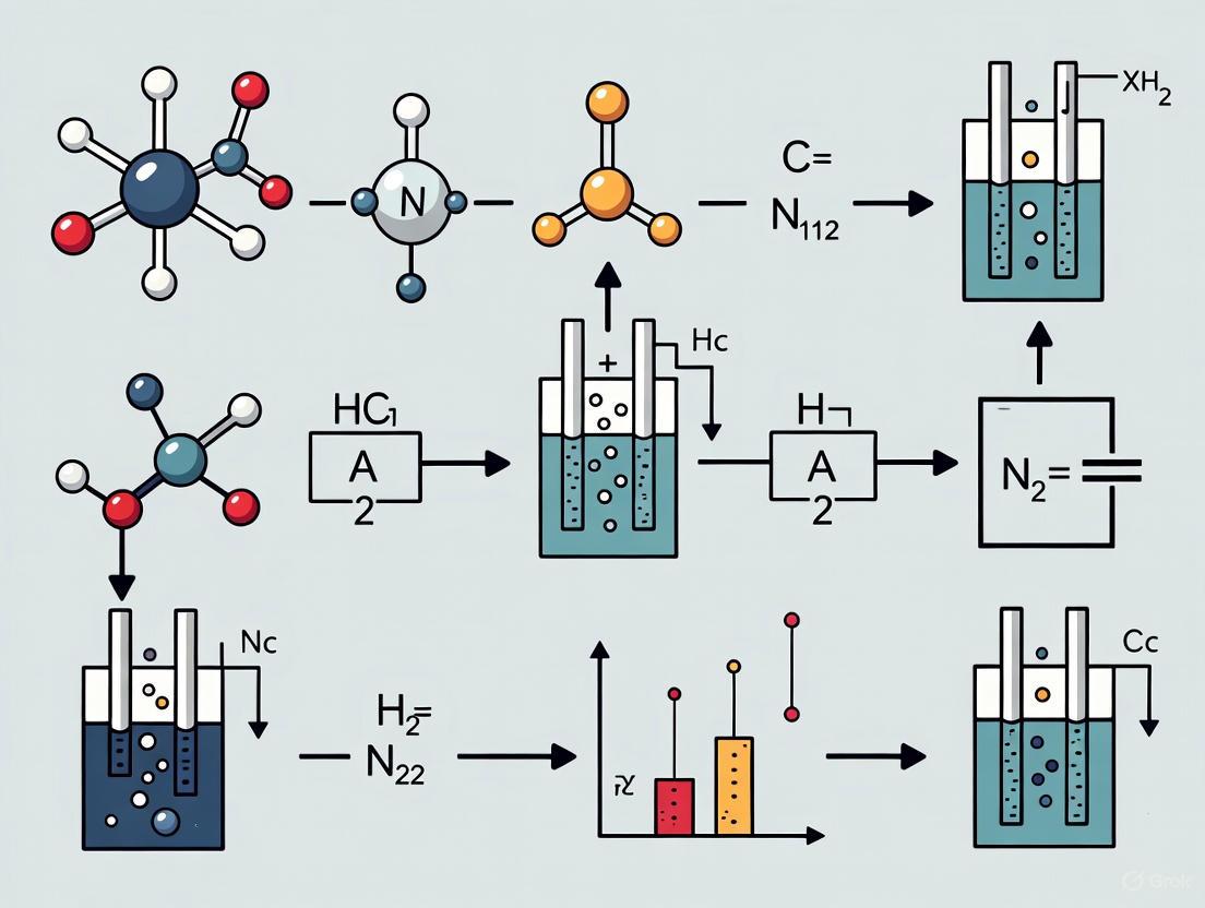
The logical workflow for applying the Nernst equation in analytical research, from fundamental principles to final application, is visualized below.
Diagram 1: Nernst Equation Application Workflow
Experimental Protocols and Methodologies
Protocol A: Determination of an Unknown Ion Concentration
This protocol outlines the standard procedure for using an Ion-Selective Electrode (ISE) to determine the concentration of an ion in a solution, a common practice in pharmaceutical quality control labs.
Principle: The potential of the ISE is measured versus a reference electrode in standard solutions of known concentration. A calibration curve of potential vs. log(concentration) is plotted, which should be linear as per the Nernst equation. The potential of an unknown sample is then measured and its concentration is determined from the calibration curve.
Materials:
- Ion-selective electrode for target ion (e.g., Na⁺, K⁺, Ca²⁺)
- Reference electrode (e.g., Ag/AgCl with double junction)
- Potentiometer (high-impedance voltmeter) or pH/mV meter
- Magnetic stirrer and stir bars
- Volumetric flasks, beakers, pipettes
- Ionic Strength Adjuster (ISA) specific to the analyte ion
- Standard stock solution of the analyte ion (e.g., 1000 ppm)
- Deionized water
- Unknown sample solution
Procedure:
- Calibration Curve Preparation: a. Prepare a series of standard solutions by diluting the stock solution (e.g., 10⁻¹ M, 10⁻² M, 10⁻³ M, 10⁻⁴ M). b. Add an equal volume of ISA to each standard solution and to the unknown sample to maintain constant ionic strength. c. Immerse the ISE and reference electrode in the most dilute standard. d. Under gentle stirring, record the stable potential reading in millivolts (mV). e. Rinse the electrodes with deionized water and blot dry. f. Repeat steps c-e for each standard solution in order of increasing concentration.
- Sample Measurement: a. Immerse the electrodes in the prepared unknown sample. b. Under gentle stirring, record the stable potential reading in mV. c. Rinse the electrodes thoroughly with deionized water after measurement.
Data Analysis:
- Plot the measured potential (mV) on the Y-axis against the logarithm of the concentration (log[C]) of the standard solutions on the X-axis.
- Perform a linear regression to obtain the equation of the best-fit line (Y = slope * X + intercept). The slope should be close to the theoretical Nernstian slope (e.g., ~59.2/n mV/decade at 25°C).
- Substitute the measured potential of the unknown sample (Y) into the linear equation and solve for X (log[C]).
- The antilog of X gives the concentration of the analyte in the unknown sample.
Protocol B: Calculation of an Equilibrium Constant
This protocol describes the use of the Nernst equation to determine the equilibrium constant (( K_{eq} )) of a redox reaction, which is valuable for characterizing APIs (Active Pharmaceutical Ingredients) prone to redox degradation.
Principle: A galvanic cell is constructed from the redox reaction of interest. The standard cell potential (( E^o{cell} )) is calculated from standard reduction potentials. The cell potential (( E{cell} )) is measured at known concentrations. The Nernst equation is then used with ( E{cell} = 0 ) at equilibrium to solve for ( K{eq} ).
Materials:
- Electrodes (e.g., Zn rod, Cu rod)
- Salt bridge (e.g., KNO₃ in agar)
- Solutions of known concentration (e.g., 1.0 M ZnSO₄, 0.001 M CuSO₄)
- Voltmeter (potentiometer)
- Beakers, wires
Procedure (Example for Zn | Zn²⁺ || Cu²⁺ | Cu):
- Construct the electrochemical cell: In one beaker place a Zn electrode in 1.0 M ZnSO₄. In a second beaker, place a Cu electrode in 0.001 M CuSO₄. Connect the two half-cells with a salt bridge.
- Connect the Zn electrode (anode) and Cu electrode (cathode) to a voltmeter.
- Measure the initial cell potential (( E_{cell} )).
- Allow the cell to discharge until the potential reaches 0 V (equilibrium). The concentrations at this point can be used to calculate ( K ), though the calculation is more straightforward using the standard potential.
Data Analysis:
- Calculate the standard cell potential: ( E^o{cell} = E^o{cathode} - E^o_{anode} ). For Zn/Cu, this is +0.337 V - (-0.763 V) = +1.10 V [4].
- At equilibrium, ( E{cell} = 0 ) and ( Q = K{eq} ). The Nernst equation becomes: [ 0 = E^o{cell} - \frac{0.0592}{n} \log{10} K{eq} ] [ \log{10} K{eq} = \frac{nE^o{cell}}{0.0592} \label{5} \tag{5} ]
- For the Zn/Cu reaction where ( n=2 ) and ( E^o{cell} = +1.10 V ): [ \log{10} K{eq} = \frac{2 \times 1.10}{0.0592} \approx 37.2 ] [ K{eq} = 10^{37.2} \approx 1.6 \times 10^{37} ] This very large value indicates the reaction strongly favors products at equilibrium [4] [1].
The relationship between cell potential and the reaction progress towards equilibrium is conceptualized in the following diagram.
Diagram 2: Cell Potential Evolution to Equilibrium
Advanced Concepts and Current Research Frontiers
Three-Dimensional Visualization of Nernstian Behavior
Advanced graphical representations, such as 3-D trend surfaces ("topos"), have been developed to visualize redox equilibria governed by the Nernst equation [7]. These plots map the electrode potential (z-axis) against the activities of both the oxidized and reduced species (x and y-axes). The resulting surfaces characteristically show a steep "cliff" where one species is depleted and a broad "plateau" where the potential is close to the standard potential ( E^0 ) [7]. These visualizations are powerful tools for teaching and for intuitively understanding how potential evolves along a reaction path in a galvanic cell until the cell "dies" when the potentials of the two half-cells equalize [7].
Limitations and Considerations
While powerful, the Nernst equation has limitations that researchers must consider:
- Activity vs. Concentration: The equation is rigorously defined in terms of ion activity, not concentration. In solutions with high ionic strength (>0.001 M), the activity coefficient can deviate significantly from unity, and concentrations must be corrected for accurate results [6] [5].
- Current Flow: The equation assumes equilibrium conditions with zero current flow. When a current flows, effects such as resistive losses and overpotential occur, which the Nernst equation does not account for [5].
- Non-Nernstian Behavior: Some sensor materials may not exhibit the ideal Nernstian slope (59.2/n mV at 25°C), or their response may be affected by interfering ions. Careful characterization is essential.
Future Directions in Potentiometry Research
Current research is pushing the boundaries of Nernstian potentiometry. Key areas include:
- Miniaturization and Solid-State Electrodes: Development of robust, solid-state ion-selective electrodes and miniaturized sensors for point-of-care diagnostics and in-vivo monitoring [3].
- Nanotechnology: Integration of nanomaterials to enhance electrode performance, sensitivity, and detection limits [3].
- Advanced Materials: Exploration of new sensing materials with improved selectivity, longer lifetimes, and reduced susceptibility to biofouling.
- Multiplexed Sensor Arrays: Combining multiple potentiometric sensors into arrays for simultaneous analysis of several ions, creating "electronic tongues" for complex mixture analysis.
The Nernst equation stands as a cornerstone of electrochemistry, providing a critical bridge between the thermodynamic driving force of redox reactions and the practical experimental conditions under which they occur. In the context of potentiometry research, particularly in drug development where precise measurements of ion concentrations and reaction equilibria are paramount, a thorough understanding of this equation is non-negotiable. It elegantly describes the relationship between the electrochemical potential of a cell and the composition of the solution, enabling researchers to predict cell voltages under non-standard conditions and determine critically important equilibrium constants, including solubility constants vital for pharmaceutical solubility studies [4]. This guide deconstructs the equation into its fundamental parameters, providing researchers and scientists with a detailed technical reference for application in advanced potentiometric research.
Fundamental Theory and Derivation
The Nernst Equation is derived from the principles of thermodynamics, linking the measurable cell potential to the Gibbs free energy change of the redox reaction. The derivation begins with the relationship between the Gibbs free energy change under non-standard conditions (ΔG) and the standard Gibbs free energy change (ΔG°):
[ \Delta G = \Delta G^o + RT \ln Q \label{1} ]
where (Q) is the reaction quotient. The electrical work done by a galvanic cell is given by (-nFE), which, under reversible conditions, equals the change in Gibbs free energy, (\Delta G) [8]. Substituting the Gibbs free energy terms with their electrochemical equivalents ((\Delta G = -nFE) and (\Delta G^o = -nFE^o)) yields:
[ -nFE = -nFE^o + RT \ln Q \label{2} ]
Dividing through by (-nF) provides the most general form of the Nernst Equation:
[ E = E^o - \frac{RT}{nF} \ln Q \label{3} ]
This form can be adapted for base-10 logarithms, which is more convenient for practical calculations:
[ E = E^o - \frac{2.303 RT}{nF} \log_{10} Q \label{4} ]
At standard temperature (298.15 K or 25 °C), the constants can be consolidated, simplifying the equation for laboratory use [4]:
[ E = E^o - \frac{0.0592\, \text{V}}{n} \log_{10} Q \label{5} ]
This expression is indispensable for predicting cell potentials outside of standard-state conditions, a scenario routinely encountered in experimental research.
Parameter Deconstruction
A deep understanding of each variable in the Nernst Equation is crucial for its correct application in potentiometric experiments. The following table summarizes these core parameters and their physical significance.
Table 1: Core Parameters of the Nernst Equation
| Parameter | Symbol | Definition & Role | Standard Units |
|---|---|---|---|
| Cell Potential | (E) | The measured electromotive force (EMF) or voltage of an electrochemical cell under non-standard conditions. It is the primary observable in potentiometric measurements. | Volts (V) |
| Standard Cell Potential | (E^o) | The intrinsic EMF of a cell under standard state conditions (all activities = 1, T = 298.15 K, P = 1 atm). It is a constant for a given redox reaction and indicates thermodynamic spontaneity. | Volts (V) |
| Universal Gas Constant | (R) | A fundamental physical constant that relates energy and temperature scales. It is the proportionality constant in the ideal gas law and thermodynamic equations. | 8.314 J·K⁻¹·mol⁻¹ |
| Temperature | (T) | The absolute temperature at which the electrochemical reaction occurs. It directly influences the thermal energy available to the system and the value of the pre-logarithmic term. | Kelvin (K) |
| Moles of Electrons | (n) | The number of moles of electrons transferred in the balanced redox reaction. It quantifies the stoichiometry of the electron transfer process. | Dimensionless (mol) |
| Faraday's Constant | (F) | The magnitude of electric charge per mole of electrons. It converts between chemical moles of electrons and electrical charge. | 96,485 C·mol⁻¹ |
| Reaction Quotient | (Q) | The ratio of the activities (approximated by concentrations) of the reaction products to reactants, each raised to the power of its stoichiometric coefficient. It describes the instantaneous composition of the system. | Dimensionless |
In-Depth Parameter Analysis
The Potential Terms ((E) and (E^o)): While (E^o) is a fixed property of a reaction, obtained from reference tables, the measured potential (E) is a dynamic variable. It reflects the system's departure from standard conditions as dictated by the reaction quotient (Q) and temperature (T). A positive (E) indicates a spontaneous reaction, while a negative (E) signifies non-spontaneity [4]. In potentiometry, (E) is the direct signal from which analyte concentration is derived.
The Constants ((R) and (F)): (R) and (F) are fundamental constants. Their combination in the term (RT/nF) has units of volts and represents the thermal voltage, scaling the logarithmic term's influence on the cell potential. At room temperature, (2.303RT/F \approx 0.0592\, V) [4] [9].
The Stoichiometric and Compositional Terms ((n) and (Q)): The value of (n) must be determined from a fully balanced redox reaction. An error in (n) propagates directly into the calculated potential. The reaction quotient (Q) for a general reaction (aA + bB \rightarrow cC + dD) is (Q = \frac{{aC}^c \cdot {aD}^d}{{aA}^a \cdot {aB}^b}). For dissolved species, activities are approximated by molar concentrations; for gases, by partial pressures; and for pure solids or liquids, their activity is 1 [9].
Experimental Methodology in Potentiometry
The practical application of the Nernst equation in research, such as determining equilibrium constants or quantifying analyte concentrations, requires rigorous experimental protocols.
Protocol: Determination of an Equilibrium Constant via Cell Potential Measurement
1. Principle: The equilibrium constant (K) for a redox reaction can be determined electrochemically by measuring the standard cell potential (E^o) and using the relationship derived from the Nernst equation at equilibrium, where (E = 0) and (Q = K) [4]: [ 0 = E^o - \frac{RT}{nF} \ln K \quad \Rightarrow \quad \log K = \frac{nE^o}{0.0592\, \text{V}} \quad (\text{at } 298 \text{ K}) ]
2. Materials and Reagents: Table 2: Essential Research Reagents and Materials
| Item | Function in Experiment |
|---|---|
| Potentiostat/Galvanostat | A precision instrument for applying potential and accurately measuring the resulting cell voltage with high impedance to minimize current draw. |
| Electrochemical Cell | A multi-port vessel (e.g., H-cell) to house the working, reference, and counter electrodes and the analyte solution. |
| Reference Electrode | Provides a stable, known reference potential (e.g., Saturated Calomel Electrode, Ag/AgCl). The Standard Hydrogen Electrode (SHE) defines the zero point [10]. |
| Working Electrode | The electrode at which the reaction of interest occurs (e.g., Pt, Au, Glassy Carbon). Material is selected for inertness and relevant electrochemical window. |
| Counter Electrode | Completes the circuit, often made of inert platinum wire. |
| High-Purity Salts & Solvents | To prepare analyte solutions with precisely known concentrations. |
| Salt Bridge | An ionic connection (e.g., KCl-agar) between half-cells to complete the circuit while preventing solution mixing [2]. |
3. Procedure: a. Cell Assembly: Construct a galvanic cell where the reaction of interest is the cell reaction. For example, to find (K) for (Zn{(s)} + Cu^{2+}{(aq)} \rightleftharpoons Zn^{2+}{(aq)} + Cu{(s)}), a Zn electrode in a Zn²⁺ solution and a Cu electrode in a Cu²⁺ solution are connected via a salt bridge [4]. b. Potential Measurement: Using a high-impedance voltmeter, measure the cell potential (E) at a controlled temperature of 25 °C. Ensure reactants and products are at standard concentrations (1 M for solutions, 1 atm for gases). c. Data Analysis: Under these standard conditions, the measured potential (E) is equal to (E^o). Use the simplified relationship (\log K = \frac{nE^o}{0.0592}) to calculate the equilibrium constant.
Workflow and Parameter Relationships
The following diagram visualizes the logical workflow for a potentiometric experiment and the interplay of Nernst equation parameters, from experimental setup to final result.
Diagram 1: Potentiometric Experiment Workflow
Advanced Considerations
Activity vs. Concentration
The thermodynamically correct form of the Nernst equation uses the chemical activities (a) of the species involved, not their concentrations. Activity accounts for non-ideal behavior in solutions, especially at medium and high concentrations where electrical interactions between ions become significant. The activity of a dissolved species i is defined as (ai = γi Ci), where (γi) is its activity coefficient and (Ci) is its molar concentration [9]. For dilute solutions ((< 0.001 M)), (γi ≈ 1), and concentrations can be used directly. In more concentrated pharmaceutical solutions, this approximation breaks down, and activity coefficients must be considered for high-precision work.
The Formal Potential ((E^{o'}))
To simplify work with real-world solutions where activity coefficients are unknown or difficult to determine, electrochemists use the formal potential ((E^{o'})), also called the conditional potential [9]. It is defined as:
[ E^{o'} = E^{o} - \frac{RT}{zF} \ln\left(\frac{\gamma{\text{Red}}}{\gamma{\text{Ox}}}\right) ]
This incorporates the activity coefficients into the standard potential, yielding a modified Nernst equation:
[ E = E^{o'} - \frac{RT}{zF} \ln\left(\frac{C{\text{Red}}}{C{\text{Ox}}}\right) ]
The formal potential is the experimentally observed potential when the concentrations of the oxidized and reduced species are equal ((C{Red}/C{Ox} = 1)) and all other solution conditions (ionic strength, pH, presence of complexing agents) are specified. It is highly dependent on the medium and is more practical for analytical applications than the standard potential [9].
Parameter Interrelationships
The following diagram maps the complex dependencies and relationships between the core parameters of the Nernst equation, illustrating how they collectively determine the cell's behavior.
Diagram 2: Nernst Equation Parameter Relationships
A meticulous, parameter-level understanding of the Nernst equation transcends academic exercise and is a fundamental prerequisite for robust potentiometric research in fields like drug development. The deconstruction of (E), (E^o), (R), (T), (n), (F), and (Q) reveals the elegant synergy between thermodynamics and experimental electrochemistry. Mastering these parameters, along with advanced concepts like formal potential and activity, empowers scientists to design more precise experiments, from determining critical equilibrium constants for drug solubility to developing novel electrochemical sensors. This detailed guide serves as a technical foundation upon which researchers can build to advance their potentiometric analyses and contribute to the broader field of analytical chemistry.
In potentiometry and the study of electrochemical cells, the Nernst equation provides the fundamental link between the measured potential of an electrochemical cell and the activities (often approximated by concentrations) of the ionic species involved [4] [9]. For researchers and scientists in drug development, this relationship is the cornerstone of a wide array of analytical techniques, from ion-selective electrode measurements to the assessment of membrane potentials in physiological studies. The equation quantitatively describes the equilibrium potential established across a membrane or at an electrode interface when a specific ion is permeable. A key feature of this relationship is its predictable, temperature-dependent slope, with a characteristic value of 59.16 mV per decade change in concentration for a monovalent ion (z = 1) at 25°C [11] [12]. This value, universally known as the "Nernstian slope," serves as a critical benchmark for validating experimental systems, designing sensors, and interpreting biological signals. This guide delves into the origin, interpretation, and practical application of this ideal slope, with a focused comparison between monovalent and divalent ions, framed within the context of rigorous potentiometric research.
Theoretical Foundation of the Nernstian Slope
Derivation from Basic Principles
The Nernst equation is derived from the principles of thermodynamics, relating the electrical work of an electrochemical cell to the chemical free energy change of the underlying redox reaction [4] [8]. For a general reduction half-reaction: [ \ce{Ox + ze^{-} <=> Red} ] the Nernst equation for the half-cell potential is expressed as: [ E = E^0 - \frac{RT}{zF} \ln \frac{a{\text{Red}}}{a{\text{Ox}}} ] where:
- (E) is the equilibrium potential (Volts, V)
- (E^0) is the standard electrode potential (V)
- (R) is the universal gas constant (8.314 J·mol⁻¹·K⁻¹) [11]
- (T) is the absolute temperature (Kelvin, K)
- (z) is the number of electrons transferred in the half-reaction (the charge of the ion for simple metal/metal-ion electrodes)
- (F) is the Faraday constant (96,485 C·mol⁻¹) [11]
- (a{\text{Red}}) and (a{\text{Ox}}) are the activities of the reduced and oxidized species, respectively
For a metal ion in solution ((M^{z+})) in equilibrium with its pure metal, the reduced form is the solid metal, which has an activity of 1. Assuming the activity of the oxidized form ((M^{z+})) can be approximated by its concentration ([(M^{z+})]), the equation simplifies to [13]: [ E = E^0 - \frac{RT}{zF} \ln \frac{1}{[M^{z+}]} = E^0 + \frac{RT}{zF} \ln [M^{z+}] ]
The Origin of the 59.16 mV Value
The factor ( \frac{RT}{F} ) is a fundamental constant in electrochemistry, representing the "thermal voltage." At standard temperature (25°C or 298.15 K), its value is: [ \frac{RT}{F} = \frac{(8.314 \, \text{J·mol}^{-1}\text{·K}^{-1}) \times (298.15 \, \text{K})}{96,485 \, \text{C·mol}^{-1}} \approx 0.02569 \, \text{V} = 25.69 \, \text{mV} ] Most practical measurements use base-10 logarithms (log) rather than natural logarithms (ln). The conversion is given by ( \ln(x) = 2.303 \log(x) ). Substituting this into the Nernst equation gives: [ E = E^0 + \frac{2.303 RT}{zF} \log [M^{z+}] ] The pre-logarithmic term ( \frac{2.303 RT}{F} ) at 25°C is: [ \frac{2.303 RT}{F} = 2.303 \times 0.02569 \, \text{V} \approx 0.05916 \, \text{V} = 59.16 \, \text{mV} ] Thus, the final, widely-used form of the Nernst equation for a cation (M^{z+}) at 25°C becomes: [ E = E^0 + \frac{0.05916}{z} \log [M^{z+}] ] This equation reveals that the measured potential changes by ( \frac{59.16}{z} ) millivolts for every tenfold change in the ion concentration [4] [12]. The slope of a plot of (E) versus (\log [M^{z+}]) is therefore ( \frac{59.16}{z} ) mV/decade.
The Critical Role of Ion Valency (z)
The valency of the ion (z) is the decisive factor in the slope of the Nernstian response, as it is inversely proportional to the term (59.16/z).
For Monovalent Ions (
z = 1) such as K⁺, Na⁺, Li⁺, H⁺, and Cl⁻, the ideal Nernstian slope is: ( \frac{59.16}{1} = 59.16 \, \text{mV/decade} ) This means a tenfold increase in the concentration of a monovalent cation will cause the equilibrium potential to increase by 59.16 mV.For Divalent Ions (
z = 2) such as Ca²⁺, Mg²⁺, and Zn²⁺, the ideal slope is: ( \frac{59.16}{2} = 29.58 \, \text{mV/decade} ) The higher charge means the electrical driving force is twice as effective per decade of concentration change, resulting in a slope that is exactly half of that for a monovalent ion [11].
The table below summarizes the ideal Nernstian slopes for different ion types at 25°C.
Table 1: Ideal Nernstian Slopes for Different Ion Valencies at 25°C
Ion Valency (z) |
Example Ions | Ideal Nernstian Slope (mV per decade) |
|---|---|---|
| +1 (Monovalent Cations) | K⁺, Na⁺, H⁺, NH₄⁺ | +59.16 |
| -1 (Monovalent Anions) | Cl⁻, I⁻, NO₃⁻ | -59.16 |
| +2 (Divalent Cations) | Ca²⁺, Mg²⁺, Cu²⁺, Zn²⁺ | +29.58 |
| -2 (Divalent Anions) | SO₄²⁻, CO₃²⁻ | -29.58 |
The following diagram illustrates the logical and mathematical relationships that lead to the ideal Nernstian slope.
Diagram 1: Derivation Path of the Nernstian Slope. This flowchart outlines the logical sequence from fundamental thermodynamics to the practical equation used to calculate the ideal Nernstian slope.
Experimental Protocols for Slope Validation
Validating that an electrochemical system (e.g., an ion-selective electrode) exhibits the ideal Nernstian slope is a critical step in confirming its proper function and measurement accuracy.
Calibration of an Ion-Selective Electrode (ISE)
Objective: To determine the response slope of an Ion-Selective Electrode (ISE) for a target ion and verify its conformity to the ideal Nernstian slope.
Materials and Reagents:
- Ion-Selective Electrode: Specific to the ion of interest (e.g., Ca²⁺-ISE).
- Reference Electrode: Double-junction or single-junction (e.g., Ag/AgCl).
- Potentiometer/Millivoltmeter: High-impedance voltmeter capable of measuring mV with 0.1 mV resolution.
- Standard Solutions: A series of standard solutions with known concentrations of the target ion, covering a range of at least 3 decades (e.g., 10⁻⁵ M, 10⁻⁴ M, 10⁻³ M, 10⁻² M, 10⁻¹ M).
- Ionic Strength Adjuster (ISA): A high-strength salt solution (e.g., KNO₃ or NaCl) added to all standards and samples to maintain a constant and high ionic background. This minimizes the variation in the activity coefficient, ensuring that concentration can be used in place of activity [12].
Procedure:
- Setup: Connect the ISE and reference electrode to the potentiometer. Immerse the electrodes in a beaker containing a low-ionic-strength rinse solution (e.g., deionized water).
- Conditioning: If required by the manufacturer, condition the ISE by soaking it in a solution of the target ion (e.g., 10⁻³ M) for 30 minutes prior to the first use.
- Measurement: a. Start with the most dilute standard solution. Add the prescribed volume of ISA to the standard and stir gently and consistently. b. Thoroughly rinse the electrodes with deionized water and blot dry with a laboratory wipe. c. Immerse the electrodes in the standard solution, allow the potential reading to stabilize (typically 1-2 minutes), and record the millivolt value. d. Repeat steps 3a-3c for all standard solutions in order of increasing concentration.
- Data Analysis:
a. Plot the recorded potential (mV, y-axis) against the logarithm (base 10) of the ion concentration (x-axis).
b. Perform a linear regression analysis on the data points to obtain the equation of the best-fit line (y = mx + b, where m is the slope).
c. Compare the experimentally determined slope (
m) to the ideal Nernstian slope (59.16/zmV). A slope within ±5% of the ideal value is often considered indicative of a well-functioning electrode.
Measurement of Membrane Equilibrium Potentials in Physiology
Objective: To demonstrate the Nernst potential across a biological or artificial membrane permeable to a specific ion.
Background: In physiology, the Nernst equation calculates the equilibrium potential for an ion across a semi-permeable membrane. This potential is a result of the concentration gradient of the permeant ion. Researchers often simulate this using artificial lipid bilayers or cultured cells.
Materials and Reagents:
- Recording Setup: Voltage-clamp or current-clamp amplifier.
- Microelectrodes: Fine-tipped glass microelectrodes (for intracellular recording) or patch-clamp pipettes.
- Cell or Vesicle Preparation: A single cell (e.g., neuron, oocyte) or an artificial lipid vesicle.
- Bath and Pipette Solutions: Solutions with precisely defined concentrations of the ion of interest (e.g., K⁺). A classic experiment involves varying extracellular K⁺ concentration ([K⁺]ₒ) while measuring membrane potential.
Procedure:
- Solution Preparation: Prepare a set of extracellular solutions where the concentration of K⁺ is varied (e.g., 1 mM, 3 mM, 10 mM, 30 mM, 100 mM), while maintaining osmolarity and the concentrations of other ions.
- Impaling the Cell: Using a micromanipulator, carefully impale a single cell with the microelectrode to measure its intracellular potential relative to the bath ground.
- Perfusion and Recording: a. Continuously perfuse the cell with the control (low K⁺) solution. Record the resting membrane potential. b. Switch the perfusion to a solution with a higher known [K⁺]ₒ. c. Allow the membrane potential to stabilize and record the new value. d. Repeat for all K⁺ solutions.
- Data Analysis: a. Plot the measured membrane potential (mV, y-axis) against log₁₀([K⁺]ₒ) (x-axis). b. Fit a linear regression to the data. The slope of this line should approach the ideal Nernst slope for K⁺ (-59.16 mV/decade at 25°C, negative as the potential becomes more positive with higher external K⁺). Deviations indicate that the membrane is not exclusively permeable to K⁺, which is often the case in real cells.
The workflow for a typical calibration experiment is summarized below.
Diagram 2: Workflow for Validating Nernstian Slope. This chart outlines the standard operational procedure for calibrating an ion-selective electrode to validate its Nernstian response.
The Researcher's Toolkit: Essential Reagents and Materials
Successful experimentation with the Nernst equation requires precise materials and an understanding of their function.
Table 2: Key Research Reagent Solutions and Materials
| Item | Function / Explanation | Example in Use |
|---|---|---|
| Standard Solutions | A series of solutions with known, precise concentrations of the analyte ion. Serve as the calibration curve for potential vs. log(concentration). | 10⁻⁵ M to 10⁻¹ M KCl solutions for calibrating a K⁺-ISE. |
| Ionic Strength Adjuster (ISA) | A high-concentration inert electrolyte added to all standards and samples. Swamps out the variable sample background, making the activity coefficient constant. This allows concentration to be used directly in the Nernst equation [12]. | 4 M KNO₃ for Ca²⁺ or K⁺ measurements. |
| Reference Electrode | Provides a stable, fixed reference potential against which the indicator electrode's potential is measured. Critical for a stable mV reading. | Ag/AgCl electrode with KCl filling solution. |
| High-Impedance Potentiometer | Measures voltage without drawing significant current. Prevents polarization of the electrodes and ensures an accurate reading of the equilibrium potential. | pH/mV meter with >10¹² Ω input impedance. |
| Formal Potential (E⁰') | The measured standard potential under a defined set of solution conditions (e.g., specific ionic strength). It is used in place of the thermodynamic E⁰ when concentrations are used instead of activities for more accurate practical calculations [9] [12]. | The y-intercept of the calibration curve is the formal potential. |
Advanced Considerations and Non-Ideal Behavior
While the ideal slope is a crucial benchmark, several factors can cause experimental results to deviate.
Temperature Dependence
The Nernst slope is directly proportional to the absolute temperature T [11]. For experiments conducted at temperatures other than 25°C, the slope must be recalculated using the formula (2.303RT)/F. For example, at physiological temperature (37°C or 310.15 K), the ideal slope for a monovalent ion is approximately 61.54 mV/decade.
Activity vs. Concentration
The Nernst equation is rigorously defined in terms of ion activity, not concentration. Activity (a) is related to concentration (C) by the activity coefficient (γ): a = γC. In dilute solutions, γ≈1, but in concentrated or complex matrices (e.g., biological fluids), γ can be significantly less than 1. The use of an ISA is the primary methodological approach to mitigate this issue [9] [12].
Non-Nernstian Response and its Implications
A measured slope significantly lower than the ideal value (e.g., 50 mV/decade for a monovalent ion) indicates a non-ideal response. This can be caused by:
- Electrode Drift or Aging: A degraded or fouled ion-selective membrane.
- Insufficient Selectivity: Interference from other ions in the solution.
- Incorrect Ionic Strength: Leading to variable activity coefficients.
- Junction Potentials: Unstable liquid junction potentials in the reference electrode. Identifying and troubleshooting the cause of a sub-Nernstian response is essential for obtaining reliable potentiometric data.
The Nernstian slope of 59.16/z mV is not merely a theoretical constant but a practical gold standard in potentiometric research. Its derivation from first principles provides a solid thermodynamic foundation, while its dependence on ion valency offers a clear, quantifiable prediction for system behavior. For researchers in drug development and the broader life sciences, a deep understanding of this relationship is indispensable. It enables the calibration of critical analytical tools like ion-selective electrodes, informs the interpretation of electrophysiological data, and provides a rigorous framework for assessing the performance of novel sensor technologies. Mastery of the concepts and experimental protocols surrounding the Nernstian slope ensures that scientific conclusions drawn from potentiometric measurements are both accurate and reliable.
In potentiometry, the fundamental relationship between the measured potential of an electrochemical cell and the analyte of interest is governed by the Nernst equation. This equation, in its fundamental form, describes the potential as a function of the logarithm of ion activity, not concentration [14] [9]. For an ion with charge ( z ), the electrode potential is given by: [ E{\mathrm{cell}} = K + \frac{0.05916}{z} \log(aA) ] where ( E{\mathrm{cell}} ) is the cell potential, ( K ) is a constant, and ( aA ) is the activity of the ion A [14]. The distinction between activity and concentration is therefore not merely academic; it is foundational to interpreting potentiometric signals accurately, especially in complex matrices like pharmaceutical formulations or biological fluids.
Activity can be defined as the effective concentration of an ion in a solution, accounting for its interactions with all other species in the solution [15]. It is related to the measured concentration ( [M^{n+}] ) by the activity coefficient ( \gamma{M^{n+}} ): [ a{M^{n+}} = [M^{n+}] \gamma_{M^{n+}} ] The activity coefficient, and thus the activity, is influenced by the ionic strength of the solution, a function of the concentrations and charges of all ions present [14] [15]. In ideal, infinitely dilute solutions, inter-ionic interactions are negligible, ( \gamma \approx 1 ), and activity equals concentration. However, in the real-world solutions analyzed by researchers and development professionals, this is rarely the case.
Table 1: Key Differences Between Activity and Concentration
| Feature | Activity | Concentration |
|---|---|---|
| Definition | Effective, thermodynamically active concentration | Analytical amount per unit volume |
| Governing Factor | Ion activity coefficient (( \gamma )) and concentration | Preparation and dilution |
| Dependence | Ionic strength and solution matrix | Independent of solution matrix |
| Nernst Equation | Directly related to potential | Related only if ( \gamma \approx 1 ) |
| Practical Use | Measures free, bioavailable ions | Measures total content |
The Nernst Equation: Bridging Theory and Practice
The Central Role in Potentiometry
The Nernst equation is the cornerstone of potentiometric analysis, providing the mathematical link between an electrochemical potential and the composition of a solution [4] [2]. For a half-cell reduction reaction of the form [ \text{Ox} + ze^- \longrightarrow \text{Red} ] the Nernst equation is expressed as: [ E = E^{\ominus} - \frac{RT}{zF} \ln \frac{a{\text{Red}}}{a{\text{Ox}}} ] where ( E ) is the half-cell potential, ( E^{\ominus} ) is the standard electrode potential, ( R ) is the universal gas constant, ( T ) is the absolute temperature, ( F ) is the Faraday constant, and ( a{\text{Red}} ) and ( a{\text{Ox}} ) are the activities of the reduced and oxidized species, respectively [9] [4]. At room temperature (25 °C), this equation simplifies using the thermal voltage approximation, and for a metal/metal ion electrode, it reduces to the form shown in Section 1 [9] [4].
From Activity to Concentration in Practice
To make potentiometry a practical tool for determining concentration, the Nernst equation must be reconciled with the activity-concentration relationship [14]. Substituting ( a{M^{n+}} = [M^{n+}] \gamma{M^{n+}} ) into the Nernst equation yields: [ E{\mathrm{cell}} = K + \frac{0.05916}{n} \log \gamma{M^{n+}} + \frac{0.05916}{n} \log [M^{n+}] ] The first two terms on the right are often combined into a new constant, ( K' ), simplifying the equation to: [ E{\mathrm{cell}} = K' + \frac{0.05916}{n} \log [M^{n+}] ] This simplification is only valid if the activity coefficient is constant [14]. This is a critical consideration for experimental design: by ensuring that the standards and samples have an identical, or very similar, ionic matrix, the value of ( \gamma{M^{n+}} ) remains fixed, and the measured potential becomes a direct function of the logarithm of the concentration [14]. This is the principle upon which most quantitative potentiometric methods are built.
The following diagram illustrates the logical pathway from a sample to a concentration measurement, highlighting the central role of the Nernst equation and the activity-concentration relationship.
Diagram 1: From Sample to Concentration in Potentiometry
Experimental Protocols: Navigating the Distinction
Calibration and Matrix-Matching Protocol
The primary methodology for ensuring that potentiometric measurements accurately reflect concentration is through careful calibration with matrix-matched standards [14].
Objective: To construct a calibration curve that relates the measured cell potential to the analyte concentration, thereby accounting for the constant activity coefficient. Procedure:
- Preparation of Standard Solutions: Prepare a series of standard solutions of the analyte with known concentrations that bracket the expected concentration in the sample.
- Matrix Matching: Add an inert electrolyte (e.g., KNO₃, NaCl) to each standard solution to adjust the ionic strength to a high and constant value. The chosen ionic strength should approximate or exceed that of the sample solutions. This swamps out variations in ionic strength between standards and samples, ensuring a constant activity coefficient [14] [15].
- Potential Measurement: Measure the cell potential for each standard solution using the appropriate ion-selective electrode and reference electrode.
- Calibration Curve: Plot the measured potential (E) versus the logarithm of the known concentration (log [Mⁿ⁺]). The slope of the linear region should be close to the Nernstian value (59.16/z mV at 25 °C).
- Sample Measurement: Measure the potential of the sample solution, which must contain the same inert electrolyte at the same concentration as the standards. Determine the unknown concentration from the calibration curve.
Determination of Free vs. Total Concentration
Potentiometry uniquely measures the activity of the free, uncomplexed ion, which is a key advantage in speciation and bioavailability studies [14] [16]. This protocol can be used to investigate metal ion speciation.
Objective: To determine the concentration of free metal ions in a solution containing complexing agents. Procedure:
- Total Concentration: Determine the total metal ion concentration (e.g., using atomic absorption spectroscopy) on an aliquot of the sample.
- Free Concentration: Measure the potential of the sample directly using a calibrated ion-selective electrode for the metal ion. The measured potential corresponds to the activity of the free metal ion. Using the calibration curve, this activity is converted to the free metal concentration, acknowledging that the activity coefficient is accounted for in the calibration [14] [16].
- Data Analysis: The difference between the total concentration and the free concentration represents the complexed fraction of the metal ion. This is crucial in pharmaceutical development for understanding drug-protein binding or the speciation of active ingredients.
Table 2: Comparison of Analytical Techniques for Trace Metal Analysis
| Technique | Measured Quantity | Key Advantage | Key Limitation |
|---|---|---|---|
| Potentiometry | Activity of free ion | Direct information on bioavailability/speciation | Requires careful control of ionic strength |
| Voltammetry | Concentration of electrochemically labile species | High sensitivity | Complexed or inert species not detected |
| Atomic Spectrometry | Total elemental concentration | Insensitive to chemical form | No information on speciation or bioavailability |
The Scientist's Toolkit: Essential Reagents and Materials
Successful implementation of potentiometric methods relies on a set of key reagents and materials designed to manage the activity-concentration relationship and ensure measurement integrity.
Table 3: Key Research Reagent Solutions for Potentiometry
| Reagent/Material | Function | Practical Consideration |
|---|---|---|
| Ionic Strength Adjuster (ISA) | Swamps out variable ionic strength in samples and standards, ensuring a constant activity coefficient for accurate concentration measurements [14]. | Typically a high concentration of an inert electrolyte (e.g., 1 M KNO₃). Must not contain ions that interfere with the electrode. |
| Ion-Selective Electrode (ISE) | The indicator electrode whose potential is selectively sensitive to the activity of a specific ion in solution [17]. | Selectivity varies; check selectivity coefficients for expected interferents. Requires proper storage and conditioning. |
| Reference Electrode | Provides a stable, constant potential against which the ISE potential is measured [18] [17]. | Ag/AgCl or saturated calomel electrodes (SCE) are common. Requires periodic refilling with correct filling solution. |
| Standard Solutions | Used to construct the calibration curve that relates measured potential to analyte concentration [14]. | Must be prepared with high-purity materials and matrix-matched with the ISA to the samples. |
| Liquid Junction / Salt Bridge | Completes the electrical circuit between the ISE and reference electrode while minimizing liquid junction potential [18]. | Often an integral part of the reference electrode. Uses an electrolyte (e.g., KCl, KNO₃) that does not cause precipitation. |
Advanced Applications and Research Implications
The distinction between activity and concentration, when properly navigated, opens doors to powerful analytical applications. In environmental chemistry, potentiometric sensors with sub-nanomolar detection limits are used to measure free copper ions in seawater, providing critical data on metal bioavailability and toxicity that total concentration measurements cannot offer [16]. In clinical chemistry, ion-selective electrodes measure electrolytes like Na⁺ and K⁺ in blood serum. The reported value is a concentration, but this is only valid because the measurement system is calibrated with standards that mimic the serum matrix, effectively controlling for activity coefficients [17].
In pharmaceutical research, this principle is applied to study the binding of drug candidates to proteins or other macromolecules. By using a potentiometric sensor to monitor the free drug ion concentration before and after adding the binding partner, the binding constant can be determined, as the electrode only responds to the free, unbound ion [16]. The following workflow visualizes a typical experiment for studying ion speciation or binding using potentiometry.
Diagram 2: Workflow for Speciation/Binding Analysis
Navigating the distinction between activity and concentration is not a theoretical obstacle but a practical necessity in potentiometric research. A deep understanding of the Nernst equation reveals that the electrode signal is fundamentally tied to ion activity. Through rigorous experimental protocols—primarily the use of ionic strength adjustment and matrix-matched calibration—researchers can transform this activity-based signal into an accurate and reliable measure of concentration. This careful approach enables scientists across pharmaceutical, clinical, and environmental disciplines to leverage the unique advantage of potentiometry: the ability to detect the biologically and chemically active free ion, providing insights that are completely lost when only the total concentration is measured.
In potentiometric research, particularly when applying the Nernst equation to complex biological environments, the selection of the correct reference potential is paramount for obtaining accurate, reliable data. The fundamental Nernst equation, E = E⁰ - (RT/nF)ln(Q), relates the measured potential (E) to the reaction quotient (Q), with E⁰ representing the standard reference potential under defined conditions [19] [20]. However, researchers confront a critical decision: whether to use the standard potential (E°), which applies only to ideal standardized conditions, or the formal potential (E°'), which accounts for the real-world complexities of biological matrices. This distinction becomes especially crucial in pharmaceutical and clinical applications where measurements occur in saliva, blood, or other biofluids containing numerous interfering species, variable pH, and complex matrices that significantly alter electrochemical behavior [21] [22].
The formal potential is not merely a theoretical adjustment but a practical necessity for accurate in-situ and point-of-care measurements. It is defined as the potential of a redox couple under a specific set of experimental conditions, including pH, ionic strength, and presence of complexing agents, where the activity coefficients for the oxidized and reduced species are incorporated into the resulting potential E°' [19]. Unlike the standard potential, which is a universal constant tabulated for standard conditions (1 M concentrations, 1 atm pressure for gases, 298.15 K), the formal potential is environment-dependent and must be determined for each experimental setup [19]. This technical guide explores the theoretical foundations, practical implications, and methodological approaches for selecting and applying the correct potential in biologically-relevant potentiometric research, framed within the broader context of Nernst equation application in modern electroanalytical science.
Theoretical Foundations: Standard vs. Formal Potential
Standard Electrode Potential (E°)
The standard electrode potential provides the fundamental reference point for all electrochemical measurements. By definition, E° represents the inherent tendency of a redox species to acquire electrons relative to the Standard Hydrogen Electrode (SHE), which is assigned a value of 0 V under standard conditions: 298.15 K, 1 atm pressure for gases, and 1 M concentrations for solutes [23] [20]. These idealized conditions establish a reproducible baseline that enables comparison of different redox couples across experimental systems. The SHE consists of a platinum electrode immersed in a 1 M H⁺ solution with hydrogen gas bubbled at 1 atm pressure, creating the reference half-reaction: 2H⁺(aq, 1 M) + 2e⁻ ⇌ H₂(g, 1 atm) [23].
When determining standard potentials for other half-cells, galvanic cells are constructed with the SHE as one electrode and the half-cell of interest as the other. For instance, to establish the standard potential for the Cu²⁺/Cu redox couple, the cell Pt(s) | H₂(g, 1 atm) | H⁺(aq, 1 M) || Cu²⁺(aq, 1 M) | Cu(s) yields a measured potential of +0.337 V, which is assigned as E° for the Cu²⁺/Cu couple [23]. This systematic approach has generated comprehensive tables of standard reduction potentials that serve as starting points for predicting reaction spontaneity and cell potentials under idealized conditions.
Formal Potential (E°')
The formal potential represents a practical correction to the standard potential that accounts for non-ideal experimental conditions. According to PalmSens, a knowledgeable source in electrochemistry, "The two activity coefficients fOx and fRed are included in the resulting potential E⁰', which is called the formal potential" [19]. Since these activity coefficients depend on the chemical environment, the formal potential incorporates the effects of variables such as ionic strength, pH, complexation, and temperature that diverge from standard conditions.
The mathematical relationship between standard and formal potential emerges from the Nernst equation. For a generalized reduction reaction: Ox + ne⁻ → Red, the Nernst equation is:
Where a_Red and a_Ox represent the activities of the reduced and oxidized species, respectively. Substituting activity coefficients (γ) and concentrations (C) (a = γC) yields:
The combination E° - (RT/nF) * ln(γ_Red/γ_Ox) constitutes the formal potential E°', resulting in the practical form of the Nernst equation:
This formulation is particularly valuable in biological systems where activity coefficients deviate significantly from unity due to high ionic strength and specific molecular interactions [19].
Comparative Analysis: Key Distinctions
Table 1: Comparison between Standard Potential and Formal Potential
| Characteristic | Standard Potential (E°) |
Formal Potential (`E°') |
|---|---|---|
| Definition | Potential under standard conditions (1 M, 1 atm, 298.15 K) | Potential under specific experimental conditions |
| Reference | Standard Hydrogen Electrode (SHE) | Standard Hydrogen Electrode (SHE) |
| Activity Coefficients | Assumed to be 1 (ideal behavior) | Incorporated into the value |
| Environmental Factors | Ignores pH, ionic strength, complexation | Accounts for pH, ionic strength, complexation |
| Tabulation | Universally tabulated for redox couples | Must be determined for each experimental system |
| Applications | Predicting spontaneity, theoretical calculations | Quantitative analysis in real matrices |
The fundamental distinction lies in their applicability: while E° provides a theoretical benchmark, E°' offers practical utility for quantitative work in complex media. As noted in the research literature, "Since it contains parameters that depend on the environment, such as temperature and activity coefficients, E⁰' cannot be listed but needs to be determined for each experiment, if necessary" [19]. This requirement for experimental determination makes formal potential both context-dependent and mathematically convenient for analytical applications.
The Challenge of Biological Matrices
Biological matrices such as blood, saliva, urine, and cellular lysates present particularly challenging environments for potentiometric measurements due to their complex and variable composition. These systems contain numerous electroactive interferents, proteins, lipids, and electrolytes that can foul electrode surfaces, alter activity coefficients, and participate in secondary reactions [21] [22]. The ISE (ion-selective electrode) design must overcome these challenges to achieve accurate readings in clinical and pharmaceutical contexts.
Saliva, for instance, contains various components including sodium chloride, magnesium bicarbonate, calcium bicarbonate, sodium phosphate, urea, and ammonium oxide, all of which can potentially interfere with measurements of target analytes [22]. Similarly, blood plasma exhibits variable electrolyte balances and contains numerous biomolecules that can adsorb to electrode surfaces. A study of electrolyte disorders found that 15% of hospitalized patients suffer from at least one electrolyte imbalance, with hyponatremia (7.7%) and hypernatremia (3.4%) being most prevalent [21]. Even slight abnormalities in electrolyte balance can significantly affect potential measurements if not properly accounted for in the calibration approach.
The pH variability in biological systems particularly impacts the formal potential of pH-dependent redox couples. A notable example is the NAD⁺/NADH couple, where the standard reduction potential is -0.358 V, but at physiological pH (7.0), the formal potential shifts to approximately -0.56 V due to the involvement of H⁺ in the reduction reaction: NAD⁺ + 2e⁻ + H⁺ → NADH [24]. This substantial difference of over 0.2 volts demonstrates why using standard potentials without adjustment for biological conditions would lead to significant analytical errors.
Table 2: Impact of Biological Matrix Components on Potential Measurements
| Matrix Component | Effect on Potential Measurement | Consequence for E⁰ Selection |
|---|---|---|
| Variable pH | Shifts equilibrium for H⁺-dependent reactions | Requires use of pH-adjusted formal potential |
| High Ionic Strength | Alters activity coefficients (γ ≠ 1) | Formal potential essential for accurate quantification |
| Electroactive Interferents | Compete for electron transfer | Increases importance of selectivity coefficients |
| Macromolecules (proteins, lipids) | Surface fouling, reduced electrode responsiveness | Necessitates frequent calibration or formal potential verification |
| Complexing Agents | Shift effective concentration of free ions | Formal potential incorporates complexation equilibria |
Determining Formal Potential for Biological Applications
Experimental Methodology
The determination of formal potential for a specific biological application requires a systematic experimental approach centered around the Nernst equation. The general methodology involves constructing a calibration curve under conditions that closely mimic the target biological matrix. The following protocol outlines a comprehensive approach for formal potential determination:
Matrix-Matched Standard Preparation: Prepare standard solutions of the target analyte across a concentration range relevant to the biological application (typically 3-5 orders of magnitude). The standard matrix should approximate the ionic strength, pH, and protein content of the target biological fluid using appropriate buffers and additives [22]. For saliva analysis, Britton-Robinson buffer (BRB) adjusted to pH 7 has been effectively employed [22].
Potentiometric Measurement: Immerse the indicator and reference electrodes in each standard solution and measure the equilibrium potential once a stable reading is obtained (typically within 1-2 mV drift per minute). The reference electrode selection should be appropriate for biological measurements, with Ag/AgCl being particularly common due to its stability and biocompatibility [21] [22].
Data Analysis: Plot the measured potential (E) against the logarithm of the analyte concentration (log C). Perform linear regression to determine the slope and intercept of the calibration curve. The formal potential (
E°') corresponds to the potential value when the concentration ratioC_Red/C_Ox = 1(or whenlog C = 0for a single species), which is the y-intercept of the regression line [19].Validation: Confirm the determined formal potential by measuring potentials in spiked biological samples with known analyte additions. The recovery should approach 100% if the formal potential is correctly established for the matrix.
Case Study: NAD⁺/NADH Formal Potential at pH 7
The calculation of formal potential for pH-dependent systems demonstrates the critical adjustment needed for biological applications. For the NAD⁺/NADH couple with the reaction:
NAD⁺ + 2e⁻ + H⁺ → NADH
The Nernst equation is:
E = E° - (RT/2F) * ln([NADH]/([NAD⁺][H⁺]))
Which can be rearranged as:
E = E° - (RT/2F) * ln(1/[H⁺]) - (RT/2F) * ln([NADH]/[NAD⁺])
Recognizing that E°' = E° - (RT/2F) * ln(1/[H⁺]), and substituting [H⁺] = 10^(-7) for pH 7, the formal potential becomes:
E°' = E° - (0.05916/2) * log(1/10^(-7)) at 25°C
E°' = E° - (0.02958) * 7
E°' = -0.358 V - 0.207 V = -0.565 V
This significant shift of -0.207 V from the standard potential of -0.358 V to a formal potential of -0.565 V at physiological pH highlights the essential nature of this adjustment for accurate biological redox measurements [24].
Addressing Matrix Effects in Complex Biofluids
In particularly complex matrices like saliva or blood, additional strategies may be necessary to account for matrix effects:
Standard Addition Method: When the matrix composition is unknown or highly variable, the standard addition method can be employed where small volumes of concentrated standard are added directly to the sample, and the potential change is measured to determine the original concentration without explicit knowledge of the formal potential.
Matrix-Matching: For routine analysis, calibration standards can be prepared in artificial saliva or simulated plasma that approximates the major ionic components of the biological fluid [22].
Internal References: For some applications, incorporating an internal reference redox couple of known formal potential in the specific matrix can provide an in-situ calibration point.
The Scientist's Toolkit: Essential Research Reagents and Materials
Successful implementation of formal potential measurements in biological matrices requires careful selection of materials and reagents. The following table outlines essential components for such research:
Table 3: Research Reagent Solutions for Formal Potential Determination in Biological Matrices
| Reagent/Material | Function/Application | Example Specifications |
|---|---|---|
| Ion-Selective Electrode (ISE) | Target analyte recognition | PVC membrane with ionophore, MWCNT-modified for enhanced sensitivity [22] |
| Reference Electrode | Stable potential reference | Double-junction Ag/AgCl electrode (prevents contamination) [22] |
| Buffer Systems | pH control and ionic strength adjustment | Britton-Robinson buffer (pH 2-11 range) [22] |
| Ion-to-Electron Transducers | Signal enhancement in solid-contact ISEs | Multi-walled carbon nanotubes (MWCNTs), conducting polymers (PEDOT) [21] [22] |
| Matrix Modifiers | Reduction of nonspecific binding | Salt modifications to paper substrates for heavy metal detection [25] |
| Selectivity Enhancers | Minimize interferent effects | Ionophores with high specificity for target ions [21] |
The development of solid-contact ion-selective electrodes (SC-ISEs) with advanced transducing materials represents a significant advancement for biological measurements. As noted in recent research, "Various types of transducers have been used in SC-ISEs based largely on conducting polymers and carbon-based materials" including "polyaniline, poly(3-octylthiophene), and poly(3,4-ethylenedioxythiophene)" as common conducting polymers, while "colloid-imprinted mesoporous carbon, MXenes, multi-walled carbon nanotubes have all been explored as the SC" [21]. These materials improve potential stability and reduce drift in complex biological matrices.
Experimental Workflow for Formal Potential Application
The following diagram illustrates the systematic decision process for selecting and applying the correct potential in biological potentiometric measurements:
Diagram 1: Decision workflow for potential selection
Recent Advances and Future Perspectives
The field of potentiometric sensing in biological matrices continues to evolve with several promising trends enhancing the accuracy and application of formal potential measurements. Recent research has focused on addressing the challenges of point-of-care testing through innovative sensor designs and materials [21] [25].
Emerging Technologies
Wearable potentiometric sensors represent a growing application area where formal potential calibration is essential for accurate continuous monitoring. These devices allow for tracking of electrolytes, biomarkers, and even pharmaceuticals in biological fluids, particularly those with narrow therapeutic indices [21]. The development of 3D-printed electrodes offers improved flexibility and precision in manufacturing ion-selective electrodes, enabling rapid prototyping and optimization of electrochemical parameters [21]. Similarly, paper-based sensors provide cost-effective, versatile platforms for in-field point-of-care analysis, permitting rapid determination of various analytes in biological samples [21].
Nanocomposite materials have shown particular promise for enhancing formal potential stability in biological measurements. Recent research demonstrates that "electron transfer kinetics, sensitivity, selectivity and response times could be improved by combining nanomaterials such as metal nanoparticles, graphene, and carbon nanotubes as the transducer layer" [21]. For example, "tubular gold nanoparticles with Tetrathiafulvalene (Au-TFF) solid contact layer that was used for the determination of potassium ions and showed high capacitance and great stability" [21].
Calibration-Free Approaches
A significant challenge in point-of-care potentiometry is the need for frequent calibration, which has prompted research into calibration-free sensors. These designs aim for high reproducibility in standard potential (E°) from sensor to sensor, allowing a single calibration curve to be used for an entire batch of sensors [25]. As noted in recent literature, "The term 'calibration-free' has been used to refer to sensors with a low batch-to-batch standard deviation in the value of E0" [25]. However, the acceptable level of reproducibility depends on application requirements, particularly for diagnostic tests where clinical decision thresholds dictate tolerable errors.
The distinction between standard potential and formal potential is not merely academic but fundamentally practical for researchers working with biological systems. While standard potential provides a theoretical foundation for understanding redox thermodynamics, formal potential offers the necessary correction for accurate quantitative work in complex matrices like blood, saliva, and cellular environments. The experimental determination of formal potential through matrix-matched calibration represents a critical step in method development for clinical, pharmaceutical, and biological potentiometric applications.
As potentiometric sensors continue to evolve toward miniaturized, wearable, and point-of-care formats, the appropriate selection and application of formal potential will remain essential for translating raw potential measurements into clinically and scientifically meaningful data. By understanding the theoretical basis, methodological approaches, and practical considerations outlined in this technical guide, researchers can more effectively navigate the complexities of electrochemical measurements in biological environments, ultimately leading to more reliable data and robust analytical conclusions.
From Principle to Practice: Implementing Potentiometric Sensors in Biomedical Assays
Potentiometry is a fundamental electrochemical technique for determining the activity of ions in solution by measuring the potential (voltage) difference between two electrodes under conditions of zero current flow [26] [21]. This method relies on the Nernst equation, which describes the relationship between the electrochemical potential of an electrode and the activity of ionic species in the surrounding solution [12] [2]. For researchers in drug development and analytical sciences, understanding the precise roles and interactions of the three core components—the ion-selective electrode (ISE), the reference electrode, and the high-impedance voltmeter—is crucial for designing accurate and reliable sensing systems, particularly for applications such as therapeutic drug monitoring and physiological ion measurement [27] [21].
The Nernst equation for a general reduction reaction (aA + ne⁻ ⇔ bB) is expressed as:
E = E⁰ - (RT/nF) ln([B]ᵇ/[A]ᵃ)
where E is the electrode potential, E⁰ is the standard electrode potential, R is the universal gas constant, T is temperature in Kelvin, n is the number of electrons transferred, F is the Faraday constant, and [A] and [B] are the activities of the oxidized and reduced species, respectively [12]. At 25°C, this simplifies to E = E⁰ - (0.0592/n) log([B]ᵇ/[A]ᵃ), providing the theoretical foundation for all potentiometric measurements [12].
Core Component 1: The Ion-Selective Electrode (ISE)
The Ion-Selective Electrode (ISE) serves as the primary sensing element in the potentiometric cell. Its fundamental purpose is to generate an electrical potential that varies predictably with the activity (effective concentration) of a specific target ion in the sample solution [28] [26]. This potential development occurs across a specialized ion-selective membrane, which is the heart of the ISE [26].
Operational Mechanism
The ISE functions by establishing a phase boundary potential at the interface between its ion-selective membrane and the sample solution [26]. This potential arises from an ion-exchange or ion-transport process that occurs selectively for the target ion [26]. The membrane is designed to be permeable only to the target ion, creating a charge separation across the membrane-solution interface [28]. The resulting electrical potential follows a Nernstian response, meaning it changes by approximately 59.2 mV per tenfold change in ion activity for a monovalent ion at 25°C [28] [12]. The key principle is that the voltage developed across the membrane depends on the logarithm of the specific ionic activity, as predicted by the Nernst equation [28].
Membrane Types and Composition
The selectivity of an ISE is determined almost entirely by the composition of its membrane. Different membrane types have been developed to target specific ions across various application domains [28].
Table 1: Types of Ion-Selective Membranes and Their Characteristics
| Membrane Type | Composition | Target Ions | Selectivity Mechanism | Common Applications |
|---|---|---|---|---|
| Glass Membranes | Silicate or chalcogenide glass [28] | H⁺ (pH), Na⁺, Ag⁺, other single-charged cations [28] | Ion-exchange properties of the glass matrix [28] | pH electrodes, sodium analysis in blood/urine [28] |
| Crystalline Membranes | Monocrystalline or polycrystalline salts (e.g., LaF₃ for fluoride) [28] | F⁻, Cl⁻, Br⁻, I⁻, CN⁻, S²⁻, Cd²⁺, Pb²⁺ [28] | Ions that can integrate into the crystal lattice [28] | Fluoride detection in water, heavy metal monitoring [28] |
| Ion-Exchange Resin Membranes | Polymer membranes (e.g., PVC) with incorporated ionophore [28] [29] | Wide range of single- and multi-atom ions [28] | Selective complexation by the ionophore [28] | Clinical chemistry, environmental analysis [28] [27] |
| Enzyme Electrodes | Enzyme-containing layer over a standard ISE [28] | Substrates like glucose, urea, creatinine [28] | Enzyme reaction produces a detectable ion (e.g., H⁺) [28] | Biomedical sensing, bioreactor monitoring [28] |
For polymer-based membranes, the typical composition includes [29]:
- Polymer Matrix: Usually poly(vinyl chloride) (PVC) or similar polymers that provide structural integrity.
- Plasticizer: An organic solvent that gives the membrane the proper flexibility and influences the dielectric constant.
- Ionophore: A selective ion carrier molecule that dictates the electrode's selectivity (e.g., BAPTA for Ca²⁺) [29].
- Ionic Additives: Lipophilic salts added to reduce membrane resistance and optimize potential response.
Core Component 2: The Reference Electrode
The reference electrode provides a stable, constant potential against which the potential of the ISE is measured [30]. Its key characteristic is that its potential remains unaffected by the composition of the sample solution, creating a reproducible reference point for the potentiometric cell [26] [30].
Design and Electrolyte Systems
A reference electrode maintains its stable potential through a redox couple at equilibrium within a fixed-concentration electrolyte solution [30]. The most common systems include:
- Silver/Silver Chloride (Ag/AgCl): A wire of silver coated with solid silver chloride (AgCl) is immersed in a solution containing chloride ions (typically KCl) [28] [30]. The half-cell reaction is AgCl(s) + e⁻ ⇌ Ag(s) + Cl⁻(aq). This system is non-toxic, has a wide temperature range (up to 140°C), and is widely used in commercial electrodes [30].
- Calomel (Hg/Hg₂Cl₂): Historically significant but less used today due to the toxicity of mercury [30].
- Iodine/Iodide: A newer system with low temperature sensitivity and no metal ions, making it ideal for use with Tris buffers and protein solutions [30].
The internal electrolyte solution, most commonly potassium chloride (KCl), must have good electrical conductivity, be chemically neutral, and contain ions with similar mobility (like K⁺ and Cl⁻) to minimize diffusion potentials [30]. This electrolyte connects to the sample solution via a reference junction (or diaphragm), which completes the electrical circuit while minimizing the mixing of solutions [30].
Diagram 1: Reference electrode components and functions.
Core Component 3: The High-Impedance Voltmeter
The high-impedance voltmeter (pH/mV meter) completes the potentiometric system by measuring the potential difference between the ISE and the reference electrode [26] [21]. The critical requirement for this instrument is that it must operate with negligible current flow (typically less than 10⁻¹² A) to prevent electrochemical reactions at the electrode surfaces and avoid disturbing the equilibrium potential established at the ISE membrane [21].
Technical Requirements and Measurement Principle
The high input impedance (typically 10¹² to 10¹⁵ Ω) is necessary because the ion-selective membrane itself has a very high electrical resistance (often 1-100 MΩ for glass electrodes) [30]. If a standard voltmeter with lower input impedance were used, it would draw significant current, leading to polarization effects and inaccurate readings [21]. By ensuring minimal current draw, the voltmeter accurately captures the true potential difference generated by the ion activity in the sample [21]. The measured cell potential (Ecell) is the difference between the potentials of the ISE (Eise) and the reference electrode (Eref): Ecell = Eise - Eref [28]. Since Eref is constant, changes in Ecell directly reflect changes in Eise, which is governed by the Nernst equation response to the target ion activity [28].
Integrated System Operation and Nernstian Response
When combined, these three components form a complete potentiometric cell capable of precise ion activity measurements. The operational principle can be summarized by the following relationship:
Ecell = Eise - Eref = Constant + (RT/nF) ln(aI)
where aI is the activity of the target ion I [28] [12]. This equation demonstrates the direct link between the measured cell potential and the logarithm of the ion activity, which is the foundation of quantitative potentiometric analysis.
Diagram 2: Signal pathway in a potentiometric sensor system.
Experimental Protocol: Calibration and Measurement
To obtain accurate concentration measurements with ISEs, a rigorous calibration protocol must be followed to account for the relationship between the measured potential and ion activity defined by the Nernst equation.
Step-by-Step Calibration Procedure
Preparation of Standard Solutions: Prepare at least three standard solutions of known concentration spanning the expected measurement range. For example, for nitrate measurement, prepare standards at 10⁻⁴ M, 10⁻³ M, and 10⁻² M [28].
Electrode Conditioning: Immerse the ISE in the lowest concentration standard for 1-2 minutes before beginning measurements to hydrate the membrane and stabilize the response [28].
Potential Measurement: Immerse both the ISE and reference electrode in each standard solution while stirring gently. Record the stable millivolt reading for each standard, progressing from lowest to highest concentration [28].
Calibration Curve Construction: Plot the measured potential (mV) versus the logarithm of the ion activity (log a). The plot should yield a straight line with a slope close to the theoretical Nernstian value (approximately 59.2 mV/decade for monovalent ions at 25°C) [28].
Sample Measurement: Measure the potential of unknown samples under identical conditions and determine their concentration from the calibration curve using the equation derived from the Nernst relationship [28].
Table 2: Example Calibration Data for a Nitrate Ion-Selective Electrode
| Standard Solution Concentration (M) | Log(Concentration) | Measured Potential (mV) | Theoretical Nernstian Slope (mV) |
|---|---|---|---|
| 1.00 × 10⁻⁴ | -4.00 | +220 | +236.8 |
| 1.00 × 10⁻³ | -3.00 | +178 | +177.6 |
| 1.00 × 10⁻² | -2.00 | +120 | +118.4 |
| 1.00 × 10⁻¹ | -1.00 | +59 | +59.2 |
Key Considerations for Accurate Measurements
- Temperature Control: Maintain constant temperature during calibration and measurement as the Nernst equation is temperature-dependent [12].
- Ionic Strength Adjustment: Use an ionic strength adjustment buffer (ISAB) in both standards and samples to maintain constant activity coefficients and liquid junction potential [28].
- Interference Assessment: Evaluate potential interfering ions using the separate solution method or fixed interference method to determine selectivity coefficients [29].
Advanced Applications in Research and Drug Development
The precise application of the Nernst equation in potentiometric sensor design has enabled significant advancements in biomedical research and pharmaceutical development.
Wearable Potentiometric Sensors
Recent research has focused on developing solid-contact ISEs (SC-ISEs) that eliminate the internal filling solution, making them ideal for wearable applications [27]. These sensors utilize materials like conducting polymers (PEDOT, polyaniline, polypyrrole) and carbon-based nanomaterials as ion-to-electron transducers, providing stable potential readings even during physical activity [27]. The mechanism involves either a redox capacitance mechanism (for conducting polymers) or an electric-double-layer capacitance mechanism (for carbon nanomaterials) [27]. Applications include continuous monitoring of electrolytes (Na⁺, K⁺, Ca²⁺, Cl⁻) in sweat to assess athletic performance, hydration status, and detect early signs of fatigue or muscle spasms [27].
Implantable Sensors for Clinical Diagnostics
Advanced ISE designs are being developed for implantable applications to monitor physiological changes in real-time. For example, researchers have created a potentiometric sensor using a conductive copolymer of 2,2'-bithiophene and BAPTA (a calcium chelator) for detecting Ca²⁺ ions in extracellular interstitial fluids [29]. This sensor demonstrates Nernstian behavior (20 ± 0.3 mV per decade) in the physiologically relevant range of 0.1 mM to 1 mM and aims at early detection of inflammation or infection around implants, where local calcium concentration becomes elevated [29].
Pharmaceutical Analysis and Therapeutic Drug Monitoring
Potentiometric sensors are increasingly employed in pharmaceutical analysis for determining drug concentrations in various formulations and biological fluids [21]. Their importance is particularly evident in therapeutic drug monitoring (TDM) for pharmaceuticals with narrow therapeutic indices, where the difference between effective and toxic concentrations is small [21]. The ability of ISEs to provide rapid, selective measurements without extensive sample preparation makes them ideal for point-of-care testing and clinical diagnostics [21].
The Scientist's Toolkit: Essential Research Reagents and Materials
Table 3: Key Materials for Potentiometric Sensor Development and Experimentation
| Material/Reagent | Function | Example Applications |
|---|---|---|
| Ionophores | Selective ion recognition elements embedded in the membrane [28] [26] | BAPTA for Ca²⁺ sensing [29]; valinomycin for K⁺ sensing [26] |
| Polymer Matrices | Structural support for the sensing membrane [28] [29] | PVC membranes; plasticizer-free poly(methyl methacrylate-co-decyl methacrylate) [29] |
| Conducting Polymers | Solid-contact ion-to-electron transducers [27] [29] | PEDOT, polyaniline, polypyrrole in wearable sensors [27] |
| Carbon Nanomaterials | High-surface-area solid contacts [27] | Carbon nanotubes, graphene in all-solid-state ISEs [27] |
| Ionic Additives | Lipophilic salts for optimizing membrane properties [28] | Tetradodecylammonium tetrakis(4-chlorophenyl)borate (ETH-500) [28] |
| Reference Electrolytes | Stable electrolyte solutions for reference electrodes [30] | 3 M KCl for conventional electrodes; 0.6 M K₂SO₄ for chloride-free applications [30] |
Potentiometric sensors represent a cornerstone of modern analytical chemistry, enabling the selective quantification of ionic species in environments ranging from industrial process streams to biological systems. The operation of these sensors is universally governed by the Nernst equation, which describes the relationship between the measured electrical potential and the activity of the target ion in solution. For a general reduction reaction, (aA + n e^- \rightleftharpoons bB), the Nernst equation is expressed as:
[E = E^{0'} - \frac{0.0592}{n} \log \frac{[B]^b}{[A]^a}\quad \text{(at 25 °C)}]
where (E) is the measured potential, (E^{0'}) is the formal potential, (n) is the number of electrons transferred, and ([A]) and ([B]) are the concentrations of the oxidized and reduced species, respectively [12]. The sensing interface—the membrane through which this potentiometric signal is generated—is the critical component determining the sensor's selectivity, sensitivity, and longevity. This review provides a comprehensive technical examination of the three principal membrane classes used in potentiometric sensing: glass, crystalline, and polymeric liquid membranes, with a specific focus on their design principles, operational mechanisms, and implementation protocols relevant to research and drug development applications.
Theoretical Foundation: The Nernst Equation in Potentiometry
The Nernst equation is the fundamental principle underpinning all potentiometric measurements. It allows for the calculation of the relative activities of species in a redox reaction based on the measured electrode potential and the standard reduction potential of the half-reaction. In practical potentiometry, the technique involves measuring cell potentials under conditions of zero current flow, which allows for the determination of ion activities without altering the composition of the sample solution [12].
The potential developed across an ion-selective membrane is a direct function of the difference in activity (or concentration) of the target ion on either side of the membrane. In the specific case of pH measurement using a glass electrode, the Nernst equation simplifies to:
[V = \frac{2.303 RT}{F} (7 - \text{pH}_1)]
where (V) is the voltage produced across the glass membrane, (R) is the universal gas constant, (T) is the absolute temperature, (F) is the Faraday constant, and (\text{pH}_1) is the pH of the measured solution [31]. This formulation assumes the probe's internal buffer is maintained at pH 7.0, resulting in no potential generation when the process solution is also at pH 7.0. A Nernstian response is typically characterized by a linear potential change of approximately 59.16 mV per unit of pIon (e.g., pH, pCa) activity change for a monovalent ion at 25 °C [12] [32].
Figure 1: Fundamental relationship between the Nernst equation, membrane properties, and overall sensor performance.
Glass Membranes
Composition and Sensing Mechanism
Glass membranes represent the historical foundation of potentiometric sensing, with their ion-selective properties first observed by Cremer in the early 20th century and subsequently developed into the modern glass electrode [32]. These membranes are composed of specialized glass formulations, typically consisting of silicon dioxide (SiO₂) networks modified with metal oxides such as lithium oxide (Li₂O) or sodium oxide (Na₂O), and incorporating lanthanum oxide (La₂O₃) to enhance chemical durability and ion-selectivity [31] [32]. The precise manufacturing processes for pH-sensitive glass remain highly guarded trade secrets, reflecting the critical importance of composition to performance.
The sensing mechanism relies on the ion-exchange principle at the solution-glass interface. When the hydrated glass membrane contacts an aqueous solution, hydrogen ions from the solution interact with metal ions in the hydrated gel layer of the glass surface, creating a phase boundary potential. This potential develops according to the Nernst equation as hydrogen ions selectively migrate through the selectively permeable glass membrane, which is specifically formulated to be permeable primarily to hydrogen ions [31]. The voltage generated across the glass membrane thickness is directly proportional to the difference between the pH of the internal reference solution (typically pH 7.0) and the external sample solution.
Experimental Implementation and Protocols
Apparatus Setup: A standard combination electrode incorporates both the glass measurement electrode and a reference electrode (e.g., Ag/AgCl) in a single body [31]. The reference electrode maintains a stable potential via a constant-concentration filling solution, typically containing KCl.
Critical Maintenance Procedures:
- Hydration: The glass membrane must remain fully hydrated for proper operation. Dehydration irreversibly damages the hydrated gel layer. Electrodes should be stored in a recommended storage solution (often a dilute KCl solution with buffering components) when not in use [31].
- Lifetime Consideration: The ion-exchange process itself causes gradual deterioration of the glass membrane. Layers of glass slowly "slough off" over time, establishing a finite operational lifetime even for properly stored electrodes [31].
Measurement Protocol:
- Calibration: Perform using at least two standard buffer solutions (e.g., pH 4.01, 7.00, 10.01) spanning the expected sample pH range.
- Stabilization: Allow adequate time for the electrode potential to stabilize after immersion in a new solution, particularly after large pH changes.
- Temperature Recording: Note the sample temperature, as the Nernstian slope is temperature-dependent.
- Sample Measurement: Immerse the electrode in the sample, ensuring the glass bulb is fully submerged. Stir gently and consistently, then record the stable potential or direct pH reading once stabilized.
Crystalline Membranes
Composition and Sensing Mechanism
Crystalline membranes are solid-state sensors typically composed of single crystals, polycrystalline pellets, or mixed crystal compounds that exhibit selective ion conductivity. A classic and widely implemented example is the fluoride-selective electrode utilizing a lanthanum fluoride (LaF₃) crystal membrane, often doped with europium fluoride (EuF₂) to enhance ionic conductivity and reduce electrical resistance [33] [32]. Another established configuration employs silver sulfide (Ag₂S)-based membranes for the detection of sulfide (S²⁻) or silver (Ag⁺) ions [32].
The sensing mechanism in crystalline membranes involves ionic conduction within the crystal lattice. The membrane is effectively an ionically conducting solid where only the target ion (or an ion of similar size and charge) can migrate through specific lattice sites or defects. When the membrane interfaces with a solution containing the target ion, a potential develops across the membrane phase according to the Nernst equation, proportional to the logarithm of the target ion's activity. For instance, in a nitrate-selective sensor with a crystalline membrane, a linear Nernstian response is typically observed across a concentration range of 1 to 6 pNO₃ [33].
Experimental Implementation and Protocols
Membrane Fabrication: For polycrystalline membranes, the process typically involves:
- Precipitation: The active material (e.g., Ag₂S, AgCl) is precipitated from solution.
- Pelletization: The washed and dried precipitate is compressed under high pressure (e.g., 5-10 tons) in a die to form a dense pellet.
- Conditioning: The pellet is mounted in an electrode body and conditioned in a solution containing the target ion (e.g., NaF for fluoride electrode) before use.
Measurement Protocol:
- Calibration: Prepare a series of standard solutions encompassing the expected concentration range of the sample. For a fluoride electrode, standards might range from 10⁻¹ M to 10⁻⁶ M NaF, with a constant ionic strength background maintained using an Ionic Strength Adjustment Buffer (ISAB) such as TISAB (Total Ionic Strength Adjustment Buffer).
- Measurement Order: Measure standards from the most dilute to the most concentrated to minimize carry-over effects, or rinse thoroughly between measurements.
- Stirring: Stir solutions consistently during measurement, unless specified otherwise.
- Interference Management: Note that crystalline membranes can be susceptible to interference from ions that either react with the membrane material or fit the crystal lattice sites.
Polymeric Liquid Membranes
Composition and Sensing Mechanism
Polymeric liquid membranes, also known as liquid membrane electrodes or ion-selective electrode (ISE) membranes, represent the most versatile and widely researched category of modern potentiometric sensors. These membranes typically consist of a polymer matrix—most commonly plasticized poly(vinyl chloride) (PVC)—that serves as an inert skeleton, hosting several critical active components [32]. The key components include:
- Ionophore: A selective ion-complexing agent (neutral or charged) that dictates the sensor's selectivity. Examples include valinomycin for potassium selectivity and hydrogen ionophore V for pH sensing [32].
- Plasticizer: A high-molecular-weight solvent (e.g., 2-nitrophenyl octyl ether, o-NPOE) that provides a suitable liquid environment for the ionophore, influences its selectivity, and determines the membrane's mechanical properties.
- Ionic Additive (Lipophilic Salt): A compound such as sodium tetrakis(4-fluorophenyl)borate or potassium tetrakis(4-chlorophenyl)borate that controls the membrane's ionic properties, reduces membrane resistance, and mitigates interference from lipophilic sample ions [32].
The sensing mechanism involves the selective extraction of the target ion from the aqueous sample into the organic membrane phase. The ionophore complexes with the target ion at the sample-membrane interface, creating a phase boundary potential. This potential is governed by the distribution of the target ion between the two phases and follows the Nernst equation. The membrane potential is thus established by the selective ion-exchange process facilitated by the ionophore.
Experimental Implementation and Protocols
Membrane Fabrication Procedure [32]:
- Cocktail Preparation: Precisely weigh the membrane components. A typical formulation might be:
- 1.10% (w/w) potassium ionophore I (valinomycin)
- 65.65% (w/w) o-NPOE (plasticizer)
- 33.00% (w/w) PVC (polymer matrix)
- 0.25% (w/w) potassium tetrakis(4-chlorophenyl)borate (ionic additive) Dissolve these components in a volatile organic solvent, typically tetrahydrofuran (THF), to create a homogeneous "cocktail."
Membrane Casting: For a coated-disc electrode, pipette a specific volume (e.g., 30 µL) of the membrane cocktail onto a polished solid-contact surface (e.g., glassy carbon disc). Allow the THF to evaporate completely at room temperature, leaving a thin, uniform polymeric film. Repeat the casting process if a thicker membrane is required.
Conditioning: Soak the prepared electrode in a solution containing the target ion (e.g., 0.01 M KCl for a potassium sensor) for several hours or overnight to hydrate the membrane and establish a stable potential.
Sensor Body Configurations:
- Coated-Disc Electrode: The simplest solid-contact design, where the membrane is applied directly to a conductive disc [32].
- Planar Sensor: A recent innovation featuring two glassy carbon discs on opposite sides of an electrode body, each coated with an identical membrane. This configuration demonstrates improved electrical properties, including lower resistance and higher capacitance, resulting in a wider linear range (e.g., 10⁻⁶ to 10⁻¹ M for K⁺) and faster response times compared to single-membrane designs [32].
Figure 2: Generalized experimental workflow for the fabrication and use of polymeric liquid membrane sensors.
Comparative Analysis of Membrane Performance
Table 1: Performance characteristics of different membrane types in potentiometric sensors.
| Parameter | Glass Membranes | Crystalline Membranes | Polymeric Liquid Membranes |
|---|---|---|---|
| Primary Ion | H⁺ (Na⁺ for some specialty glasses) | F⁻, S²⁻, Ag⁺, Cu²⁺, NO₃⁻ [33] | K⁺, Na⁺, Ca²⁺, H⁺, various others [32] |
| Selectivity Mechanism | Selective H⁺ permeability in hydrated glass | Ionic conduction through crystal lattice | Selective ion complexation by ionophore |
| Typical Linear Range | pH 0-14 (for standard electrodes) | ~10⁻⁶ to 10⁻¹ M (e.g., 1-6 pNO₃) [33] | ~10⁻⁶ to 10⁻¹ M (can be wider with novel designs) [32] |
| Response Time | Seconds to tens of seconds | Seconds to minutes | Seconds (can be faster with optimized designs) [32] |
| Lifetime | 1-3 years (limited by glass hydration wear) [31] | Several years (robust crystal structure) | Months to 2 years (ionophore leaching, plasticizer loss) |
| Robustness | Fragile (glass bulb susceptible to breakage) | Generally robust (but crystals can crack) | Good mechanical flexibility |
| Key Advantages | Excellent long-term stability, well-established protocol | High selectivity for specific ions, long lifetime | High design flexibility, tunable for many ions |
| Key Limitations | Primarily for H⁺, high impedance, fragile | Limited to specific ions, can suffer from light interference | Limited lifetime, sensitive to solvent extraction |
Table 2: Essential research reagents and materials for potentiometric sensor development.
| Reagent/Material | Typical Function | Example Application/Note |
|---|---|---|
| Hydrogen Ionophore V | Selective H⁺ complexing agent in polymeric membranes [32] | H⁺-selective membrane component |
| Valinomycin (Potassium Ionophore I) | Selective K⁺ complexing agent [32] | K⁺-selective membrane component |
| Poly(Vinyl Chloride) (PVC) | Polymer matrix for liquid membranes [32] | Provides structural integrity |
| 2-Nitrophenyl Octyl Ether (o-NPOE) | Plasticizer for polymeric membranes [32] | Creates liquid phase, influences selectivity |
| Sodium Tetrakis(4-fluorophenyl)borate | Lipophilic ionic additive [32] | Controls membrane permselectivity, reduces resistance |
| Tetrahydrofuran (THF) | Solvent for membrane casting [32] | Evaporates after membrane application |
| Lanthanum Fluoride (LaF₃) | Crystalline membrane material [32] | F⁻-selective electrode |
| Specialty Glass | H⁺-selective membrane material [31] | SiO₂ modified with Li₂O, La₂O₃, etc. |
The design of the sensing interface—whether based on glass, crystalline, or polymeric liquid membranes—is a sophisticated exercise in materials science rooted in the fundamental electrochemistry described by the Nernst equation. Each membrane class offers distinct advantages and suffers from specific limitations, making them suitable for different application niches. Glass membranes remain the uncontested standard for precise pH measurement, crystalline membranes provide robust and selective detection for a limited set of ions, and polymeric liquid membranes offer unparalleled versatility for sensing a wide spectrum of ionic analytes. Recent innovations, such as the planar sensor design utilizing dual membranes, demonstrate that continued refinement of the physical sensor architecture can yield significant improvements in analytical performance, including wider linear ranges and enhanced stability. As potentiometric sensing continues to evolve, the integration of novel materials, including nanomaterials and advanced polymers, alongside sophisticated sensor designs, promises to further expand the capabilities of these indispensable analytical tools in pharmaceutical research, environmental monitoring, and clinical diagnostics.
Direct potentiometry is a well-established electrochemical technique that allows for the direct measurement of ion activities in complex biological matrices such as blood, serum, and urine. This method is founded on the principle of measuring the potential difference between an ion-selective electrode (ISE) and a reference electrode under conditions of zero current flow [17]. The measured potential is directly related to the logarithm of the target ion's activity, as described by the Nernst equation, which serves as the fundamental theoretical cornerstone for quantitative analysis [12] [17]. In clinical chemistry, direct potentiometry has become the predominant method for measuring essential electrolytes like sodium (Na⁺), potassium (K⁺), chloride (Cl⁻), and calcium (Ca²⁺), providing rapid, accurate, and reproducible results critical for diagnosing and managing disorders of fluid and electrolyte balance [17].
The Nernst equation provides the quantitative relationship between the measured electrode potential and the activity of the target ion. For a general reduction reaction: ( aA + ne^- ⇔ bB ), the Nernst equation is expressed as:
[E = E^0 - \frac{RT}{nF} \ln \frac{aB^b}{aA^a}]
Where (E) is the measured potential, (E^0) is the standard electrode potential, (R) is the gas constant (8.314 J·K⁻¹·mol⁻¹), (T) is the absolute temperature in Kelvin, (n) is the number of electrons transferred in the half-reaction, (F) is the Faraday constant (96,485 C·mol⁻¹), and (aA) and (aB) are the activities of the oxidized and reduced species, respectively [12]. At 25°C (298 K), for a monovalent ion (n=1), the equation simplifies to:
[E = E^0 - 0.0592 \log \frac{aB}{aA}]
In practical applications, formal potentials ((E^{0'})) are often used instead of standard potentials, as they account for the specific experimental conditions and provide a more accurate reference when working with concentration measurements [12]. For clinical ISEs, the potential developed across the ion-selective membrane ((E_{MEM})) is described by a simplified Nernstian relationship:
[E{MEM} = E^0 + \frac{0.0592}{n} \log a1]
where (a_1) represents the activity of the target ion in the sample solution [17].
Potentiometric Sensing Principles and Electrode Types
Basic Potentiometric Cell Configuration
A complete potentiometric cell consists of two half-cells: an indicator electrode that responds selectively to the target ion, and a reference electrode that maintains a constant potential regardless of sample composition [34] [17]. The overall cell potential is the sum of several components: the potential at the internal reference element, the liquid junction potential, and the potential developed across the ion-selective membrane [17]. By designing the cell to keep all potential gradients constant except for the membrane potential, the measured voltage becomes directly proportional to the target ion's activity [17].
Diagram 1: Potentiometric cell configuration.
Ion-Selective Electrode Types for Clinical Analysis
Ion-selective electrodes are classified based on the composition and properties of their sensing membranes. Each type offers distinct advantages for specific clinical applications.
Glass Membrane Electrodes are primarily used for measuring H⁺ (pH) and Na⁺ ions [17]. These electrodes are fabricated from specially formulated glass melts containing silicon dioxide with added metal oxides. The composition is critical for determining selectivity; for instance, Corning 015 glass (72.2% SiO₂, 21.4% Na₂O, 6.4% CaO) is used for pH measurements, while sodium-selective glass typically contains silicon dioxide, sodium oxide, and aluminum oxide in a ratio of 71:11:18 [17]. Glass electrodes offer excellent reproducibility and longevity but have high electrical resistance and require careful maintenance.
Polymer Membrane Electrodes represent the most versatile category of ISEs for clinical applications, used to measure K⁺, Na⁺, Ca²⁺, Cl⁻, Li⁺, and Mg²⁺ directly in blood [17]. These electrodes incorporate either charged ion-exchangers or neutral ionophores (ion carriers) embedded in a plasticized polymer matrix, typically polyvinyl chloride (PVC) [17]. The membrane composition includes the polymer matrix (≈30% w/w), plasticizer (≈60% w/w), ionophore (2-5% w/w), and lipophilic ion exchanger (1-3% w/w) [35]. The ionophore is the key component that determines selectivity through molecular recognition of the target ion.
Solid-State Electrodes feature a solid ion-selective material in direct contact with the sample solution, eliminating the need for an internal filling solution [34]. Examples include chalcogenide glass electrodes for heavy metal ions and LaF₃ crystal electrodes for fluoride determination [34]. Recent advances have focused on developing all-solid-state sensors with improved stability and biocompatibility for wearable and implantable applications [35].
Table 1: Ion-Selective Electrode Types for Clinical Electrolyte Measurement
| Electrode Type | Target Ions | Membrane Composition | Clinical Applications | Key Characteristics |
|---|---|---|---|---|
| Glass Membrane | H⁺ (pH), Na⁺ | Silicon dioxide with metal oxides (Na₂O, CaO, Al₂O₃) | Blood pH analysis, Sodium determination | Robust, requires etching maintenance, high resistance |
| Polymer Membrane | K⁺, Na⁺, Ca²⁺, Cl⁻, Li⁺, Mg²⁺ | PVC with plasticizer, ionophore, and ion exchanger | Blood electrolyte panels, Lithium therapeutic monitoring | Versatile, customizable selectivity, moderate cost |
| Solid-State | Various ions | Crystalline materials (LaF₃) or chalcogenide glasses | Fluoride detection, heavy metal screening | No internal solution, suitable for miniaturization |
Experimental Protocols and Methodologies
Electrode Selection and Preparation
The selection of appropriate ISEs depends on several factors: ion selectivity for the target analyte, sensitivity to concentration changes, stability over time, and resistance to interference from other ions in biological samples [36]. For clinical measurements of Na⁺, K⁺, Cl⁻, and Ca²⁺, polymer membrane electrodes are typically employed [17].
Electrode Preparation Protocol:
- Conditioning: New or stored electrodes must be conditioned before use by soaking in a solution containing the target ion (typically 0.01-0.1 M) for several hours to establish a stable equilibrium at the membrane surface [36].
- Cleaning: Electrodes should be gently rinsed with deionized water between measurements to prevent carryover contamination. Avoid abrasive cleaning that could damage the sensitive membrane surface.
- Storage: When not in use, electrodes should be stored in a recommended solution, typically a dilute concentration of the target ion, to maintain membrane hydration and stability [36].
Calibration and Measurement Procedures
Proper calibration is essential for accurate potentiometric measurements. The calibration procedure establishes the relationship between the measured potential and the ion activity/concentration.
Standard Calibration Protocol:
- Preparation of Standard Solutions: Prepare at least three standard solutions bracketing the expected concentration range in samples. For blood electrolytes: Na⁺ (120-160 mM), K⁺ (2.0-8.0 mM), Cl⁻ (80-120 mM), Ca²⁺ (0.5-2.5 mM) [17].
- Measurement Sequence: Immerse the electrodes in each standard solution in order of increasing concentration, measuring the potential after stabilization.
- Calibration Curve: Plot the measured potential (E) against the logarithm of ion activity. The plot should yield a straight line with slope close to the theoretical Nernstian value (59.2 mV/decade for monovalent ions, 29.6 mV/decade for divalent ions at 25°C) [34].
- Validation: Verify calibration with quality control solutions before analyzing unknown samples.
Sample Measurement Protocol:
- Temperature Control: Maintain samples and standards at constant temperature (typically 25°C or 37°C) as temperature fluctuations affect the Nernst slope [36].
- Stirring: Stir solutions gently and consistently during measurement to ensure homogeneity but avoid turbulent stirring that may introduce noise [36].
- Measurement: Immerse electrodes in the sample solution and record the potential after stabilization (typically 30-60 seconds for modern ISEs).
- Rinsing: Rinse thoroughly with deionized water between samples to prevent cross-contamination.
Diagram 2: Potentiometric measurement workflow.
Quality Control and Troubleshooting
Maintaining quality in potentiometric measurements requires regular assessment of electrode performance:
Performance Monitoring:
- Slope Check: Daily verification that the calibration slope is within ±5% of theoretical Nernstian value [17].
- Response Time: Document the time required to reach a stable potential (<2 minutes for most clinical ISEs) [34].
- Precision: Repeated measurement of control solutions should yield a coefficient of variation <2%.
Common Issues and Solutions:
- Drifting Potential: Often caused by reference electrode instability or membrane degradation. Check reference electrode filling solution and replace aged membranes [36].
- Slow Response: May indicate membrane fouling or need for conditioning. Clean membrane surface according to manufacturer instructions [36].
- Non-Nernstian Response: Suggests membrane deterioration, incorrect calibration, or significant interference. Replace electrode or use standard addition method [36].
Performance Evaluation and Data Analysis
Analytical Performance Parameters
The effectiveness of potentiometric sensors for clinical measurements is evaluated through several key performance characteristics:
Selectivity is arguably the most critical parameter for ISEs used in complex biological matrices. It quantifies the electrode's ability to respond preferentially to the target ion in the presence of interfering ions [34]. Selectivity is numerically expressed by the selectivity coefficient ((K_{ij}^{pot})), typically determined using the separate solution method or fixed interference method [17]. The modified Nernst equation accounting for interference is:
[E = E^0 + \frac{0.0592}{n} \log(ai + K{ij}aj^{ni/n_j})]
Where (ai) is the activity of the primary ion, (aj) is the activity of the interfering ion, and (K{ij}) is the selectivity coefficient [17]. Lower values of (K{ij}) indicate better selectivity.
Sensitivity refers to the change in electrode potential per decade change in ion activity, ideally approaching the theoretical Nernstian slope (59.2/z mV/decade at 25°C) [34]. Significant deviation from the Nernstian slope indicates potential issues with membrane composition or measurement conditions.
Detection Limit is typically defined as the ion activity where the measured potential deviates by 18/z mV from the extrapolated linear portion of the calibration curve [17]. For clinical ISEs, detection limits should be well below the physiological concentrations of target electrolytes.
Response Time is clinically important for high-throughput analyzers and is defined as the time required to reach a stable potential (typically ±1 mV of final value) after a step change in ion concentration [34]. Response times depend on membrane thickness, sample stirring, and temperature.
Table 2: Performance Characteristics of Clinical Ion-Selective Electrodes
| Analyte | Physiological Range | Theoretical Slope (25°C) | Typical Response Time | Major Interferents | Required Selectivity (log K ≤) |
|---|---|---|---|---|---|
| Na⁺ | 135-145 mM | 59.2 mV/decade | 10-30 seconds | K⁺, H⁺, Li⁺ | -3.0 for K⁺ |
| K⁺ | 3.5-5.0 mM | 59.2 mV/decade | 10-30 seconds | Na⁺, Ca²⁺, Mg²⁺ | -3.5 for Na⁺ |
| Cl⁻ | 98-107 mM | -59.2 mV/decade | 10-30 seconds | HCO₃⁻, I⁻, SCN⁻ | -3.0 for HCO₃⁻ |
| Ca²⁺ | 1.1-1.3 mM (ionized) | 29.6 mV/decade | 10-30 seconds | Mg²⁺, Zn²⁺, H⁺ | -4.0 for Mg²⁺ |
Data Interpretation and Nernst Equation Application
The primary data analysis in direct potentiometry involves converting the measured potential into ion activity or concentration using the Nernst equation. For a monovalent cation (M⁺) with a solid contact ISE, the working equation is:
[E = E^{0'} + 0.0592 \log a_M]
Where (E^{0'}) is the formal potential determined from calibration. For biological fluids, it is crucial to distinguish between ion activity (the thermodynamically effective concentration) and ion concentration (the total amount present) [17]. Most clinical ISEs respond to ion activity, though results are typically reported as concentration equivalents for clinical interpretation.
For samples with significant matrix effects or electrode drift, the standard addition method can improve accuracy. This involves measuring the sample potential before and after adding a known amount of standard, then calculating the original concentration using the potential change:
[Cx = \frac{Cs Vs}{Vx} \left( 10^{\frac{\Delta E}{S}} - \frac{Vx}{Vx + V_s} \right)^{-1}]
Where (Cx) is the unknown concentration, (Cs) is the standard concentration, (Vx) and (Vs) are sample and standard volumes, (\Delta E) is the potential change, and (S) is the electrode slope.
The Scientist's Toolkit: Essential Materials and Reagents
Successful implementation of direct potentiometry for clinical electrolyte analysis requires specific materials and reagents, each serving distinct functions in the analytical process.
Table 3: Research Reagent Solutions for Potentiometric Analysis
| Reagent/Material | Composition/Type | Function in Analysis | Application Notes |
|---|---|---|---|
| Ion-Selective Membrane Components | |||
| Ionophore | Valinomycin (K⁺), ETH 157 (Ca²⁺), monensin (Na⁺) | Selective molecular recognition of target ion | 1-2% in membrane; determines selectivity [35] |
| Polymer Matrix | Polyvinyl chloride (PVC), polyurethane (PU) | Structural support for membrane components | 30-33% w/w; affects diffusion coefficients [35] |
| Plasticizer | DOS, oNPOE, TEHP | Provides membrane fluidity and dissolves components | 60-65% w/w; influences dielectric constant [35] |
| Ion Exchanger | KTpClPB, NaTFPB | Maintains electroneutrality during ion transport | 0.5-1% w/w; critical for proper functioning [35] |
| Electrode Systems | |||
| Reference Electrode | Ag/AgCl with KCl electrolyte | Provides stable reference potential | Requires periodic filling solution replacement [17] |
| Solid Contact Layer | PEDOT, graphene, carbon nanotubes | Ion-to-electron transduction in solid-state ISEs | Prevents water layer formation; enhances stability [35] |
| Calibration & Standards | |||
| Primary Standards | Certified reference materials (NIST) | Establishing traceability and accuracy | Used for method validation and verification |
| Working Standards | NaCl, KCl, CaCl₂ solutions in physiological range | Daily calibration and quality control | Matrix-matched to samples when possible |
Recent Advances and Future Perspectives
The field of potentiometric sensing continues to evolve with several emerging trends enhancing the capabilities for clinical electrolyte measurement.
Biocompatible Sensors represent a significant advancement for in vivo and wearable applications. Traditional ISE membranes contain potentially toxic components including plasticizers like bis(2-ethylhexyl sebacate) (DOS) and 2-nitrophenyl octyl ether (oNPOE), along with some ionophores [35]. Recent research focuses on developing alternative materials with improved biocompatibility, including covalent attachment of membrane components to prevent leaching, use of green solvents for membrane fabrication, and implementation of biopolymers as membrane matrices [35].
Wearable Potentiometric Sensors enable continuous monitoring of electrolytes in sweat, interstitial fluid, or blood, providing dynamic information beyond snapshot measurements [37]. These devices integrate ISEs with flexible electronics and wireless data transmission, allowing for real-time monitoring of electrolyte balance during exercise, drug therapy, or disease states [37]. Key challenges include maintaining stability against biofouling, calibration drift, and ensuring sufficient selectivity in complex biological fluids [35].
3D Printing and Miniaturization technologies are revolutionizing sensor fabrication by enabling rapid prototyping of customized electrode designs and fluidic systems [37]. Additive manufacturing allows creation of complex geometries optimized for specific applications, while reducing production costs and time [37]. Paper-based potentiometric sensors offer an alternative platform for point-of-care testing, providing low-cost, disposable options for field use [37].
All-Solid-State and Solid-Contact ISEs represent the current state-of-the-art in clinical potentiometry, eliminating the internal filling solution and reducing maintenance requirements [35]. These sensors incorporate conductive polymer layers or nanostructured materials as ion-to-electron transducers, enhancing long-term stability and enabling miniaturization [35]. Recent innovations focus on improving the reproducibility and potential drift of these systems through novel transducer materials and optimized membrane compositions.
The integration of potentiometric sensors with artificial intelligence and digital data processing is shaping the future of electrolyte analysis, enabling automatic calibration, drift correction, and advanced pattern recognition in continuous monitoring scenarios [38]. These developments, combined with the fundamental principles of the Nernst equation, continue to expand the applications and capabilities of direct potentiometry in clinical chemistry and biomedical research.
Potentiometric biosensors represent a powerful class of analytical devices that combine the specificity of biological recognition elements with the simplicity of potential difference measurements. These sensors operate on the fundamental principle of measuring the potential difference between a working electrode (ion-selective electrode) and a reference electrode with a constant potential, which develops when the biological recognition element interacts with its target analyte [34] [39]. This potential difference provides quantitative information about the concentration of the target species in the sample.
The theoretical foundation of all potentiometric biosensors is the Nernst equation, which precisely describes the relationship between the measured electrode potential and the activity of the target ion [34]:
E = E⁰ + (RT/zF) ln aᵢ
Where:
- E = measured electrode potential (V)
- E⁰ = standard electrode potential (V)
- R = gas constant (8.314 J·mol⁻¹·K⁻¹)
- T = absolute temperature (K)
- z = charge of the ion
- F = Faraday constant (96,485 C·mol⁻¹)
- aᵢ = activity of the target ion (dimensionless)
At 25°C, the Nernst equation simplifies to a practical form where the electrode potential changes by 59.2/z mV per decade change in ion activity [34]. This predictable logarithmic relationship enables precise quantification of biomarker concentrations across multiple orders of magnitude, making potentiometric biosensors particularly valuable for detecting physiological biomarkers that can vary significantly in concentration under different pathological conditions.
Fundamental Principles and Biosensor Architecture
Core Components of Potentiometric Biosensors
All potentiometric biosensors share three essential components that work in concert to transform a biological recognition event into a quantifiable electrical signal [39]:
Biological Recognition Element: This component provides specificity through enzymes, aptamers, antibodies, nucleic acids, or whole cells that selectively interact with the target analyte.
Transducer: A potentiometric transducer converts the biochemical reaction into a measurable potential difference, typically using ion-selective electrodes (ISEs) with specialized membranes.
Signal Processing System: This unit amplifies, processes, and displays the output in an appropriate format for interpretation.
The architecture of these biosensors can be classified based on their transduction mechanism and biological recognition element. Table 1 summarizes the key characteristics of the main potentiometric biosensor types relevant to biomarker detection.
Table 1: Classification of Potentiometric Biosensors for Biomarker Detection
| Biosensor Type | Recognition Element | Transduction Mechanism | Key Biomarkers | Detection Limit |
|---|---|---|---|---|
| Enzyme-Based | Glucose oxidase, Urease, Lactate oxidase | H⁺ or ion concentration change from enzymatic reaction | Glucose, Urea, Lactate, Glutamate | nM to μM range [40] |
| Aptamer-Based | DNA/RNA oligonucleotides | Conformational change-induced surface potential modulation | Ofloxacin, ATP, Ochratoxin A | pM to nM range [41] [42] |
| Immunosensor | Antibodies/Antigens | Binding-induced charge distribution changes | Proteins, Pathogens, Cancer antigens | nM range [39] |
| Ion-Selective Electrode | Ion-selective membrane | Direct potentiometric measurement | Na⁺, K⁺, Ca²⁺, F⁻ | μM range [34] |
Ion-Selective Electrodes in Potentiometric Biosensing
Ion-selective electrodes (ISEs) form the transducer core of most potentiometric biosensors and come in several configurations [34]:
- Membrane-based ISEs: Incorporate an ion-selective membrane (glass, crystalline, or polymer), internal filling solution, and internal reference electrode
- Solid-state ISEs: Feature solid-state ion-selective material in direct contact with the sample solution, eliminating the need for internal filling solutions
- Gas-sensing electrodes: Measure partial pressure of dissolved gases through gas-permeable membranes with internal pH electrodes
The performance of these electrodes is critically dependent on membrane composition and selectivity coefficients, which determine their ability to distinguish target ions from interfering species in complex biological matrices [34].
Enzyme-Based Potentiometric Biosensors
Working Principles and Enzyme Types
Enzyme-based potentiometric biosensors operate on the principle that enzymatic reactions often produce or consume ions, leading to measurable potential changes. When a specific substrate interacts with its corresponding enzyme, the reaction generates products that alter the local ion concentration, which is detected by an ion-selective electrode [40] [39].
The most common enzymatic biosensors utilize the following reaction schemes:
- Substrate detection: Enzyme catalyzes conversion of target substrate to products including H⁺, NH₄⁺, or other ions
- Inhibitor detection: Target inhibitor reduces enzymatic activity, decreasing ion production in a concentration-dependent manner
Table 2: Key Enzymes Used in Potentiometric Biosensors for Biomarker Detection
| Enzyme | Target Analyte | Reaction | Application Domain |
|---|---|---|---|
| Glucose Oxidase (GOx) | Glucose | β-D-glucose + O₂ → Gluconic acid + H₂O₂ | Diabetes management, Biotechnology [40] [43] |
| Urease | Urea | Urea + H₂O → 2NH₃ + CO₂ | Kidney function diagnostics, Environmental monitoring [40] |
| Lactate Oxidase (LOx) | Lactate | L-lactate + O₂ → Pyruvate + H₂O₂ | Sports medicine, Critical care, Sepsis monitoring [40] [44] |
| Cholesterol Oxidase (ChOx) | Cholesterol | Cholesterol + O₂ → Cholest-4-en-3-one + H₂O₂ | Cardiovascular health monitoring, Food science [40] |
| Acetylcholinesterase (AChE) | Organophosphates | Acetylcholine → Choline + Acetate (inhibited by pesticides) | Pesticide detection, Neurotoxin monitoring [40] |
Experimental Protocol: Potentiometric Urea Biosensor
Principle: Urease catalyzes hydrolysis of urea to ammonia and carbon dioxide, causing a pH change detectable by a pH electrode [40].
Materials and Reagents:
- Urease enzyme (source: Canavalia ensiformis)
- Urea standards (1-100 mM in phosphate buffer, pH 7.4)
- pH-selective electrode with glass membrane
- Ag/AgCl reference electrode
- Potentiometer with high input impedance
- Immobilization matrix (polyacrylamide gel or glutaraldehyde-crosslinked bovine serum albumin)
Procedure:
- Enzyme Immobilization: Mix 5 mg urease with 100 μL polyacrylamide precursor solution. Polymerize at room temperature for 1 hour to form a thin membrane.
- Sensor Assembly: Fix the urease-containing membrane onto the sensitive surface of the pH-selective electrode using a nylon mesh.
- Calibration: Immerse the biosensor in urea standards (1, 5, 10, 25, 50, 100 mM) while stirring continuously.
- Measurement: Record the steady-state potential reached at each concentration (typically 2-4 minutes per measurement).
- Data Analysis: Plot potential (mV) versus logarithm of urea concentration. The slope should approach Nernstian behavior (≈59 mV/decade for monovalent ions).
Performance Characteristics:
- Linear range: 1-100 mM urea
- Response time: 2-4 minutes
- Shelf life: 2-4 weeks with proper storage at 4°C
Figure 1: Working principle of an enzyme-based potentiometric urea biosensor
Aptamer-Based Potentiometric Biosensors
Aptamer Selection and Sensor Design
Aptamers are short, single-stranded DNA or RNA oligonucleotides that bind to specific targets with high affinity and selectivity, making them ideal recognition elements for biosensors [45] [46]. These molecules are developed through Systematic Evolution of Ligands by Exponential Enrichment (SELEX), an iterative process that selects high-affinity binders from random sequence libraries containing up to 10¹⁴ different molecules [45].
The SELEX process involves:
- Library Incubation: Incubating the random oligonucleotide library with the target molecule
- Partitioning: Separating bound sequences from unbound sequences
- Amplification: PCR amplification of target-binding sequences
- Iteration: Repeating the process 6-15 times to enrich high-affinity aptamers [45]
Compared to traditional antibodies, aptamers offer significant advantages as listed in Table 3, including better stability, easier modification, lower production costs, and reduced batch-to-batch variation [45] [46].
Table 3: Comparison of Aptamers versus Antibodies as Recognition Elements
| Characteristic | Aptamers | Antibodies |
|---|---|---|
| Molecular Weight | 5-15 kDa | 150-170 kDa |
| Production Process | SELEX (in vitro) | Immune response (in vivo) |
| Generation Time | Weeks to months | Several months |
| Production Scalability | Highly scalable (chemical synthesis) | Limited scalability |
| Stability | Reversible denaturation, long shelf life | Irreversible denaturation, limited shelf life |
| Modification | Easily modified with functional groups | Limited modification options |
| Cost | Lower production cost | Higher production cost |
| Batch Variation | Low | High |
Signaling Mechanisms in Aptasensors
Aptamer-based potentiometric biosensors employ several mechanisms to convert target binding into measurable potential changes:
Conformational Change-Induced Charge Redistribution: Target binding causes aptamer folding, altering the charge distribution at the electrode interface and generating a potential shift.
Ion-Blocking Effects: Aptamer folding upon target binding can block or permit access of specific ions to the electrode surface.
Enzyme-Linked Amplification: Aptamer binding triggers enzymatic reactions that produce measurable ions, combining aptamer specificity with enzymatic amplification [41].
Experimental Protocol: Potentiometric ATP Aptasensor
Principle: This protocol adapts the fluorescent AOSAC (ATP Output Sensor Activated by CRISPR) biosensor for potentiometric detection [41]. ATP binding to its specific aptamer triggers a conformational change that releases a trigger strand, ultimately leading to ion concentration changes detectable potentiometrically.
Materials and Reagents:
- ATP-specific aptamer (sequence: 5'-ACCTGGGGGAGTATTGCGGAGGAAGGT-3')
- CRISPR-Cas12a enzyme and crRNA complex
- Exonuclease III (Exo III)
- ATP standards (0-20 μM in HEPES buffer)
- Cation-selective electrode (for H⁺ or NH₄⁺ detection)
- Ag/AgCl reference electrode
- Potentiometric measurement system
Procedure:
- Aptamer Immobilization: Thiol-modified ATP aptamer is self-assembled on a gold electrode surface via gold-thiol chemistry overnight at room temperature.
- Sensor Activation: The aptamer-functionalized electrode is treated with 6-mercaptohexanol (1 mM) for 1 hour to block non-specific binding sites.
- Assay Assembly: The sensing interface is incubated with Cas12a-crRNA complex and Exo III in appropriate reaction buffer.
- Measurement: Introduce ATP standards to the electrochemical cell. ATP binding initiates the cleavage cascade, releasing ions that generate a potentiometric signal.
- Signal Recording: Monitor the potential change until stabilization (typically 10-15 minutes for complete reaction).
- Calibration: Plot steady-state potential versus logarithm of ATP concentration.
Performance Characteristics:
- Linear range: 0-20 μM ATP
- Detection limit: 44.2 nM
- Total assay time: 15-20 minutes
- Specificity: Excellent discrimination against ATP analogs (CTP, GTP, UTP) [41]
Figure 2: Signaling mechanism of aptamer-based potentiometric biosensor for ATP detection
The Scientist's Toolkit: Essential Research Reagents and Materials
Successful development of potentiometric biosensors requires carefully selected materials and reagents. The following table summarizes essential components and their functions for researchers in this field.
Table 4: Essential Research Reagents and Materials for Potentiometric Biosensor Development
| Category | Specific Examples | Function | Key Characteristics |
|---|---|---|---|
| Biological Recognition Elements | Glucose oxidase, Urease, Lactate oxidase | Target-specific recognition and catalytic reaction initiation | High specificity, catalytic efficiency, stability [40] |
| Aptamers | ATP-specific aptamer, Ofloxacin-binding AWO-06 | Molecular recognition through conformational change | High affinity (nM-pM range), target-induced folding [41] [42] |
| Immobilization Matrices | Polyacrylamide gel, Glutaraldehyde-BSA, Nafion | Enzyme/aptamer stabilization on transducer surface | Biocompatibility, permeability, mechanical stability [40] |
| Transducer Materials | pH-selective glass membranes, Polymeric ISE membranes | Signal transduction from biochemical to electrical domain | Nernstian response, high selectivity, low detection limits [34] |
| Reference Electrodes | Ag/AgCl (3M KCl), Double-junction reference electrodes | Stable reference potential unaffected by sample composition | Stable potential, minimal liquid junction potential [34] |
| Signal Amplification Elements | CRISPR-Cas12a, Exonuclease III, Nanozymes | Signal enhancement for improved sensitivity | High catalytic efficiency, compatibility with biosensor format [41] |
| Nanomaterials | Graphene oxide, Carbon nanotubes, Gold nanoparticles | Enhanced electron transfer, increased surface area | High conductivity, large surface-to-volume ratio [40] |
Applications in Biomarker Detection and Future Perspectives
Clinical and Biomedical Applications
Potentiometric biosensors have found significant applications across multiple domains of biomarker detection:
Medical Diagnostics:
- Glucose monitoring for diabetes management using glucose oxidase-based sensors [40] [43]
- Lactate detection in critical care and sports medicine using lactate oxidase [40]
- Urea and creatinine measurement for renal function assessment [40]
- Neurotransmitter detection (dopamine, serotonin, glutamate) for neurological disorder diagnosis [44]
Environmental Monitoring:
- Pesticide detection using acetylcholinesterase inhibition-based sensors [40]
- Heavy metal monitoring using specific aptamers or enzyme inhibition [40]
- Antibiotic residue detection in water supplies, such as ofloxacin sensors [42]
Food Safety:
- Mycotoxin detection (ochratoxin A, aflatoxins) using aptamer-based sensors [46]
- Antibiotic residue monitoring in meat and dairy products [42]
- Food freshness indicators through biogenic amine detection [40]
Current Challenges and Future Directions
Despite significant advances, several challenges remain in the widespread implementation of potentiometric biosensors:
Current Challenges:
- Enzyme instability under varying environmental conditions [40]
- Interference from complex biological matrices [40] [44]
- Limited operational lifespan of biological recognition elements [40]
- Signal drift requiring frequent recalibration [34]
Emerging Solutions and Future Trends:
- Nanozymes and artificial enzymes with enhanced stability and tunable properties [40]
- Advanced immobilization techniques using nanomaterials like graphene and carbon nanotubes [40]
- Wearable and implantable biosensors for continuous health monitoring [44] [43]
- Multiplexed sensing platforms for simultaneous detection of multiple biomarkers [44]
- CRISPR-integrated biosensing for enhanced specificity and signal amplification [41]
- Machine learning integration for drift compensation and improved data analysis [40]
The convergence of nanotechnology, biotechnology, and artificial intelligence is poised to address current limitations and unlock the full potential of potentiometric biosensors for personalized medicine, point-of-care testing, and environmental surveillance.
The development of miniaturized and wearable sensors is fundamentally rooted in the application of the Nernst equation for potentiometric analysis. Solid-Contact Ion-Selective Electrodes (SC-ISEs) represent a transformative advancement in this field, enabling the decentralization of ion concentration measurements from clinical laboratories to real-time, on-body monitoring [47] [48]. The core potentiometric response of an ISE is described by the Nernst equation:
[ E = E^0 + \frac{RT}{zF} \ln a_I ]
where (E) is the measured potential, (E^0) is the standard potential, (R) is the universal gas constant, (T) is the absolute temperature, (z) is the charge of the ion, (F) is the Faraday constant, and (a_I) is the activity of the ion (I) in the sample [49]. This relationship forms the theoretical foundation for all potentiometric sensors, confirming that the measured potential is directly proportional to the logarithm of the target ion's activity [50] [51]. The transition from traditional liquid-contact ISEs to all-solid-state configurations has been crucial for miniaturization, eliminating the internal filling solution and creating a two-phase system with more robust detection limits [52] [49]. This evolution has opened up emerging opportunities in wearable sensing for medical, fitness, and environmental applications [47] [48].
Transduction Mechanisms and Material Selection in SC-ISEs
In SC-ISEs, the ion-to-electron transduction mechanism is critical for stabilizing the potential at the substrate/ISM interface. Two primary mechanisms have been identified: the redox capacitance mechanism and the electric-double-layer (EDL) capacitance mechanism [48].
Redox Capacitance Mechanism
This mechanism employs materials with highly reversible redox behavior, such as conducting polymers (e.g., PEDOT, PPy) or redox molecules (e.g., ferrocene). These materials possess both electronic and ionic conductivity, converting the target ion concentration into an electron signal through oxidation or reduction reactions [48] [52]. For example, with a PEDOT-based transducer doped with Y⁻ anions responding to K⁺ ions, the overall ion-to-electron reaction can be represented as:
[ \text{PEDOT}^+ \text{Y}^- (\text{SC}) + \text{K}^+ (\text{aq}) + e^- (\text{GC}) \rightleftharpoons \text{PEDOT} (\text{SC}) + \text{Y}^- (\text{ISM}) + \text{K}^+ (\text{ISM}) ]
The potential is thermodynamically defined and stable, as it is governed by the well-defined redox equilibrium of the solid-contact material [48].
Electric-Double-Layer (EDL) Capacitance Mechanism
This approach utilizes materials with high surface area, such as nanostructured carbon materials (e.g., multi-walled carbon nanotubes-MWCNTs, 3D-ordered mesoporous carbon), which form a capacitive interface at the boundary between the ion-selective membrane and the electron-conducting substrate [48] [52]. The ion-to-electron transduction occurs through the formation of an electric double layer, effectively creating an asymmetric capacitor where one side consists of ions in the ISM and the other side consists of electrons (or holes) in the solid-contact layer [48]. This mechanism significantly increases the interfacial capacitance, thereby reducing potential drift and enhancing electrode stability [52].
The table below summarizes the key characteristics of these transduction mechanisms:
Table 1: Comparison of Transduction Mechanisms in Solid-Contact ISEs
| Feature | Redox Capacitance Mechanism | Electric-Double-Layer Capacitance Mechanism |
|---|---|---|
| Representative Materials | Conducting polymers (PEDOT, PANi, PPy), Ferrocene | Carbon nanomaterials (MWCNTs, mesoporous carbon) |
| Transduction Principle | Reversible redox reaction | Capacitive charging at the interface |
| Key Advantage | Thermodynamically defined potential | High interfacial stability and hydrophobicity |
| Typical Capacitance | High redox capacitance | High double-layer capacitance |
| Potential Drift | Low (e.g., 34.6 µV/s for MWCNTs [52]) | Very low |
Figure 1: Signal Transduction Pathway in SC-ISEs. This diagram illustrates the sequential process from ion recognition at the membrane to electronic signal measurement.
Research Reagent Solutions: Essential Materials for SC-ISE Development
The development of high-performance SC-ISEs requires carefully selected materials for each component. The following table outlines essential research reagents and their functions in constructing reliable sensors:
Table 2: Essential Research Reagents for SC-ISE Development
| Component | Example Materials | Function | Specific Example Composition |
|---|---|---|---|
| Ion-Selective Membrane | PVC, o-NPOE plasticizer, Ionophores, Ionic additives | Selective recognition of target ions; provides primary response mechanism | Membrane M1: 1.0% Na X ionophore, 0.2% KClTPB, 0.6% ETH 500, 65.5% oNPOE, 32.7% PVC [53] |
| Solid Contact Transducer | PEDOT, PANi, MWCNTs, Ferrocene, Mesoporous carbon | Ion-to-electron transduction; stabilizes potential | MWCNTs for EDL mechanism [52]; PEDOT for redox mechanism [48] |
| Electrode Substrate | Glassy carbon, Screen-printed electrodes, Flexible polymers | Provides electronic conduction; platform for sensor integration | Flexible substrates for wearable sensors [47] |
| Reference Electrode | Ag/AgCl, KCl electrolyte | Maintains constant reference potential | Ag/AgCl in 3.5 M KCl with salt bridge [53] |
Experimental Protocols for SC-ISE Fabrication and Characterization
Sensor Fabrication Protocol
A standardized protocol for fabricating reproducible SC-ISEs involves sequential layer deposition [47] [52]:
Substrate Preparation: Begin with cleaning and pretreatment of the conductive substrate (e.g., screen-printed electrodes, glassy carbon). For flexible wearable sensors, use appropriate flexible substrates [47].
Transducer Layer Deposition: Apply the solid-contact material using appropriate methods:
Ion-Selective Membrane Application: Prepare membrane cocktail according to Table 2 specifications:
- Dissolve PVC, plasticizer (o-NPOE), ionophore, and additives in tetrahydrofuran (THF) [53] [52].
- Use roller-mixer for 30 minutes to ensure complete dissolution and homogeneity [53].
- Apply cocktail over transducer layer using drop-casting, spin-coating, or printing techniques.
- Allow slow solvent evaporation over 48 hours under controlled conditions (covered with filter paper) [53].
Conditioning: Condition fabricated electrodes in primary ion solution (e.g., 0.01 M NaCl for Na+-ISEs) for 24 hours to establish stable potential [53] [49].
Electrochemical Characterization Protocols
Comprehensive characterization is essential to validate SC-ISE performance:
Potentiometric Measurement Protocol [53] [52]:
- Use high-impedance potentiometer (e.g., 16-channel potentiometric station).
- Measure against appropriate reference electrode (e.g., Ag/AgCl).
- Perform calibration from high to low concentrations (e.g., 10⁻¹ M to 10⁻⁸ M) using automated burette system.
- Record potential values once stable (typically <2 minutes response time for wearable sensors [54]).
- Plot EMF vs. log a₁ to determine slope, linear range, and detection limit.
Electrochemical Impedance Spectroscopy (EIS) Protocol [52]:
- Setup: Frequency range 0.1 Hz to 100 kHz, amplitude 10 mV.
- Parameters to extract: Bulk resistance (R₆), double-layer capacitance (Cₐₗ), geometric capacitance (Cɡ).
- Analysis: Use equivalent circuit modeling to characterize interfacial properties.
Chronopotentiometric Protocol [52]:
- Apply small constant current (typically ±1 nA).
- Record potential vs. time.
- Analyze potential drift (∆E/∆t) - lower values indicate better stability.
- Example: MWCNT-based sensors showed drift of 34.6 µV/s [52].
Cyclic Voltammetry Protocol [52]:
- Scan rate: 50-100 mV/s.
- Window: Appropriate for transducer material.
- Assess redox behavior and capacitance.
Figure 2: SC-ISE Fabrication and Characterization Workflow. This experimental workflow outlines the sequential process from substrate preparation to performance validation.
Performance Validation and Analytical Figures of Merit
Rigorous validation ensures SC-ISEs meet requirements for specific applications. The following performance characteristics must be established:
Table 3: Analytical Performance Characteristics of Representative SC-ISEs
| Parameter | Target Value | Experimental Results | Validation Method |
|---|---|---|---|
| Nernstian Slope | Theoretical Nernstian (59.2 mV/dec for z=1) | 56.1 ± 0.8 mV/dec for VEN-TPB ISE [52] | Potentiometric calibration |
| Detection Limit | Low micromolar to nanomolar | 3.8 × 10⁻⁶ M for VEN [52] | Calibration curve extrapolation |
| Response Time | <2 minutes for wearables | <2 minutes for pH and Na+ [54] | Dynamic potential measurement |
| Potential Drift | <50 µV/s | 34.6 µV/s for MWCNT-based SC-ISE [52] | Chronopotentiometry |
| Working Range | 10⁻² - 10⁻⁶ M | 10⁻² - 10⁻⁷ M for VEN [52] | Potentiometric calibration |
| Selectivity | log Kₚₒₜ < -2 for interferents | Excellent for VEN in complex matrices [52] | Separate solution method/Matched potential method |
For medical applications, additional validation against reference methods is crucial. For example, a recent study comparing SC-ISE results with HPLC demonstrated no significant difference between methods, confirming reliability for pharmaceutical analysis [52].
Applications in Wearable Sensing and Medical Diagnostics
SC-ISEs have found significant applications in wearable sensors for continuous physiological monitoring:
Sweat Analysis: Approximately 80% of reported wearable potentiometric sensors focus on sweat analysis, measuring ions like Na⁺, K⁺, Cl⁻, and pH for fitness and hydration monitoring [47]. The high ion content (mM range) and non-invasive sample collection make sweat ideal for wearable applications.
Medical Diagnostics: While most applications target sports performance, medical implementations are emerging, particularly for cystic fibrosis diagnosis through sweat chloride analysis [47]. Research is expanding to other biofluids including saliva, tears, and urine for broader medical applications [47].
Therapeutic Drug Monitoring: SC-ISEs enable monitoring of charged drugs like lithium (for bipolar disorder) and venlafaxine (antidepressant) [47] [52], allowing personalized dosing regimens.
The miniaturization of SC-ISEs and their integration into wearable platforms represents a significant advancement in potentiometric sensing, enabling real-time, continuous monitoring of physiological parameters outside traditional laboratory settings [47] [54]. These developments perfectly illustrate the practical application of the Nernst equation in modern analytical science, demonstrating how fundamental electrochemical principles can be translated into cutting-edge diagnostic technologies.
The integration of additive manufacturing into electrochemical sensor development has revolutionized prototyping processes, enabling the creation of highly customizable, low-cost potentiometric sensors with complex geometries and rapid iteration capabilities. This whitepaper examines how 3D printing technologies—particularly fused deposition modeling (FDM) and stereolithography (SLA)—are transforming sensor fabrication for research and drug development applications. By leveraging the Nernst equation foundation of potentiometry, these advanced manufacturing techniques allow for precise control over sensor design parameters, significantly enhancing performance characteristics including sensitivity, detection limits, and stability. The following sections provide a comprehensive technical overview of fabrication methodologies, material selections, experimental protocols, and performance evaluations that demonstrate the transformative potential of 3D-printed sensors in analytical chemistry and pharmaceutical applications.
Potentiometric sensors represent a cornerstone of electrochemical analysis, operating on the fundamental principle of the Nernst equation which describes the relationship between ionic activity and measured potential under zero-current conditions. For a target ion with activity a and charge z, the electrode potential E is given by E = E⁰ + (RT/zF)ln(a), where E⁰ is the standard electrode potential, R is the universal gas constant, T is temperature, and F is Faraday's constant [55]. This theoretical foundation enables the quantitative determination of specific ions across diverse applications from clinical diagnostics to environmental monitoring.
The advent of additive manufacturing has addressed critical limitations in traditional sensor fabrication, particularly the challenges of miniaturization, customization, and cost-effective production. 3D printing facilitates the layer-by-layer construction of complex sensor architectures that would be difficult or impossible to achieve with conventional manufacturing methods [55] [56]. This approach enables researchers to rapidly prototype and iterate designs, testing geometrical parameters and material compositions with unprecedented flexibility. The synergy between potentiometric principles and additive manufacturing has thus opened new frontiers in sensor technology, particularly for applications requiring specialized form factors or operating conditions.
Key advantages of employing 3D printing for sensor prototyping include:
- Design Flexibility: Creation of complex geometries including electrode housings, solid contacts, reference electrodes, and microfluidic systems [55]
- Rapid Iteration: Significantly reduced prototyping timeline from design conception to functional testing [57]
- Cost Reduction: Elimination of expensive tooling and molds, particularly beneficial for low-volume specialized applications [56]
- Material Versatility: Compatibility with diverse materials including conductive composites, ceramics, and polymers [58]
- Integration Capabilities: Monolithic fabrication of multi-component systems reduces assembly requirements and potential failure points [56]
3D Printing Technologies for Sensor Fabrication
Fundamental Additive Manufacturing Techniques
The landscape of additive manufacturing for sensor applications is dominated by two principal technologies, each offering distinct advantages for specific sensor components and performance requirements:
Fused Deposition Modeling (FDM): This extrusion-based technique employs thermoplastic materials that are heated and deposited layer-by-layer to build three-dimensional structures. For electrochemical sensors, carbon-infused polylactic acid (PLA) has emerged as a particularly valuable FDM material, creating conductive components that function effectively as solid-contact transducers in ion-selective electrodes [56]. The relatively low resolution of FDM is offset by its material versatility, ease of use, and capacity for producing robust functional components.
Stereolithography (SLA): Utilizing photopolymer resins that are selectively cured by ultraviolet light, SLA offers significantly higher resolution than FDM, enabling the fabrication of intricate features essential for miniaturized sensors and microfluidic components. SLA is particularly suitable for producing ion-selective membranes with precise geometrical control and surface characteristics that directly influence sensor performance [55] [56]. The superior surface finish achievable with SLA makes it ideal for components requiring exact dimensional tolerances.
Advanced and Specialty Printing Methods
Beyond these core technologies, specialized printing methods have been developed to address specific sensor fabrication challenges:
Extrusion-Based Ceramic Printing: Advanced applications requiring high-temperature stability or specific ionic conduction pathways utilize specialized printers capable of extruding ceramic slurries. For instance, the Delta Wasp 2040 Clay system has been successfully employed to fabricate proton-conducting barium cerate-zirconate electrolytes for potentiometric hydrogen sensors [58]. This approach enables the creation of complex ceramic geometries that would be challenging with conventional pressing or casting techniques.
Multi-Material Printing: Emerging systems offer the capability to deposit multiple materials within a single print job, enabling the monolithic fabrication of complete sensor systems with integrated conductive traces, insulating structures, and functionalized sensing elements [55]. This capability significantly reduces assembly requirements and potential failure points in complex sensor architectures.
Materials for 3D-Printed Sensor Components
Conducting Materials for Transducers and Contacts
The selection of conductive materials represents a critical consideration in 3D-printed sensor design, directly influencing electron transfer efficiency and signal stability:
Carbon-Infilled Thermoplastics: Composites such as carbon-infused PLA serve as effective solid-contact materials in ion-selective electrodes, providing the crucial interface between ion-selective membranes and electrical measurement systems [56]. These materials offer an optimal balance of electrical conductivity, printability, and chemical compatibility with sensing membranes.
Conductive Polymers: Materials such as polypyrrole (PPy) and poly(3-octylthiophene) have been successfully integrated into 3D-printed sensors as intermediate layers that enhance signal stability by facilitating the conversion between ionic and electronic conductivity [55]. These polymers address the historical instability challenges of early coated wire electrodes by providing a reversible redox interface.
Sensing and Membrane Materials
Ion-selective membranes represent the core recognition element in potentiometric sensors, with material composition directly determining sensor selectivity and sensitivity:
Ion-Selective Resins: Photocurable resins formulated with ionophores, plasticizers, and lipophilic additives create membranes with tailored selectivity for target ions. For sodium ion detection, specially formulated resins exhibit the requisite selectivity against interfering ions like potassium, ammonium, magnesium, and calcium present in biological fluids [56].
Ceramic Electrolytes: For high-temperature applications such as hydrogen sensing, perovskite-structured ceramics like BaCe₀.₆Zr₀.₃Y₀.₁O₃₋α (BCZY) offer exceptional proton conductivity and thermal stability [58]. These advanced materials enable sensor operation in demanding environments including solid oxide fuel cells and nuclear fusion reactors.
Structural and Support Materials
Non-conductive materials provide the structural framework and environmental protection essential for sensor functionality and longevity:
Standard Polymers: Materials including ABS, PLA, and TPU serve as housings, encapsulation, and protective elements that shield sensitive components from environmental factors while providing mechanical stability [55] [59]. Thermoplastic polyurethane (TPU) offers particular advantages for applications requiring flexibility or impact resistance.
Specialty Composites: For applications demanding specific chemical resistance or thermal properties, advanced composites including peek materials and ceramic-filled resins provide enhanced performance characteristics for challenging operational environments [55].
Table 1: Essential Research Reagent Solutions for 3D-Printed Potentiometric Sensors
| Material Category | Specific Examples | Function in Sensor System |
|---|---|---|
| Conductive Composites | Carbon-infused PLA, Graphene-doped filaments | Solid-contact transducer, Electron transfer pathway |
| Ion-Selective Formulations | Ionophore-doped resins, Plasticized PVC membranes | Selective target ion recognition, Nernstian response generation |
| Reference Electrode Materials | Ag/AgCl composites, Salt-impregnated polymers | Stable reference potential, Ionic junction formation |
| Ceramic Electrolytes | BaCe₀.₆Zr₀.₃Y₀.₁O₃₋α (BCZY) | High-temperature proton conduction, Solid-state ion sensing |
| Structural Polymers | ABS, PLA, TPU, Photopolymer resins | Sensor housing, Mechanical support, Environmental protection |
Experimental Protocols and Fabrication Workflows
Fully 3D-Printed Solid-Contact Sodium Ion-Selective Electrode
The fabrication of a fully 3D-printed solid-contact ion-selective electrode for sodium determination exemplifies the integrated approach enabled by additive manufacturing [56]. This protocol demonstrates the sequential fabrication of individual sensor components into a functional analytical device:
Diagram 1: Sensor Fabrication Workflow
Step 1: Transducer Fabrication - The solid-contact transducer is fabricated using FDM printing with carbon-infused PLA filament. Critical parameters include print orientation (angle) and layer thickness, which directly influence the resulting material's hydrophobicity and water layer formation, ultimately affecting potential stability. Printing at specific angles (e.g., 45°) with layer heights of 0.2-0.3 mm produces structures with optimal performance characteristics.
Step 2: Membrane Printing - The sodium ion-selective membrane is printed using SLA technology with a specially formulated resin containing the sodium ionophore, plasticizer, and lipophilic additives. The high resolution of SLA enables precise control over membrane thickness (typically 200-500 μm), ensuring uniform ionophore distribution and consistent potentiometric response.
Step 3: Sensor Assembly - The printed membrane is integrated with the carbon/PLA transducer using a chemically compatible adhesive or thermal bonding process. The complete sensor is then housed in a custom-printed enclosure that provides mechanical protection and standardized connection interfaces.
Step 4: Electrochemical Conditioning - The assembled sensor is conditioned in a 0.1 M NaCl solution for 12-24 hours to establish stable potential baselines and equilibrate the ion-selective membrane. This critical step ensures the development of a stable phase boundary potential at the membrane-electrolyte interface.
Step 5: Performance Validation - Sensor performance is characterized through comprehensive calibration using standard solutions with sodium concentrations spanning the physiologically relevant range (240 μM–250 mM). Validation includes determination of response slope, detection limit, selectivity coefficients against interfering ions, and potential stability assessment.
Ceramic Hydrogen Sensor Fabrication via Extrusion Printing
For high-temperature hydrogen sensing applications, extrusion-based ceramic printing enables the fabrication of robust proton-conducting electrolytes with complex geometries [58]. This specialized protocol demonstrates the adaptation of additive manufacturing for advanced material systems:
Step 1: Slurry Preparation - A printable slurry is formulated by mixing BCZY ceramic powder with polyethylene glycol 400 (PEG-400) and deionized water in a rotational ball mill. The optimal composition of 83 wt.% ceramic loading provides sufficient solids content for dimensional stability while maintaining appropriate rheological properties for extrusion.
Step 2: Printing Process - The ceramic slurry is loaded into a pressurized tank (2 bar nitrogen pressure) and extruded through a 1 mm PTFE nozzle onto a wooden build plate. Critical printing parameters include a layer height of 0.5 mm, print speed of 100 mm/s, and specific infill patterns (zig-zag for pellets, spiralized contour for crucibles) to ensure structural continuity.
Step 3: Post-Processing - Printed green bodies are dried overnight at room temperature to prevent cracking, followed by a controlled thermal treatment program. The sintering process ramps to 1700°C at 5°C/min with a 1-hour hold to achieve densification while maintaining structural integrity and proton conductivity.
Step 4: Sensor Assembly - Electrodes are applied to opposing faces of the sintered ceramic using platinum ink, forming the working and reference electrodes necessary for potentiometric measurement. Electrical connections are established using platinum wires embedded in the electrode material.
Step 5: High-Temperature Testing - Sensor performance is evaluated at operational temperatures (500°C) using calibrated hydrogen/nitrogen gas mixtures. Response characteristics including sensitivity, response time, recovery time, and detection limits are quantified across the relevant concentration range.
Performance Characterization and Analytical Metrics
Quantitative Performance Comparison
The analytical performance of 3D-printed potentiometric sensors has demonstrated remarkable capabilities comparable to traditionally fabricated devices. The following table summarizes key performance metrics for representative 3D-printed sensors documented in recent literature:
Table 2: Performance Metrics of 3D-Printed Potentiometric Sensors
| Sensor Type | Linear Response Range | Slope (mV/decade) | Detection Limit | Stability (Drift) | Response Time | Reference |
|---|---|---|---|---|---|---|
| Na+-ISE | 240 μM – 250 mM | 57.1 mV | 2.4 μM | ~20 μV/hour | <30 seconds | [56] |
| Hydrogen Sensor (BCZY) | 0.5% – 4% H₂ (500°C) | Not specified | <100 ppm | Stable at high temperature | <5 minutes | [58] |
| Atenolol CWE | 45 nM – 10 mM | 56.23 mV | 13 nM | 34 days lifetime | 26 seconds | [60] |
| Atenolol CGE | 6.2 μM – 10 mM | 52.95 mV | 1.8 μM | 41 days lifetime | 38 seconds | [60] |
Critical Performance Parameters
The evaluation of 3D-printed potentiometric sensors encompasses multiple analytical parameters that collectively define their operational capabilities:
Nernstian Response: The response slope quantifies how effectively the sensor translates changes in ion activity to measurable potential differences. Ideal Nernstian behavior manifests as slopes of approximately 59.16 mV/decade for monovalent ions and 29.58 mV/decade for divalent ions at 25°C. 3D-printed sodium ISEs demonstrate slopes of 57.1 mV/decade, confirming nearly ideal behavior [56].
Detection Limit and Sensitivity: The lower limit of detection (LOD) represents the smallest analyte concentration that can be reliably distinguished from background noise. Advanced 3D-printed sensors achieve remarkable detection limits extending to nanomolar concentrations for pharmaceutical compounds like atenolol [60].
Selectivity Coefficients: Sensor selectivity against interfering ions is quantified using the separate solution method or fixed interference method following IUPAC guidelines. Properly formulated 3D-printed ion-selective membranes exhibit excellent discrimination against common interferents, with logarithmic selectivity coefficients often better than -3.0 for primary interfering ions [56].
Response Time and Stability: The dynamic response characteristics of 3D-printed sensors, including response time and potential drift, are critically influenced by printing parameters. Optimized print angles and layer thickness enhance hydrophobicity, resulting in potential drifts as low as 20 μV/hour – comparable to commercially available sensors [56].
Applications in Pharmaceutical and Clinical Research
The implementation of 3D-printed potentiometric sensors has demonstrated particular utility in pharmaceutical analysis and clinical monitoring applications:
Drug Formulation Analysis: Sensors specifically designed for pharmaceutical compounds like atenolol (a beta-blocker medication) enable direct determination in commercial products without extensive sample preparation. The coated wire electrode configuration exhibits exceptional sensitivity with a detection limit of 13 nM and linear response across four orders of magnitude concentration [60].
Clinical Ion Monitoring: Sodium ion-selective electrodes have been successfully applied to the analysis of human saliva samples, providing measurements within the physiologically relevant concentration range without dilution or pretreatment. This capability demonstrates the robustness of 3D-printed sensors in complex biological matrices with inherent fouling resistance [56].
Wearable Health Monitoring: The design flexibility of 3D printing facilitates the development of specialized sensor geometries compatible with wearable form factors for continuous health monitoring. These devices enable real-time assessment of physiological parameters in point-of-care and remote monitoring scenarios [55] [25].
Future Perspectives and Concluding Remarks
The continued advancement of 3D printing technologies for sensor fabrication promises to address current limitations while expanding application possibilities. Future developments will likely focus on several key areas:
Material Innovation: Expanded libraries of functional materials with tailored electrochemical properties will enhance sensor performance and enable new detection capabilities. Development of specialized composites with improved conductivity, selectivity, and stability represents an active research frontier [55] [61].
Multi-Sensor Integration: The capacity for monolithic fabrication of complete analytical systems incorporating multiple sensor elements, microfluidic networks, and electronic components will enable highly integrated "lab-on-a-chip" platforms for comprehensive sample analysis [55] [25].
Standardization and Quality Control: As the technology matures, establishing standardized protocols for printer calibration, material characterization, and performance validation will be essential for translating research prototypes into reliable analytical tools suitable for regulated environments [56] [25].
The role of 3D printing in potentiometric sensor development represents a paradigm shift in analytical device fabrication, offering unprecedented capabilities for customization, rapid prototyping, and performance optimization. By leveraging the fundamental principles of the Nernst equation within innovative manufacturing frameworks, researchers can now create highly specialized sensing platforms with exceptional analytical performance. As material options expand and printing technologies advance, additive manufacturing is poised to become the dominant approach for sensor prototyping and production across pharmaceutical, clinical, and environmental applications.
Achieving Precision: Troubleshooting Non-Ideal Responses and Optimizing Sensor Performance
Diagnosing and Correcting Non-Nernstian Response Slopes
In potentiometry, the Nernst equation provides the fundamental relationship between the measured potential of an electrochemical cell and the activity of an ion in solution. A Nernstian response, characterized by a specific, predictable slope (e.g., approximately 59.2 mV per log unit for a monovalent ion at 25°C), is the ideal for ion-selective electrodes (ISEs). However, researchers frequently encounter non-Nernstian slopes—deviations from this theoretical value—which can compromise the accuracy and reliability of their data. This guide details the common origins of these deviations, from fundamental thermodynamic reasons to experimental pitfalls, and provides a systematic framework for their diagnosis and correction, framed within the context of advancing potentiometric research and application.
Theoretical Background: The Nernstian Ideal
Potentiometry measures the potential of an electrochemical cell under static conditions where no current, or only negligible current, flows. This potential is used to determine the activity (and thus concentration) of an analyte [62].
The cornerstone of potentiometry is the Nernst equation, which for a general reduction reaction is expressed as: [ E = E^0 - \frac{RT}{zF} \ln Q ] where (E) is the cell potential, (E^0) is the standard cell potential, (R) is the universal gas constant, (T) is the temperature, (z) is the number of electrons transferred in the reaction, (F) is the Faraday constant, and (Q) is the reaction quotient [63].
For practical use with ISEs, this equation is adapted to relate the measured potential to the activity of the primary ion ((a_i)). At 25°C, the equation simplifies to the well-known form where the slope is 59.2/z mV per decade [63]. Any significant, reproducible deviation from this theoretical slope is termed a non-Nernstian response and warrants investigation.
Diagnosing Non-Nernstian Slopes
Non-Nernstian behavior can manifest as slopes that are either too high (super-Nernstian), too low (sub-Nernstian), or exhibit a non-linear progression. Diagnosing the root cause is the first critical step. The following workflow provides a systematic diagnostic pathway, with the core causes and their characteristics summarized in the table below.
Common Causes and Characteristics of Non-Nernstian Slopes
| Primary Cause | Typical Slope Manifestation | Underlying Mechanism | Key Diagnostic Tests |
|---|---|---|---|
| Multiple Complex Stoichiometries [64] | Super-Nernstian (Slope > Theoretical) | Single primary ion simultaneously forms multiple complexes (e.g., 1:1 and 1:2 ionophore:ion) in the membrane phase. | Determine complex formation constants in the membrane; fit response curves with a phase boundary model that accounts for multiple complexes. |
| Incorrect Membrane Formulation [64] | Sub- or Super-Nernstian | Molar ratio of ionophore to ionic sites is not optimal for the stoichiometry of the primary and interfering ion complexes. | Re-calibrate with membranes of varying ionophore/site ratios; determine selectivity coefficients. |
| Currentless Ion Fluxes [64] | Super-Nernstian in low activity range | Sample contamination or depletion due to passive ion fluxes across the membrane, altering ion activities at the phase boundary. | Measure response times; use a well-stirred, large sample volume; employ a controlled background electrolyte. |
| Donnan Failure (High Conc.) [63] [64] | Sub-Nernstian plateau at high activity | At very high sample concentrations, the membrane's ionic sites become saturated, and the phase boundary potential fails to respond further. | Perform calibration over a very wide concentration range to identify the upper detection limit. |
| Interfering Ions [64] | Sub-Nernstian, erratic, or non-linear | Interfering ions with complex stoichiometries different from the primary ion co-exist in the membrane over a wide activity range. | Use the Separate Solution Method (SSM) or Fixed Interference Method (FIM) to determine selectivity coefficients [64]. |
| Extreme Concentrations [63] | Sub-Nernstian or erratic | In very concentrated solutions, the assumption of ideal behavior breaks down; in very dilute solutions, the model can predict unrealistic potentials. | Re-measure using diluted/concentrated samples; use an appropriate ionic strength adjuster. |
Correction Strategies and Experimental Protocols
Once a likely cause is identified, targeted corrective actions can be applied.
Protocol: Optimizing Membrane Composition
Objective: To correct for non-Nernstian slopes caused by an improper balance of ionophore and ionic sites [64].
- Prepare Membrane Cocktails: Prepare a series of membrane cocktails with a constant total mass but varying molar ratios of ionophore to ionic sites (e.g., from 1:2 to 3:1).
- Fabricate Electrodes: Cast these cocktails into ISEs using a consistent procedure (e.g., using porous PTFE disks as supports [64]).
- Conditioning: Condition all electrodes in a solution of the primary ion (e.g., 100 mmol L⁻¹ KCl for several hours [64]).
- Calibration: Calibrate each electrode in a series of standard solutions of the primary ion, covering the desired measurement range (e.g., from 10⁻⁶ M to 10⁻¹ M). Use a constant, inert background electrolyte (e.g., 0.1 M KNO₃) to maintain a consistent ionic strength [65].
- Data Analysis: Plot the EMF vs. log(a_i) for each electrode. The formulation that yields a slope closest to the theoretical Nernstian value and the widest linear range should be selected for further use.
Protocol: Investigating Multiple Complexes
Objective: To diagnose and model super-Nernstian responses caused by the simultaneous formation of 1:1 and 1:2 ionophore-primary ion complexes [64].
- Potentiometric Titration: Perform a detailed calibration of the ISE from high to low primary ion activities, paying close attention to the low-activity region where super-Nernstian slopes often appear. Note that response times may be longer in this region [64].
- Determine Complex Stability Constants: Use established methods (e.g., the sandwich membrane method [64]) to determine the cumulative formation constants for the 1:1 (( \beta{1:1} )) and 1:2 (( \beta{1:2} )) complexes within the membrane phase.
- Phase Boundary Model Fitting: Input the experimentally determined stability constants into a phase boundary model that accounts for both complexation equilibria. Software such as Mathematica can be used for this calculation [64].
- Validation: Compare the modeled response curve to the experimental calibration data. A good fit confirms that the multiple complexation is the source of the non-Nernstian slope.
General Best Practices for Reliable Potentiometry
- Minimize Ion Fluxes: Use high-volume, well-stirred samples and ensure the membrane is properly conditioned to establish stable equilibrium [64].
- Control Ionic Strength: Use a constant, relatively high concentration of an inert electrolyte (e.g., KNO₃, NaClO₄) in all standards and samples to maintain a consistent ionic strength. This simplifies the reaction quotient ((Q)) by making activity coefficients constant, allowing concentration to be used in place of activity in the Nernst equation [62].
- Ensure Proper Electrode Care: Follow manufacturer guidelines for storage and conditioning. Replace electrodes showing signs of degradation or irreproducible response.
The Scientist's Toolkit: Key Research Reagents and Materials
The following table details essential materials used in the development and troubleshooting of ion-selective electrodes.
| Item | Function / Rationale | Example / Specification |
|---|---|---|
| Ionophore | The key membrane component that selectively binds the target ion, determining the electrode's selectivity and sensitivity. | e.g., Fluorophilic crown ether 4,4',5,5'-tetrakis(heptadecafluoroundecyl)dibenzo-18-crown-6 for K⁺ [64]. |
| Ionic Sites (Lipophilic Salts) | Incorporated into the membrane to impart permselectivity and reduce interference from co-ions; the ionophore-site ratio is critical for optimal performance. | e.g., Sodium tetrakis[3,5-bis(perfluorohexyl)phenyl]borate [64]. |
| Membrane Matrix/Solvent | The inert, low-polarity medium that hosts the ionophore and ionic sites. | e.g., Perfluoroperhydrophenanthrene (fluorous phase) [64] or traditional plasticizers like o-NPOE for PVC membranes. |
| Inert Electrolyte | Used in sample and standard solutions to maintain a constant, high ionic strength, minimizing activity coefficient variations and liquid junction potentials. | e.g., 0.1 M Potassium Nitrate (KNO₃) [65] or 1 M Lithium Acetate (LiOAc) in reference electrode bridges [64]. |
| Porous Membrane Support | Provides a mechanical scaffold for liquid membrane phases. | e.g., Pure PTFE Fluoropore filters (0.45 µm pore size, 50 µm thick) [64]. |
Non-Nernstian response slopes are not merely experimental nuisances; they are rich sources of information about the thermodynamic and kinetic processes occurring within an ion-selective membrane. A systematic approach to diagnosis—evaluating membrane composition, sample conditions, and complexation equilibria—allows researchers to not only correct these deviations but also to deepen their understanding of the system under study. By applying the protocols and principles outlined in this guide, scientists can enhance the reliability of their potentiometric data, ensuring its continued value in rigorous chemical analysis, drug development, and environmental monitoring.
Strategies for Lowering the Limit of Detection (LOD) in Complex Samples
In the evolving landscape of analytical chemistry, the relentless pursuit of lower limits of detection (LOD) represents a fundamental challenge with significant implications across scientific disciplines, from environmental monitoring to pharmaceutical development. The drive to detect and quantify analytes at increasingly minute concentrations in complex matrices necessitates sophisticated methodological approaches. Central to this endeavor in electrochemical sensing is the Nernst equation, which provides the theoretical foundation for potentiometric sensor response. For the general redox reaction (aA + ne^- \leftrightarrow bB), the Nernst equation takes the form:
[E = E^0 - \left(\frac{RT}{nF}\right)\ln Q]
where (E) represents the measured potential, (E^0) is the standard potential, (R) is the gas constant, (T) is temperature, (n) is the number of electrons transferred, (F) is the Faraday constant, and (Q) is the reaction quotient [12]. At 25°C, this simplifies to:
[E = E^{0'} - \left(\frac{0.0592}{n}\right)\log\left(\frac{[B]^b}{[A]^a}\right)]
where (E^{0'}) is the formal potential used when working with concentrations rather than activities [12]. This relationship establishes the fundamental connection between measured potential and analyte concentration that enables trace-level analysis.
The unique advantage of potentiometric methods lies in their ability to detect free ion activities rather than total concentrations, providing crucial information about bioavailability and chemical speciation [16]. This review explores integrated strategies for lowering LODs by examining recent advances in sample preparation, sensor design, and data processing, all framed within the context of maximizing the utility of the Nernst equation for trace analysis in complex samples.
Fundamental Concepts: Defining and Calculating Detection Limits
Statistical Foundations of LOD Determination
The limit of detection represents the minimum concentration of an analyte that can be reliably distinguished from its absence. Proper LOD determination requires careful statistical consideration of Type I (false positive) and Type II (false negative) errors [66]. The critical level ((L_C)) establishes the threshold above which a signal is considered detectable:
[LC = z{1-\alpha} \sigma_0]
where (z{1-\alpha}) is the critical value from the standardized normal distribution at the desired confidence level (typically 95%, where (z = 1.64)), and (\sigma0) is the standard deviation of the blank measurements [66]. The LOD itself ((L_D)) incorporates both error types:
[LD = LC + z{1-\beta} \sigmaD \approx 3.3\sigma_0]
when (\alpha = \beta = 0.05) and assuming constant variance [66]. This statistical framework ensures that detected concentrations have a 95% probability of being distinguished from both background noise and the critical level itself.
The Unique Potentiometric LOD Definition
Potentiometry employs a distinctive LOD definition that differs from conventional analytical techniques. Rather than using the 3σ approach, the IUPAC-defined potentiometric LOD represents the intersection of the two linear segments of the electrode's response curve [16]. As illustrated in Figure 1, this occurs where the deviation from the Nernstian slope reaches 17.8/z₁ mV (for an ion with charge z₁) [16]. This definition has mechanistic significance, corresponding to the point where approximately 50% of primary ions in the membrane phase have been replaced by interfering ions for monovalent species [16].
Table 1: Comparison of LOD Definitions Across Analytical Techniques
| Technique | LOD Definition | Measured Quantity | Key Considerations |
|---|---|---|---|
| Potentiometry | Intersection of linear response segments | Free ion activity | Unique IUPAC definition; provides speciation information |
| Chromatography | Signal-to-noise ratio of 3:1 | Total concentration | Dependent on sample clean-up and matrix effects |
| Spectrometry | 3 × standard deviation of blank | Total concentration | Requires background correction and matrix-matched standards |
| Voltammetry | 3 × standard deviation of blank | Labile concentration | Measures electrochemically available species |
Notably, the practically useful LOD for potentiometric sensors is approximately two orders of magnitude higher than what would be calculated using the conventional 3σ definition due to this specialized convention [16]. Understanding this distinction is crucial for proper method comparison and selection.
Sample Preparation Strategies for Complex Matrices
Advanced Extraction and Clean-Up Techniques
Effective sample preparation is paramount for achieving low LODs in complex samples, as matrix effects and interfering substances can significantly impact analytical sensitivity. Solid-phase extraction (SPE) remains a versatile and widely employed technique that offers selective adsorption of analytes, removal of interferences, and concentration enhancement [67]. The selectivity of SPE phases can be tailored to specific analyte classes, significantly reducing sample complexity and decreasing baseline interferences that obscure detection at trace levels.
Liquid-liquid extraction (LLE) continues to evolve as a valuable technique for purifying compounds from complex matrices based on relative solubilities in immiscible solvents [67]. Modern approaches to LLE, including supported liquid extraction (SLE), offer advantages in efficiency, easier automation, and reduced solvent consumption compared to traditional methods [67]. For biological samples, protein precipitation represents a crucial clean-up step, with common precipitating agents including ammonium sulfate, trichloroacetic acid, and organic solvents that remove interfering proteins while preserving analyte integrity [67].
Pre-Concentration Methodologies
Analyte pre-concentration represents one of the most direct approaches to lowering practical detection limits. Evaporation and reconstitution techniques, including rotary evaporation, nitrogen blowdown evaporation, and centrifugal evaporation, enable significant concentration factors by removing solvent and reconstituting samples in smaller volumes [67]. These approaches are particularly valuable when coupled with selective extraction techniques that isolate target analytes from the matrix.
On-line SPE integrates the sample preparation process directly with chromatographic or potentiometric analysis, enabling automation that reduces sample handling, minimizes contamination potential, and improves both throughput and reproducibility [67]. This approach is especially beneficial for large sample sets where consistency and robustness are paramount concerns in trace analysis.
Table 2: Comparison of Sample Preparation Techniques for LOD Improvement
| Technique | Mechanism | Best For | Typical Concentration Factor | Key Limitations |
|---|---|---|---|---|
| Solid-Phase Extraction (SPE) | Selective adsorption/elution | Broad analyte classes; moderate clean-up | 10-100x | Method development complexity |
| Liquid-Liquid Extraction (LLE) | Partitioning between immiscible phases | Non-polar analytes; simple matrices | 5-20x | Emulsion formation; solvent volume |
| Protein Precipitation | Protein denaturation and removal | Biological samples; high protein content | 2-5x | Limited selectivity; matrix effects |
| Evaporation/Reconstitution | Solvent removal | All analyte types; post-extraction | 10-100x | Loss of volatile analytes |
| On-line SPE | Automated extraction/concentration | High-throughput applications; complex matrices | 10-50x | Initial equipment investment |
Sensor Design and Material Innovations
Advanced Membrane Compositions
Substantial progress in lowering detection limits for potentiometric sensors has been achieved through innovative membrane formulations. Polymeric membranes containing selective ionophores paired with appropriate ionic sites have demonstrated remarkable performance for trace-level analysis [16]. These systems operate by establishing an equilibrium between sample ions and the membrane phase, with the resulting potential difference described by the Nernst equation [53]. Key advancements include the incorporation of ion-exchange resins or complexing agents in the inner solution to control zero-current ion fluxes, which traditionally limited detection capabilities [16].
The composition of the polymeric membrane directly impacts sensor performance characteristics. Optimized membranes typically include a polymer matrix (most commonly PVC), a plasticizer to ensure proper membrane mobility and selectivity, a selective ionophore that determines recognition properties, and ionic sites that influence the extraction properties and permselectivity [53]. Table 3 provides specific examples of advanced membrane compositions that have achieved notably low detection limits for various analytes.
Table 3: Potentiometric Sensors with Low Detection Limits [16]
| Analyte Ion | Achieved LOD (M) | Membrane Composition Features | Application Notes |
|---|---|---|---|
| Pb²⁺ | 8 × 10⁻¹¹ | Polymeric membrane with EDTA in inner solution | Environmental water analysis |
| Cd²⁺ | 1 × 10⁻¹⁰ | Polymeric membrane with NTA in inner solution | Speciation studies in biological systems |
| Cu²⁺ | 1 × 10⁻⁹ | Solid-state membrane with rotating electrode | Seawater analysis |
| Ca²⁺ | ~1 × 10⁻¹¹ | Polymeric membrane with EDTA in inner solution | Physiological studies |
| I⁻ | 2 × 10⁻⁹ | Polymeric membrane with resin in inner solution | Environmental monitoring |
| Na⁺ | 3 × 10⁻⁸ | Filled monolithic column | Biological fluids |
Solid-Contact and Miniaturized Sensors
Recent innovations in sensor design have focused on solid-contact electrodes that eliminate the traditional internal filling solution, potentially enhancing robustness and facilitating miniaturization [16]. These designs incorporate conducting polymers or carbon-based materials as ion-to-electron transducers between the sensing membrane and the electronic conductor [53]. While offering practical advantages, such electrodes require careful characterization to ensure stable potentiometric response and minimize drift that can compromise detection capabilities at trace levels.
Miniaturization approaches, including the development of sensors with microfabricated dimensions, offer advantages for small-volume samples and in vivo applications. However, maintaining low detection limits with miniaturized sensors presents unique challenges related to increased electrical resistance and potential compromises in membrane composition uniformity.
Methodological and Operational Optimizations
Experimental Protocol for Potentiometric LOD Determination
Establishing reliable detection limits for potentiometric sensors requires a systematic experimental approach:
Sensor Conditioning: Equilibrate newly prepared sensors in a solution containing the primary ion at approximately 10⁻³ M for 12-24 hours before first use [53].
Calibration: Perform measurements in a series of standard solutions from high to low concentration (typically from 10⁻¹ to 10⁻⁸ M), allowing potential stabilization at each concentration [53]. Solutions should be stirred consistently at moderate speed to ensure equilibration while minimizing streaming potentials.
Data Collection: Record the stable potential reading at each concentration, ensuring that measurements are made under zero-current conditions to maintain Nernstian equilibrium [53].
LOD Calculation: Plot potential versus logarithm of concentration and determine the LOD as the intersection of the two linear segments of the calibration curve [16].
Validation: Confirm calculated LODs through repeated measurements of independent samples at concentrations near the determined detection limit.
Non-Zero-Current Measurement Techniques
Traditional potentiometry operates at zero current to maintain Nernstian equilibrium conditions. However, recent research has explored non-zero-current techniques that can enhance sensitivity for specific applications. Chronopotentiometric and voltammetric methods with ion-selective membranes have demonstrated potential for achieving extremely small relative changes in analyte concentrations, with reported sensitivity of 0.1% for K⁺ and Ca²⁺ in blood model solutions [53].
These approaches rely on the establishment of interfacial electrochemical equilibrium despite current flow, justified by sufficiently fast ion-transfer kinetics at the membrane-solution interface [53]. Exchange current densities at Na⁺-selective membrane interfaces have been found to be significantly larger than currents typically flowing during non-zero-current measurements, supporting the maintenance of Nernstian response principles [53].
Complementary Techniques and Data Processing
Chromatographic and Mass Spectrometric Approaches
While potentiometry offers unique advantages for free ion activity measurement, complementary techniques provide additional avenues for LOD improvement in complex analyses. Chromatographic methods benefit from advanced column technologies, including sub-2μm particle columns that provide enhanced resolution and peak capacity [67]. The transition to nano-LC or micro-LC with reduced column inner diameters (75-100μm) and lower flow rates (200-500 nL/min) dramatically increases analyte concentration at detection, significantly enhancing sensitivity [67].
Mass spectrometric detection offers exceptional selectivity and sensitivity when coupled with appropriate sample introduction techniques. Key strategies for LOD improvement in MS analysis include careful mobile phase optimization with volatile additives like formic acid or ammonium acetate, fine-tuning of source parameters (spray voltage, gas flows, temperatures), and implementation of advanced acquisition modes such as parallel reaction monitoring (PRM) that offer improved selectivity and sensitivity for targeted analysis [67].
Advanced Data Processing Algorithms
Sophisticated data processing approaches can extract meaningful information from noisy signals, effectively lowering practical detection limits. Advanced peak detection and integration algorithms enhance the ability to distinguish analyte signals from background noise in chromatographic and spectrometric methods [67]. Machine learning approaches are increasingly employed for improved signal extraction from complex backgrounds, particularly in high-resolution mass spectrometry applications dealing with complex exposomics samples [68].
Intelligent data processing also enables more accurate baseline correction and peak integration, which is particularly valuable for analytes present at concentrations near traditional detection limits. These computational approaches complement experimental improvements to provide comprehensive LOD enhancement strategies.
Visualizing the LOD Optimization Workflow
The following diagram illustrates the integrated approach to lowering detection limits, highlighting the interconnected strategies across different stages of the analytical process:
Figure 1: Integrated Workflow for LOD Optimization in Complex Samples
The Scientist's Toolkit: Essential Research Reagents and Materials
Table 4: Key Research Reagents and Materials for Potentiometric Sensor Development
| Reagent/Material | Function | Example Applications | Performance Considerations |
|---|---|---|---|
| Ionophores (e.g., 4-tert-Butylcalix[4]arene-tetraacetic acid tetraethyl ester) | Selective ion recognition | Na⁺-selective electrodes [53] | Determines sensor selectivity and sensitivity |
| Ionic Additives (e.g., Potassium tetrakis-p-Cl-phenylborate) | Controls membrane permselectivity | Cation-selective electrodes [53] | Influences detection limit and linear range |
| Lipophilic Salts (e.g., Tetradodecylammonium tetrakis-p-Cl-phenylborate) | Reduces membrane resistance | Low-level measurements [53] | Minimizes ohmic distortion |
| Polymer Matrices (e.g., Polyvinyl chloride - PVC) | Provides structural support | Various ISE membranes [53] | Affects diffusion coefficients and response time |
| Plasticizers (e.g., 2-Nitrophenyl octyl ether - oNPOE) | Controls membrane mobility and dielectric constant | Optimizing extraction properties [53] | Influences selectivity and response stability |
| Inner Solution Additives (e.g., EDTA, ion-exchange resins) | Controls zero-current ion fluxes | Trace-level measurements [16] | Critical for achieving sub-nanomolar LODs |
Lowering detection limits in complex samples requires a multifaceted approach that integrates advances across sample preparation, sensor design, and measurement methodology. The Nernst equation continues to provide the fundamental theoretical framework for potentiometric trace analysis, while modern materials science and engineering innovations push practical detection capabilities to increasingly impressive levels. Successful implementation of these strategies enables researchers to address challenging analytical problems in environmental monitoring, pharmaceutical development, and clinical diagnostics that were previously beyond the scope of potentiometric methods.
Future directions in LOD improvement will likely focus on further refinement of membrane compositions to minimize ion fluxes, development of increasingly selective recognition elements, integration of nanomaterials to enhance signal transduction, and implementation of sophisticated data processing algorithms that can extract meaningful information from increasingly complex samples. As these technologies mature, the unique ability of potentiometric sensors to provide information about free ion activities and chemical speciation will continue to make them invaluable tools for understanding complex chemical systems at trace concentrations.
Combating Signal Drift and Ensuring Long-Term Potential Stability
In potentiometric research and analysis, the measured electrode potential is the primary signal, theoretically described by the Nernst equation for ideal systems under equilibrium conditions [9] [55]. However, in practical applications, this potential signal is susceptible to temporal deviations known as signal drift, which represents a fundamental challenge for the reliability of long-term measurements. Signal drift manifests as a gradual, often directional, change in the measured potential over time, even when the analyte activity remains constant [69]. This phenomenon directly compromises measurement accuracy, necessitates frequent recalibration, and diminishes the practical usability of potentiometric sensors in applications requiring sustained monitoring, such as continuous environmental sensing and pharmaceutical quality control [70] [71].
The pursuit of long-term potential stability is not merely an engineering concern but a core aspect of advancing potentiometric science. As the field moves toward miniaturized, solid-contact electrodes and emerging manufacturing techniques like 3D printing [56] [55], understanding and mitigating the root causes of drift becomes paramount. This guide examines the sources of signal drift within the framework of the Nernst equation and provides evidence-based strategies to combat it, ensuring that potentiometric sensors deliver on their promise of precise, reliable quantitative analysis.
Theoretical Foundation: The Nernst Equation and Its Practical Limitations
The Nernst equation provides the fundamental thermodynamic relationship between the measured potential of an electrochemical cell and the activity of ions in solution. For a general reduction reaction, ( \text{Ox} + ze^- \longrightarrow \text{Red} ), the equation is expressed as: [ E = E^{\ominus} - \frac{RT}{zF} \ln \frac{a{\text{Red}}}{a{\text{Ox}}} ] where (E) is the electrode potential, (E^{\ominus}) is the standard electrode potential, (R) is the universal gas constant, (T) is the absolute temperature, (z) is the number of electrons transferred in the cell reaction, (F) is the Faraday constant, and (a{\text{Red}}) and (a{\text{Ox}}) are the activities of the reduced and oxidized species, respectively [9].
In the context of ion-selective electrodes (ISEs), the equation is adapted to describe the potential difference across an ion-selective membrane, which depends on the logarithm of the target ion's activity [55]. A sensor exhibiting ideal Nernstian behavior for a monovalent ion will show a slope of approximately 59.16 mV per decade of activity change at 25 °C [4] [8]. However, this relationship holds true only for a perfectly stable and reversible system at equilibrium. In reality, the parameters represented as constants in the Nernst equation can become variables over time. The formal standard potential, (E^{\ominus '}), which incorporates activity coefficients, can drift due to physical and chemical changes within the sensor structure, leading to a measured potential that deviates from the theoretical value predicted by the sample's composition alone [9].
Physical-Chemical Instabilities in Sensor Materials
Long-term sensor stability is fundamentally linked to the physical and chemical integrity of its components. The slow leaching of active components, such as ionophores or ionic sites, from the polymeric membrane into the sample solution is a major cause of gradual drift [69]. This leaching alters the membrane's composition and, consequently, its intrinsic standard potential. Furthermore, the hydration of the membrane and the underlying solid-contact layer can create an unstable water layer that is susceptible to changes in pH and CO₂ levels, leading to a drifting potential [69]. For sensors using conducting polymers like polypyrrole as a solid-contact material, slow, irreversible redox side reactions or de-doping processes can also cause a drift in the baseline potential [69].
Manufacturing and Design Imperfections
The method of sensor fabrication plays a crucial role in its long-term performance. Traditional manufacturing can suffer from batch-to-batch inconsistencies. Conversely, modern techniques like 3D printing offer high reproducibility but introduce their own stability considerations. For instance, in fused-deposition modeling, the print angle and layer thickness have been found to directly influence the transducer's hydrophobicity, which in turn affects the stability of the solid-contact interface [56]. A poorly printed transducer with inadequate hydrophobicity can lead to increased water layer formation and potential drift. Similarly, for screen-printed electrodes, the design of the reference electrode, including its electrolyte layer and junction, is critical for achieving a stable potential over extended periods [72].
External Operational and Environmental Factors
Sensor drift is not solely an internal phenomenon. Fluctuations in environmental conditions, particularly temperature, can introduce significant drift, as the Nernst equation has an explicit temperature dependence [9] [69]. While less critical for liquid samples, temperature and humidity fluctuations are major concerns for gas sensors used in electronic noses [69]. Additionally, the sample matrix itself can be a source of instability. The fouling of the sensor membrane by proteins, lipids, or other lipophilic compounds in complex samples can poison the membrane surface, altering its response properties [69]. Exposure to samples with extreme pH values can also degrade some membrane materials over time [71].
The diagram below illustrates the interconnected causes and effects of signal drift in potentiometric sensors.
Experimental Protocols for Stability Assessment and Mitigation
Protocol for Long-Term Stability and Drift Rate Measurement
Objective: To quantitatively evaluate the temporal stability of a potentiometric sensor and determine its hourly or daily potential drift.
- Sensor Preparation: Fabricate or condition the sensor according to its standard protocol. For all-solid-state sensors, this may involve a defined period of conditioning in an electrolyte solution [70].
- Experimental Setup: Immerse the sensor in a stable, well-stirred standard solution (e.g., 0.01 M NaCl for a sodium sensor) alongside a stable reference electrode. Use a Faraday cage if possible to minimize electrical noise.
- Data Acquisition: Connect the cell to a high-impedance data acquisition system. Measure and record the potential (E) at a fixed time interval (e.g., every 10 seconds) over a long-term period, typically 24 to 48 hours for initial assessment, and up to several months for long-term studies [70].
- Data Analysis:
- Plot the measured potential (mV) versus time (hours).
- Identify a linear region of the drift after an initial stabilization period.
- Calculate the drift rate as the slope of this linear region, typically reported in μV/h or mV/day. High-performance sensors, such as the fully 3D-printed Na+-ISE, have demonstrated low drift rates of approximately 20 μV per hour [56].
Protocol for Calibration and Reproducibility Studies
Objective: To assess the stability of the sensor's sensitivity (slope) and standard potential over repeated calibration cycles and across multiple sensors.
- Calibration Procedure: Perform a full calibration of the sensor in standard solutions across a relevant concentration range (e.g., 10⁻⁵ M to 10⁻² M) [71]. Record the potential at each concentration once a stable reading is achieved.
- Long-Term Regression Analysis: Repeat the calibration procedure at regular intervals (e.g., daily or weekly) over an extended period (e.g., three months) [70].
- Data Analysis:
- For each calibration, perform a linear regression of E (mV) vs. log(a).
- Track the changes in the calibration parameters (slope and intercept) over time.
- High-stability sensors will show minimal, nearly parallel shifts between regression lines from different days [70].
- Calculate the reproducibility (between different sensors or batches) as the standard deviation of the slope and intercept.
Protocol for Conditioning and Storage Optimization
Objective: To determine the optimal storage conditions that minimize signal drift and preserve sensor performance.
- Conditioning Study: Test different conditioning solutions (e.g., a dilute solution of the primary ion vs. deionized water) and durations.
- Storage Study: Divide sensors into groups and store them under different conditions: dry storage, stored in a conditioning solution, or stored in a reference electrolyte [70].
- Performance Testing: After defined storage periods (e.g., 1 week, 1 month), recalibrate the sensors and compare their performance (slope, LOD, response time) to their initial state.
- Data Analysis: Identify the storage protocol that allows the sensor to retain its performance with the shortest re-conditioning time. Studies have shown that with a sufficiently long conditioning period, sensors can recover excellent performance even after one-month periods of dry storage [70].
The workflow for a comprehensive stability study is visualized below.
Quantitative Data on Sensor Stability and Performance
The following tables consolidate key performance metrics from recent research, providing benchmarks for sensor stability and effectiveness of mitigation strategies.
Table 1: Measured Drift Rates and Stability Performance of Modern Potentiometric Sensors
| Sensor Type / Configuration | Primary Ion | Measured Drift Rate | Stability Duration | Key Stability Feature |
|---|---|---|---|---|
| Fully 3D-Printed Solid-Contact ISE [56] | Na⁺ | ~20 μV/hour | Not specified | Tunable transducer hydrophobicity via print parameters |
| Screen-Printed ISE with PPy Solid Contact [70] | NO₃⁻ | Minimal, near-parallel calibration shifts | 3 months | Stable after dry storage with conditioning |
| Ionophore-doped PVC Membrane [71] | Palonosetron | Stable response | 25-35 °C | Calix[8]arene ionophore for enhanced selectivity & stability |
| Screen-Printed Ag/AgCl Reference [72] | N/A | Low potential drift | Extended periods | Hydrophobic junction layer & electrolyte layer design |
Table 2: Impact of Material and Design Choices on Sensor Stability
| Strategy / Material | Function / Mechanism | Impact on Stability |
|---|---|---|
| Electropolymerized Polypyrrole [70] | Solid-contact layer for ion-to-electron transduction | Demonstrated superior long-term stability, minimal drift over months |
| Print Angle/Thickness (3D Printing) [56] | Controls transducer hydrophobicity & water layer formation | Directly related to potential stability; enables fine-tuning |
| Calix[8]arene Ionophore [71] | Host-guest complexation, increases membrane lipophilicity | Reduces ionophore leaching, lowers LOD, improves selectivity & stability |
| Hydrophobic Junction Layer (REF) [72] | Slows electrolyte leakage in reference electrodes | Contributes to long-term potential stability of the reference element |
The Scientist's Toolkit: Essential Reagents and Materials
Table 3: Key Research Reagents and Materials for Stable Potentiometric Sensors
| Material / Reagent | Function in Sensor Design | Specific Role in Enhancing Stability |
|---|---|---|
| Polyvinyl Chloride (PVC) [71] | Polymeric membrane matrix | Provides an inert solid support; its composition directly affects sensor longevity. |
| Plasticizers (e.g., DOS, o-NPOE) [71] | Membrane component | Dissolves ionophore, plasticizes membrane, and controls lipophilicity to reduce leaching. |
| Ionophores (e.g., Valinomycin, Calix[n]arene) [71] | Selective ion recognition element | High lipophilicity and stable complexation kinetics prevent leaching and provide stable response. |
| Conducting Polymers (e.g., Polypyrrole, PEDOT) [70] [55] | Solid-contact transducer layer | Converts ionic to electronic signal; stable, hydrophobic polymers minimize water layer formation. |
| Tetraphenylborate Derivatives [71] | Lipophilic additive / ion-exchanger | Improves ion-exchange kinetics and membrane conductivity, contributing to a stable baseline. |
| Carbon-Infused PLA [56] | Conductive filament for 3D printing | Allows fabrication of customized, stable solid-contact transducers via fused-deposition modeling. |
Achieving long-term potential stability in potentiometric sensors is a multi-faceted challenge that requires an integrated approach, combining materials science, optimized manufacturing, and rigorous experimental validation. By understanding the deviation from ideal Nernstian behavior as a function of material degradation and interfacial instability, researchers can design sensors that are intrinsically more robust. The continued development of advanced materials like stable conducting polymers and highly lipophilic ionophores, coupled with precise fabrication techniques like 3D printing, paves the way for a new generation of potentiometric sensors. These sensors will be capable of delivering reliable, drift-free performance in demanding real-world applications, from pharmaceutical analysis to environmental monitoring and point-of-care diagnostics.
In both analytical chemistry and drug discovery, selectivity is a fundamental property that determines the reliability of a measurement or the efficacy and safety of a pharmaceutical agent. In the context of potentiometry, which relies on the Nernst equation to relate the measured potential of an electrochemical cell to the activity of target ions, selectivity quantifies an ion-selective electrode's (ISE) ability to respond to a primary ion in the presence of interfering ions [73] [74]. The potentiometric selectivity coefficient, ( K^{Pot}_{A,B} ), is the key parameter that provides this quantitative information [75] [76]. Its accurate determination is critical for validating any analytical method based on potentiometric sensors, as it defines the boundaries of a sensor's practical applicability. This guide details the theoretical basis, established and emerging methods for determination, and practical application of selectivity coefficients, framing them within the essential context of the Nernst equation for researchers and development professionals.
Theoretical Foundation: The Nernst Equation and Selectivity
The response of an ion-selective electrode is classically described by the Nikolsky-Eisenman equation, an extension of the Nernst equation that accounts for interfering ions [75] [74]. For a primary ion, ( A ), with charge ( zA ), and an interfering ion, ( B ), with charge ( zB ), the potential of the ISE is given by:
[ E = const. + \frac{RT}{zA F} \ln \left[ aA + \sum{B \neq A} K^{Pot}{A,B} (aB)^{zA/z_B} \right] ]
Here, ( E ) is the measured potential, ( const. ) is a constant potential contribution, ( R ) is the gas constant, ( T ) is the absolute temperature, ( F ) is the Faraday constant, and ( aA ) and ( aB ) are the activities of the primary and interfering ions, respectively [75] [74]. The selectivity coefficient, ( K^{Pot}{A,B} ), is a weighting factor that quantifies the relative contribution of the interfering ion to the total membrane potential. An ideal sensor would have a ( K^{Pot}{A,B} \ll 1 ), meaning it is highly selective for ion ( A ) over ion ( B ).
The following conceptual diagram illustrates the relationship between the potentiometric measurement, the Nernst equation, and the determination of the selectivity coefficient:
Diagram 1: Pathway from potentiometric measurement to selectivity coefficient determination.
Established Methods for Determining K_Pot
The IUPAC has recommended several methods for determining the selectivity coefficient. Each method has its own procedural specifics, advantages, and limitations, which are summarized in the table below.
Table 1: Comparison of Key Methods for Determining Potentiometric Selectivity Coefficients
| Method | Core Principle | Procedure Overview | Key Advantages | Reported Limitations |
|---|---|---|---|---|
| Separate Solution Method (SSM) [75] [77] | Compares electrode response in separate solutions of primary and interfering ions. | Measure EMF in a solution of primary ion (A) only, then in a solution of interfering ion (B) only. ( K^{Pot}_{A,B} ) is calculated from the difference in potential at identical activities. | Simple to perform; provides a quick estimate of selectivity. | Does not represent mixed-ion conditions; can yield unrealistic estimates for ions of different charge [75] [77]. |
| Fixed Interference Method (FIM) [75] [77] | Measures response to primary ion against a constant background of interfering ion. | Measure EMF while varying activity of primary ion (A) in a solution with a fixed, high activity of interfering ion (B). ( K^{Pot}_{A,B} ) is determined from the resulting response curve. | Represents a more realistic scenario with multiple ions present; IUPAC recommended. | Can be time-consuming; requires careful control of background interference level. |
| Matched Potential Method (MPM) [75] [77] | Defines selectivity based on the activity of interferent needed to match the potential change caused by the primary ion. | A reference activity of primary ion is added to a reference solution, and the potential change (ΔE) is recorded. Then, interfering ion is added to a fresh reference solution until the same ΔE is obtained. ( K^{Pot}_{A,B} ) is the ratio of the primary to interfering ion activities. | Considered independent of the Nikolsky-Eisenman equation; recommended for ions of different charge. | Found to be inaccurate and inconsistent in some studies; results can be difficult to interpret when using concentration instead of activity [75]. |
A generalized workflow for the Fixed Interference Method, one of the most common protocols, is detailed below:
Diagram 2: Experimental workflow for the Fixed Interference Method (FIM).
Detailed Experimental Protocol: Fixed Interference Method (FIM)
The following protocol is adapted for determining the selectivity coefficient of a commercial ammonium ion-selective electrode against sodium ion (Na⁺) interference [77].
1. Materials and Reagents
- Ion-Selective Electrode: Ammonium liquid membrane electrode (e.g., EDT Instrument QSE 334).
- Reference Electrode: Double-junction calomel electrode (e.g., EDT Instrument E8094).
- Instrumentation: High-impedance pH/mV meter (e.g., Orion SA 720).
- Chemical Reagents:
- Primary Ion Stock: 1.0 M NH₄Cl solution.
- Interfering Ion Stock: 1.0 M NaCl solution.
- Background Electrolyte: A fixed concentration of NaCl (e.g., 0.1 M) to maintain a constant ionic strength and background of interferent.
- All solutions should be prepared with analytical-grade reagents and doubly distilled water.
2. Procedure
- Background Solution: Prepare a solution containing a fixed, high activity of the interfering ion, Na⁺ (e.g., 0.1 M NaCl).
- Initial Measurement: Immerse the ISE and reference electrode in the background solution. Measure and record the stable EMF value.
- Primary Ion Addition: Add a small, known volume of the concentrated NH₄Cl stock solution to the background solution to achieve a specific, low concentration of NH₄⁺ (e.g., 10⁻⁵ M). Stir gently and allow the potential to stabilize.
- EMF Measurement: Record the new stable EMF value.
- Incremental Addition: Repeat steps 3 and 4, successively increasing the concentration of NH₄⁺ over a wide range (e.g., from 10⁻⁵ M to 10⁻² M). Ensure thorough mixing and stable readings at each point.
- Data Collection: Tabulate the EMF (mV) versus the logarithm of the NH₄⁺ activity ((a{NH4^+})). Activity coefficients can be calculated using an appropriate model, such as the Debye-Hückel equation.
3. Data Analysis
- Plot the measured EMF (y-axis) against (-\log(a{NH4^+})) (x-axis).
- The plot will typically show a non-linear region at very low (a{NH4^+}) (where the interferent dominates) and a linear Nernstian region at higher (a{NH4^+}).
- The selectivity coefficient, ( K^{Pot}{NH4,Na} ), is determined from the intersection of the extrapolated linear portion of the curve with the baseline EMF measured in the pure interfering ion solution [77].
Advanced and Emerging Concepts
Selectivity in Drug Discovery: The Gini Coefficient
In drug discovery, the concept of selectivity is paramount for minimizing off-target effects. While traditional selectivity ratios (ratio of IC₅₀ or Kᵢ values) are common, the Gini coefficient has been proposed as a single-value, standardized metric to quantify the selectivity of small molecules across large panels of targets, such as kinases [78].
The Gini coefficient is calculated from Lorenz curves generated by plotting the cumulative fraction of total inhibition against the cumulative fraction of targets, rank-ordered by the level of inhibition. A perfectly non-selective compound (equal inhibition of all targets) yields a Gini coefficient of 0, while an exquisitely selective compound (inhibiting only one target) yields a coefficient of 1. In practice, a compound is generally considered selective if its Gini coefficient is > 0.75 [78]. This metric allows for a robust and cost-effective ranking of compound selectivity from single-concentration profiling data.
Recent Trends in Potentiometric Sensors
The field of potentiometric sensors is rapidly evolving, with significant implications for how selectivity is engineered and assessed.
- Miniaturization and Solid-State Electrodes: There is a strong trend towards the development of all-solid-state reference electrodes and ion-selective electrodes for biomedical analysis and point-of-care testing. The miniaturization of these components is crucial for applications in wearable sensors and small-sample analysis [37] [79].
- Novel Manufacturing: Techniques like 3D printing are being used for the rapid prototyping and fabrication of ISEs, offering improved flexibility and precision in design [37].
- Wearable Sensors: A major application area is the development of wearable potentiometric sensors for the continuous monitoring of electrolytes and pharmaceuticals in biological fluids like sweat [37].
The Scientist's Toolkit: Essential Reagents and Materials
Table 2: Key Research Reagent Solutions for Potentiometric Selectivity Studies
| Item | Function / Description | Example / Specification |
|---|---|---|
| Ion-Selective Electrode | Sensor whose membrane generates a potential selective to a specific ion. | Commercial liquid membrane electrode (e.g., for NH₄⁺, K⁺) or solid-state membrane electrode (e.g., for Cl⁻). |
| Reference Electrode | Provides a stable, constant potential against which the ISE potential is measured. | Double-junction Ag/AgCl or calomel electrode. The double junction prevents contamination of the sample by the reference electrode's filling solution [79]. |
| Ionophore | The active component in the ISE membrane that selectively binds the target ion. | e.g., Valinomycin for K⁺-selective electrodes; ETHT 5506 for Mg²⁺-selective electrodes [75]. |
| Ionic Strength Adjuster (ISA) | A solution added to samples and standards to maintain a constant, high ionic strength, minimizing the variability of activity coefficients. | e.g., A high concentration of an inert salt like NaNO₃ or an appropriate pH buffer. |
| Primary Ion Standard Solutions | Solutions of known activity of the primary ion for sensor calibration and measurement. | Prepared by serial dilution from a certified standard stock solution. |
| Interfering Ion Stock Solutions | Solutions of potential interfering ions used in selectivity determinations. | Prepared from high-purity salts to avoid contamination. |
The accurate determination and interpretation of the potentiometric selectivity coefficient (( K^{Pot} )) is a cornerstone of reliable analytical measurements using ion-selective electrodes. From its foundation in the Nernst equation, a suite of standardized methods, including FIM and SSM, have been developed to quantify this critical parameter. While challenges remain, particularly for ions of different charge, ongoing research continues to refine these methods and develop new models. Furthermore, the fundamental principle of quantifying selectivity has proven universally important, extending from sensor design to modern drug discovery, where metrics like the Gini coefficient provide powerful tools for profiling compounds. As the field advances with trends like miniaturization, 3D printing, and wearable sensors, the precise characterization of selectivity will remain essential for developing next-generation analytical and biomedical technologies.
Optimizing Response Time through Membrane Composition and Solid-Contact Materials
The application of the Nernst equation in potentiometric research fundamentally links the measured potential of an ion-selective electrode (ISE) to the logarithm of the target ion's activity [62] [34]. However, a key performance parameter in practical applications, especially for real-time monitoring in clinical and environmental settings, is the sensor's response time. This technical guide delves into the optimization of response time in solid-contact ISEs (SC-ISEs), focusing on the synergistic engineering of the ion-selective membrane (ISM) composition and the properties of the solid-contact (SC) material. Advances in these areas directly influence the kinetic processes that underlie the establishment of the Nernstian equilibrium potential, enabling faster, more stable, and more reliable measurements.
The principle of potentiometric sensing is governed by the Nernst equation, ( E = E^0 + \frac{RT}{zF} \ln ai ), where the measured potential (E) is proportional to the logarithm of the ion activity (ai) [34]. While this provides the thermodynamic basis for sensing, the time taken for the electrode to reach a stable potential after a change in sample composition—the response time—is a critical kinetic parameter dependent on the sensor's design and materials [34].
Traditional liquid-contact ISEs face challenges in miniaturization and integration, leading to the development of all-solid-state SC-ISEs [48] [80]. In an SC-ISE, an ion-to-electron transducer layer is sandwiched between a conductive substrate and the ISM, replacing the internal filling solution [48]. The response mechanism of this solid contact, whether based on redox capacitance (e.g., conducting polymers) or electric-double-layer (EDL) capacitance (e.g., carbon nanomaterials), is paramount for defining the potential stability and response kinetics of the entire sensor [48] [80]. This guide explores the strategies to optimize these components for minimal response time.
Optimizing the Ion-Selective Membrane (ISM)
The ISM is not merely a passive, selective filter; its physical and chemical properties profoundly impact ion transport and, consequently, response time.
Membrane Composition and Fabrication
The ISM is typically a polymeric matrix comprising several key components, each playing a specific role as detailed in Table 1 [80].
Table 1: Key Components of an Ion-Selective Membrane and Their Functions
| Component | Example Materials | Primary Function |
|---|---|---|
| Polymer Matrix | Poly(vinyl chloride) (PVC), Polyurethane, Acrylic esters | Provides mechanical stability and serves as the membrane backbone. |
| Plasticizer | Bis(2-ethylhexyl) sebacate (DOS), 2-Nitrophenyl octyl ether (o-NPOE) | Imparts fluidity to the membrane, reducing resistance and facilitating ion diffusion. |
| Ionophore | Valinomycin (for K⁺), Calix[4]arene (for Ag⁺), Tridecylamine (for H⁺) | Selectively complexes with the target ion, providing selectivity. |
| Ion Exchanger | Potassium tetrakis[3,5-bis(trifluoromethyl)phenyl]borate (KTFPB) | Introduces permselectivity and reduces membrane resistance. |
Strategies for Response Time Reduction
A primary factor controlling response time is the electrical resistance of the ISM, which can be drastically reduced through specific fabrication techniques.
- Thin-Layer Membrane Fabrication: A highly effective method to lower membrane resistance is to reduce its thickness. Spin-coating has been successfully employed to create thin, uniform ISMs. For instance, one protocol involves depositing 1-3 drops of membrane cocktail onto a rotating electrode (e.g., 1500 rpm) to form a thin film, significantly shortening the response time compared to conventional drop-cast thick membranes [81].
- Membrane Composition Tuning: The choice of plasticizer affects the membrane's dielectric constant and viscosity, thereby influencing ion mobility. High concentrations of plasticizers (e.g., ~65-68% as used in multiple studies) ensure high membrane fluidity, which is conducive to fast ion transport [81] [53]. Furthermore, the incorporation of a lipophilic electrolyte (e.g., ETH-500) increases the number of intrinsic ionic sites, enhancing membrane conductivity [81] [53].
Engineering the Solid-Contact (SC) Layer
The solid-contact layer is crucial for translating an ionic current in the ISM into an electronic current in the electrode substrate. Its properties define the capacitance and stability of the potential.
Material Classes and Transduction Mechanisms
Table 2: Comparison of Solid-Contact Materials and Their Properties
| Material Class | Example Materials | Transduction Mechanism | Key Advantages |
|---|---|---|---|
| Conducting Polymers | PEDOT(PSS), Polypyrrole (PPy) | Redox Capacitance [48] | High redox capacitance, mixed ionic/electronic conductivity, well-defined potential. |
| Carbon Nanomaterials | Multi-Walled Carbon Nanotubes (MWCNTs), Reduced Graphene Oxide | Electric-Double-Layer (EDL) Capacitance [48] [82] | High hydrophobicity (prevents water layer), large specific surface area, excellent stability. |
| Ion-Selective Solid Contacts | Ag/AgCl, LiFePO₄ | Mixed Mechanism [83] | Simplicity (no ISM needed for some anions), high selectivity, stable potential. |
Impact on Sensor Performance
The choice of SC material directly influences response kinetics and signal stability.
- Capacitance and Signal Amplification: Materials with high capacitance, such as PEDOT(PSS) and MWCNTs, are essential for achieving high potential stability. This high capacitance also enables advanced signal transduction methods like coulometric transduction, where a change in ion activity produces a current transient. The integrated charge (coulometric signal) is amplified by increasing the SC layer's capacitance, allowing for the detection of minute concentration changes (as low as 0.1%) [81].
- Hydrophobicity and Water Layer Prevention: A critical challenge in SC-ISEs is the formation of a thin water layer between the ISM and the SC, which causes potential drift. Hydrophobic SC materials like MWCNTs effectively block this water layer, significantly improving the long-term stability and reproducibility of the response [82].
- Simplified Design with ISM-Free Electrodes: For certain anions, classic electrodes like Ag/AgCl can function as the sensing element without a polymeric ISM. These ISM-free SC-ISEs have demonstrated comparable selectivity, faster response times, and better stability against interferents like light and CO₂ compared to their ISM-based counterparts [83].
Experimental Protocols for Optimization
Protocol: Fabrication of a Thin-Layer SC-ISE with PEDOT(PSS) Solid Contact
This protocol is adapted from studies on high-sensitivity K⁺- and H⁺-SCISEs [81].
- Substrate Preparation: Polish a glassy carbon (GC) disk electrode (e.g., 3 mm diameter) successively with diamond paste and alumina slurry. Ultrasonicate in ethanol and water to remove residues.
- Electrodeposition of PEDOT(PSS):
- Use a three-electrode cell with the GC as the working electrode.
- Utilize an aqueous solution of 0.01 M EDOT and 0.1 M NaPSS.
- Electrodeposit galvanostatically by applying a constant current density of 0.2 mA/cm² for a defined time to achieve a desired polymerization charge (e.g., 1-100 mC).
- Spin-Coating of the ISM:
- Prepare a membrane cocktail. For a K⁺-selective membrane, a typical composition is: 1 wt% valinomycin, 0.5 wt% KTFPB, 1 wt% ETH-500, 65.3 wt% DOS, and 32.2 wt% PVC, dissolved in THF.
- Place the GC/PEDOT(PSS) electrode vertically in a spin-coater.
- While rotating at 1500 rpm, add 1-3 drops of the cocktail sequentially, allowing each drop to dry before adding the next.
- Conditioning: Condition the finished SC-ISE overnight in a solution of the primary ion (e.g., 0.01 M KCl for K⁺-SCISE) before potentiometric or coulometric measurements.
Protocol: Assembling an ISM-Free Ag/AgCl Solid-Contact Anion Sensor
This protocol outlines the construction of a simple yet robust chloride sensor [83].
- Substrate Preparation: Polish a silver wire or disk electrode with alumina powder and clean ultrasonically in water and ethanol.
- Formation of AgCl Layer: Immerse the silver electrode in a 3 M KCl solution. Apply a constant anodic current (e.g., 25 μA) for a set duration (e.g., 90 minutes) to electrochemically form a uniform AgCl layer on the surface.
- Conditioning: Condition the finished Ag/AgCl electrode in a dilute KCl solution (e.g., 10⁻⁷ M) for 2 hours before use.
The Scientist's Toolkit: Essential Research Reagents
Table 3: Key Reagents for SC-ISE Development
| Reagent/Solution | Function in Research | Example Use Case |
|---|---|---|
| PEDOT(PSS) Electrodeposition Solution | Forms the redox-capacitive solid-contact layer for cation-sensing SC-ISEs. | Ion-to-electron transducer in K⁺, Na⁺, H⁺ sensors [81] [48]. |
| Multi-Walled Carbon Nanotube (MWCNT) Dispersion | Forms a hydrophobic, EDL-capacitive solid-contact layer to prevent water layers. | Transducer layer in Ag⁺-SCISEs for pharmaceutical analysis [82]. |
| Ion-Selective Membrane Cocktail | Creates the selective sensing interface for the target ion. | The core component of any polymer-membrane-based SC-ISE [81] [80]. |
| Lipophilic Salt (e.g., ETH-500) | Adds ionic sites to the ISM, enhancing conductivity and permselectivity. | Component in Na⁺- and K⁺-selective membranes to optimize performance [81] [53]. |
Visualization of Optimization Pathways and Signaling Mechanisms
The following diagrams illustrate the core concepts and optimization workflows discussed in this guide.
SC-ISE Optimization Workflow Diagram
Ion-to-Electron Transduction Mechanisms
Optimizing the response time of solid-contact ion-selective electrodes is a multi-faceted endeavor that sits at the intersection of the thermodynamic principles of the Nernst equation and the kinetic realities of mass and charge transport. The synergistic engineering of the ion-selective membrane—through thickness reduction and composition tuning—and the strategic selection of solid-contact materials—based on their capacitance, hydrophobicity, and transduction mechanism—provides a powerful pathway to achieving high-performance sensors. As research continues to develop novel materials like advanced conducting polymers and nanostructured carbons, and to refine fabrication techniques such as spin-coating, the potential for SC-ISEs in rapid, on-site, and continuous monitoring across healthcare, environmental science, and industrial processing will be fully realized.
The relentless pursuit of miniaturization represents a paradigm shift in the design and application of potentiometric sensors, driven by demands for point-of-care diagnostics, environmental monitoring, and wearable health technologies. This technological evolution necessitates a fundamental re-examination of fabrication methodologies and material substrates, as conventional macro-scale designs face significant challenges when translated to micro-dimensional platforms. At the heart of this transition lies the Nernst equation (E = E⁰ + (RT/zF)ln(a)), which establishes the theoretical foundation for potentiometric measurements by relating the measured potential (E) to the activity (a) of the target ion [14] [3]. While this relationship remains physically constant, its practical application in miniaturized systems is profoundly influenced by substrate interactions, material compatibility, and fabrication imperfections that introduce deviations from ideal Nernstian behavior [25]. The integration of rapid prototyping technologies, particularly 3D printing, has emerged as a transformative solution, enabling customizable, low-cost, and rapid fabrication of sensor components that were previously constrained by traditional manufacturing limitations [84] [55]. This technical analysis examines the core challenges associated with substrate selection and scalable fabrication, providing a structured framework for developing next-generation miniaturized potentiometric sensors without compromising analytical performance.
Fundamental Principles: The Nernst Equation in Miniaturized Systems
The Nernst equation provides the fundamental thermodynamic basis for all potentiometric measurements, predicting a linear relationship between the measured potential and the logarithm of the target ion activity. For a monovalent ion at 25°C, this translates to a theoretical slope of 59.16 mV per decade change in activity [14] [84]. In conventional macroscopic electrodes, this ideal behavior is readily achievable with properly formulated membranes. However, miniaturization introduces significant deviations from this ideal, primarily due to increased interface resistances, uncompensated solution potentials, and substrate-membrane interactions that alter the thermodynamics and kinetics of the ion-to-electron transduction process [25].
In miniaturized potentiometric sensors, the effective application of the Nernst equation requires careful consideration of activity coefficients in complex matrices. As sensor dimensions decrease, the relationship between concentration and activity becomes increasingly influenced by the localized environment created by the substrate and adjacent components. Furthermore, the limit of detection (LOD) in miniaturized systems often degrades due to increased influence of interfacial potentials and ion fluxes that become magnified at smaller scales [25]. The ionic strength of the sample solution, which affects activity coefficients, must be carefully controlled or compensated for in calibration protocols, particularly when sensors are deployed in real-world samples with highly variable matrices [14].
Substrate Materials: Critical Impact on Sensor Performance
The selection of substrate materials represents one of the most critical factors in miniaturized potentiometric sensor design, directly influencing key performance parameters including potential reproducibility, response stability, and lower detection limits. The substrate functions not merely as a mechanical support but as an active component that interacts electrochemically with the sensing layers, potentially introducing parasitic potentials or promoting undesirable ion exchange processes [25].
Material-Specific Performance Characteristics
Table 1: Comparison of Substrate Materials for Miniaturized Potentiometric Sensors
| Substrate Material | Key Advantages | Performance Limitations | Optimal Applications |
|---|---|---|---|
| Polyethylene Terephthalate (PET) | Flexibility, low cost, commercial availability | Hydrophobicity requiring surface treatments, limited chemical resistance | Disposable strip sensors, wearable prototypes [85] |
| Paper/Cellulose | Natural wicking ability, ultra-low cost, biocompatibility | Swelling with hydration, high background impurities, limited dimensional stability | Single-use diagnostic tests, environmental field monitoring [25] [84] |
| Textiles | Conformability, breathability, integration into clothing | Fiber heterogeneity, leaching of dyes and finishes, washing durability | Wearable health monitors, sports performance tracking [25] |
| Polyvinyl Chloride (PVC) | Traditional membrane matrix, established formulation protocols | Plasticizer leaching, limited adhesion to rigid substrates | Conventional ion-selective membranes, drop-cast sensing films [85] |
| 3D-Printable Polymers (PLA, ABS, Resins) | Design flexibility, rapid prototyping, custom geometries | Layer adhesion defects, porosity, limited chemical resistance | Custom electrode housings, microfluidic integration, reference electrode bodies [84] [55] |
Substrate-Induced Performance Deviations
Direct contact between unmodified supporting substrates and ion-selective or reference membranes frequently results in non-Nernstian response slopes and elevated detection limits [25]. For instance, paper-based substrates with high cellulose content can introduce significant background interference due to inherent ions and functional groups that participate in unintended ion-exchange processes, thereby distorting the membrane potential. Hydrophobic substrates like PET or polyimide may necessitate surface activation treatments (e.g., plasma oxidation, chemical etching) to achieve adequate membrane adhesion, but these treatments can introduce surface charges that adversely affect potential stability [25] [85].
The electrical double-layer formation at substrate-membrane interfaces becomes increasingly dominant at micro-scale dimensions, where surface-to-volume ratios are substantially higher. This effect manifests as signal drift and prolonged response times, particularly in sensors with insufficiently sealed substrate boundaries. Studies have demonstrated that substrates with high cation-exchange capacity (e.g., certain modified papers) can preferentially bind specific ions, creating localized concentration gradients that deviate from bulk solution values, thereby violating a fundamental assumption of the Nernst equation [25].
Scalable Fabrication Methods: Advancements and Limitations
The transition from laboratory prototypes to commercially viable miniaturized sensors necessitates fabrication approaches that balance precision, scalability, and cost-effectiveness. Traditional sensor manufacturing methods face significant challenges in maintaining performance consistency when applied to micro-scale production, driving innovation in additive manufacturing and printing technologies.
3D Printing Technologies
Additive manufacturing, particularly fused deposition modeling (FDM) and stereolithography (SLA), has revolutionized potentiometric sensor prototyping by enabling rapid iteration of complex geometries that integrate multiple functional components [84] [55]. 3D printing facilitates the fabrication of custom electrode housings, microfluidic sample handling systems, and solid-contact transducers in unified architectures that minimize interfacial resistances and enhance mechanical stability.
Table 2: 3D Printing Techniques for Potentiometric Sensor Fabrication
| Printing Technology | Resolution | Compatible Materials | Sensor Applications | Current Limitations |
|---|---|---|---|---|
| Fused Deposition Modeling (FDM) | 50-200 μm | Thermoplastics (PLA, ABS, PETG) | Electrode housings, structural components, fluidic channels | Layer delamination, anisotropic properties, limited conductivity [84] [55] |
| Stereolithography (SLA) | 25-100 μm | Photopolymer resins | High-precision components, microfluidic networks | Limited material choices, UV sensitivity, potential bio-incompatibility [84] |
| Inkjet Printing | 10-50 μm | Conductive inks, polymer solutions | Electrode patterning, membrane deposition, multi-layer structures | Nozzle clogging, ink formulation complexity, substrate wetting issues [85] |
| Screen Printing | 50-100 μm | Paste-based materials (carbon, Ag/AgCl) | Mass-produced electrodes, disposable sensors, wearable devices | Limited design flexibility, high setup cost, minimum feature size constraints [85] |
Despite these advantages, 3D-printed sensors face challenges related to inter-print reproducibility and material-specific limitations. The layer-by-layer construction inherent to additive manufacturing can create microscopic voids and interfacial boundaries that serve as sites for ion accumulation and uncompensated diffusion potentials, particularly in hydrophilic polymers. Additionally, the limited chemical resistance of common 3D printing materials to organic solvents used in membrane formulation (e.g., tetrahydrofuran, cyclohexanone) necessitates careful material selection or post-printing treatments to ensure long-term stability [84].
Printed Solid-Contact Electrodes
The development of all-printed potentiometric sensors represents the ultimate convergence of substrate engineering and scalable fabrication. Recent demonstrations have successfully integrated printed ion-selective electrodes with printed reference electrodes on flexible substrates such as polyethylene naphthalate (PEN) and polyethylene terephthalate (PET) [85]. These fully printed systems achieve respectable performance, with reported sensitivities of -48.0 ± 3.3 mV/decade for nitrate detection between 0.62 and 6200 ppm in aqueous solutions, demonstrating the viability of printed platforms for environmental monitoring applications [85].
The critical advancement in printed solid-contact electrodes is the incorporation of conductive polymer interlayers (e.g., polypyrrole, PEDOT:PSS) between the substrate-supported electrode and the ion-selective membrane. These materials facilitate the ion-to-electron transduction while establishing a well-defined capacitive interface that stabilizes the potential and reduces drift [84]. The historical development of these materials traces back to the discovery of conductive polymers by Shirakawa and their subsequent application in ion-selective electrodes by Lewenstam's team in the early 1990s, representing a fundamental enabling technology for modern solid-contact sensors [84].
Experimental Protocols: Methodologies for Performance Validation
Rigorous characterization of miniaturized potentiometric sensors requires standardized experimental protocols to enable meaningful performance comparisons across different platforms and research groups. The following methodologies represent current best practices derived from recent literature.
Sensor Fabrication and Optimization
Protocol 1: Printed Nitrate Sensor Fabrication [85]
- Substrate Preparation: Begin with 25-μm-thick polyethylene naphthalate (PEN) or 100-μm-thick polyethylene terephthalate (PET) substrates, cleaned with isopropanol and oxygen plasma treatment (100 W, 1 minute).
- Electrode Printing: Deposit gold electrode patterns (3.5-mm diameter circles with 1-mm wide traces) using inkjet printing with gold nanoparticle ink (e.g., Harima Nanopaste). Sinter at 250°C for 50 minutes in a convection oven.
- Encapsulation: Apply 75-μm-thick laser-cut Teflon tape with 5-mm diameter windows to define active areas while preventing membrane delamination.
- ISE Membrane Formulation: Prepare membrane cocktail containing 5.2 wt% nitrate ionophore VI, 47.1 wt% dibutyl phthalate plasticizer, 0.6 wt% tetaroctylammonium chloride, and 47.1 wt% high-molecular-weight PVC. Dissolve 0.2 g of this mixture in 1.3 mL tetrahydrofuran (THF).
- Membrane Deposition: Drop-cast 16 μL of membrane solution onto the electrode active area and allow solvent evaporation for 15 minutes in a fume hood.
- Reference Electrode Fabrication: Screen print Ag/AgCl patterns followed by deposition of a reference membrane containing polyvinyl butyral (PVB), sodium chloride, and sodium nitrate to minimize sensitivity to nitrate concentration changes.
Protocol 2: 3D-Printed Electrode Housing Fabrication [84] [55]
- CAD Design: Create a multi-compartment design with separate wells for working and reference electrodes, incorporating fluidic channels for sample introduction (minimum channel width: 500 μm for FDM, 200 μm for SLA).
- Printing Parameters: For FDM printing, use PLA filament with 0.1 mm layer height, 100% infill density, and elevated bed temperature (60°C) to minimize warping. For SLA printing, use biocompatible resin with 25 μm layer thickness and post-cure under UV light for 30 minutes.
- Post-Processing: Smooth FDM-printed parts with acetone vapor treatment (30 seconds exposure) to reduce surface porosity. Rinse SLA-printed parts in isopropanol to remove uncured resin.
- Electrode Integration: Insert conventional or printed electrodes into designated compartments and secure with epoxy resin, ensuring electrical isolation between channels.
Performance Characterization Methods
Protocol 3: Comprehensive Sensor Evaluation [25] [85]
- Calibration Procedure: Immerse sensors in standard solutions with known concentrations (typically 10⁻⁵ to 10⁻¹ M) of the primary ion. Measure potential after stabilization (±0.1 mV/min) using a high-impedance voltmeter (>10¹² Ω). Maintain constant temperature (±0.5°C) and stirring conditions.
- Slope and Linear Range Determination: Plot potential versus logarithm of ion activity. Perform linear regression between 10⁻⁴ and 10⁻¹ M to determine practical slope (mV/decade) and correlation coefficient (R²). Acceptable sensors should demonstrate R² > 0.995.
- Detection Limit Calculation: Extend calibration to lower concentrations (10⁻⁷ to 10⁻⁴ M). Define detection limit as the intersection of the extrapolated linear response region with the low-concentration plateau region.
- Selectivity Assessment: Utilize the separate solution method (SSM) or fixed interference method (FIM) according to IUPAC guidelines. Prepare solutions with fixed concentration of interfering ion (e.g., 0.01 M) while varying primary ion concentration. Calculate selectivity coefficients (log Kᵖᵒᵗ) using the Nicolsky-Eisenman equation.
- Response Time Measurement: Record potential at 1-second intervals after transferring sensor from low concentration (e.g., 10⁻⁴ M) to high concentration (10⁻² M) solution. Define response time as the duration to reach 90% of the final steady-state potential.
- Stability and Drift Assessment: Continuously monitor potential in a fixed concentration solution (e.g., 0.01 M) over 24-72 hours. Calculate drift as μV/hour.
Figure 1: Experimental workflow for fabrication and characterization of miniaturized potentiometric sensors
The Scientist's Toolkit: Essential Materials and Reagents
The development and fabrication of miniaturized potentiometric sensors requires specialized materials carefully selected for their electrochemical, physical, and biocompatibility properties.
Table 3: Essential Research Reagents for Miniaturized Potentiometric Sensors
| Material Category | Specific Examples | Function/Purpose | Performance Considerations |
|---|---|---|---|
| Ionophores | Valinomycin (K⁺), nonactin (NH₄⁺), nitrate ionophore VI (NO₃⁻) | Selective target ion recognition and complexation | Determines sensor selectivity; must have appropriate binding constants and kinetic properties [85] |
| Polymer Matrices | Polyvinyl chloride (PVC), polyvinyl butyral (PVB), polyurethane | Membrane structural integrity; modulates ionophore mobility | Affects response time, adhesion, and lifetime; plasticizer compatibility is critical [85] |
| Plasticizers | Dioctyl sebacate (DOS), dibutyl phthalate (DBP), ortho-nitrophenyl octyl ether (o-NPOE) | Controls membrane flexibility and dielectric constant | Influences ionophore selectivity and detection limit; potential leaching concerns [85] |
| Conductive Polymers | Polypyrrole (PPy), poly(3,4-ethylenedioxythiophene) (PEDOT) | Solid-contact ion-to-electron transduction | Reduces impedance and potential drift; requires reproducible deposition methods [84] |
| Lipophilic Additives | Tetraphenylborate derivatives, tetradocecylammonium chloride | Minimizes membrane resistance and enhances selectivity | Critical for anion-exchanger based sensors; prevents ohmic distortion [85] |
| Substrate Materials | PEN, PET, paper, textiles, 3D-printing polymers | Mechanical support and device integration | Surface properties dictate membrane adhesion and potential stability [25] [85] |
The successful miniaturization of potentiometric sensors hinges on resolving the fundamental tension between dimensional scaling and electrochemical performance. Substrate selection and fabrication methodology collectively determine the practical applicability of these devices in real-world scenarios, where complex matrices and variable environmental conditions present additional challenges beyond laboratory characterization. The integration of advanced materials (e.g., graphene-based solid contacts, nanostructured ionophores) with scalable fabrication platforms (e.g., multi-material 3D printing, roll-to-roll processing) represents the most promising pathway forward [86] [84]. Furthermore, the development of universal calibration protocols and standardized performance metrics will accelerate the transition from research prototypes to commercially viable products. As these technological advancements mature, miniaturized potentiometric sensors will increasingly fulfill their potential as ubiquitous, low-cost analytical tools that democratize chemical sensing across healthcare, environmental science, and industrial monitoring applications.
Ensuring Analytical Rigor: Validation Protocols and Comparative Analysis with Orthogonal Techniques
Potentiometry, an electrochemical technique measuring the potential of an electrochemical cell under zero-current conditions, is fundamental to modern chemical analysis. The Nernst equation provides the theoretical backbone for this technique, directly linking the measured electrode potential to the concentration (activity) of an analyte in solution [12]. For a general redox reaction ( aA + n e^- ⇔ bB ), the Nernst equation at 25°C takes the form:
[E = E^{0'} – (0.0592 / n) \log ([B]^b / [A]^a )]
where (E) is the measured potential, (E^{0'}) is the formal potential, (n) is the number of electrons transferred, and ([A]) and ([B]) are the concentrations of the oxidized and reduced species [12]. This relationship enables the quantitative determination of ion concentrations by measuring potential, forming the basis for characterizing the key analytical parameters of sensitivity, linear range, accuracy, and reproducibility in potentiometric sensors. These parameters are critically important across numerous fields, including pharmaceutical development, environmental monitoring, and clinical analysis, where reliable quantitative data is essential [87] [3].
Core Analytical Parameters in Potentiometry
The performance of any potentiometric sensor is evaluated against a set of well-defined analytical parameters. These parameters, often validated according to established guidelines, determine the sensor's suitability for real-world applications [88].
Sensitivity and Limit of Detection
Sensitivity in potentiometry is primarily reflected in the calibration slope of the electrode. An ideal sensor exhibits a Nernstian response, meaning the slope is close to the theoretical value predicted by the Nernst equation (approximately 59.2 mV/decade for a single electron transfer at 25°C) [12]. The Limit of Detection (LOD) is the lowest analyte concentration that can be reliably distinguished from zero. It is typically calculated from the calibration curve as the concentration at the intersection of the two extrapolated linear segments [88].
Linear Range
The Linear Range is the concentration interval over which the sensor's response (measured potential) changes linearly with the logarithm of the analyte concentration. A wide linear range is desirable as it allows the measurement of the analyte across various concentrations without requiring sample dilution [88].
Accuracy, Trueness, and Bias
Accuracy is a measure of the closeness of agreement between a measured value and the true value. It encompasses trueness (the closeness of the mean of a set of results to the true value) and precision (the closeness of agreement among repeated measurements) [88]. In practice, accuracy is often assessed through recovery studies, where a known amount of standard is added to a sample, and the measured value is compared to the expected value [88]. Bias is the systematic difference between the measured and true value.
Reproducibility and Precision
Reproducibility refers to the precision obtained under different conditions, such as different days, different analysts, or different sensor assemblies. It is often expressed as the Relative Standard Deviation (RSD) or the between-days variability [88]. Precision, or within-day variability (repeatability), measures the spread of results obtained from the same sample under the same conditions within a short period [88].
Table 1: Key Performance Parameters of Exemplary Potentiometric Sensors [88]
| Analyte | Sensor Type | Slope (mV/decade) | Linear Range (M) | Limit of Detection (M) | Response Time (s) |
|---|---|---|---|---|---|
| Citicoline | Sensor I (PVC membrane) | 55.9 ± 1.8 | 6.3 × 10⁻⁶ – 1.0 × 10⁻³ | 3.16 × 10⁻⁶ | < 10 |
| Citicoline | Sensor II (PVC membrane) | 51.8 ± 0.9 | 1.0 × 10⁻⁵ – 1.0 × 10⁻³ | 7.1 × 10⁻⁶ | < 10 |
| Malachite Green | PVC Membrane (S1) | Nernstian | 2.00 × 10⁻⁷ – 1.00 × 10⁻² | 2.00 × 10⁻⁷ | ~5 |
| Malachite Green | Coated Wire (S2) | Nernstian | 2.00 × 10⁻⁷ – 1.00 × 10⁻² | 2.00 × 10⁻⁷ | ~5 |
Table 2: Validation Parameters for Citicoline Potentiometric Sensors [88]
| Parameter | Sensor I | Sensor II |
|---|---|---|
| Accuracy (%) | 98.1 ± 0.7 | 97.3 ± 1.1 |
| Within-day variability (% Cvw) | 0.9 | 1.1 |
| Between-days variability (% Cvb) | 1.2 | 1.5 |
| Lifespan (weeks) | 8 | 8 |
Experimental Protocols for Parameter Assessment
A standardized approach is required to reliably determine the analytical parameters described above. The following protocols outline key experiments for sensor characterization.
Sensor Calibration and Linearity Assessment
Purpose: To establish the relationship between the measured potential and the analyte concentration, thereby determining the slope, linear range, and limit of detection.
- Preparation of Standard Solutions: Prepare a series of standard solutions of the analyte across a wide concentration range (e.g., from 10⁻⁶ M to 10⁻² M). The matrix of the standards should match the expected sample matrix as closely as possible [88].
- Measurement of Potential: Immerse the potentiometric sensor and a reference electrode in each standard solution, starting with the most dilute. Under constant stirring, record the stable potential value for each solution [88].
- Construction of Calibration Plot: Plot the measured potential (mV) against the logarithm of the analyte concentration. The plot should yield a linear segment over the sensor's working range.
- Data Analysis: Perform linear regression on the linear segment of the plot. The slope of the line indicates the sensitivity. The LOD is calculated from the intersection of the extrapolated linear segments of the calibration curve [88].
Determination of Accuracy and Trueness
Purpose: To evaluate the systematic error of the method and its ability to recover a known amount of analyte.
- Analysis of Reference Material: Analyze a certified reference material or a sample of known concentration using the potentiometric sensor. The mean of the results (X) is compared to the accepted true value [88].
- Spiking/Recovery Experiment: To a sample with a known baseline concentration (X), add a known amount of the analyte standard (Xadd). Measure the concentration of the spiked sample (Xs). The recovery (%) is calculated as: [ \text{Accuracy (%)} = [(Xs - X) / X{add}] \times 100 ] [88]
- Bias Calculation: Bias can be calculated as the difference between the measured mean value and the true value.
Evaluation of Reproducibility and Precision
Purpose: To assess the random error and the reliability of the method under varying conditions.
- Within-day Repeatability: Using the same sensor, instrument, and analyst, perform at least six replicate measurements of a homogeneous sample on the same day. Calculate the mean, standard deviation (S), and Relative Standard Deviation (RSD %): ( \text{Precision, %} = (S / X) \times 100 ) [88].
- Between-days Reproducibility: Repeat the measurement of the same sample over several different days (e.g., 8 weeks for citicoline sensors [88]). Different analysts and sensor assemblies can also be incorporated. The standard deviation across these results (SR) is used to calculate reproducibility [88].
Figure 1: A workflow for the key experiments in the validation of a potentiometric method, showing the logical progression from calibration to the final integrated report.
The Scientist's Toolkit: Essential Research Reagents and Materials
The development and application of robust potentiometric sensors require a specific set of reagents and materials.
Table 3: Essential Research Reagent Solutions for Potentiometry
| Reagent/Material | Function/Application | Example Use Case |
|---|---|---|
| Ion Association Complex | Serves as the ionophore in polymeric membranes, providing selectivity for the target ion [88]. | Citicolinium/phosphomolybdate for citicoline sensors [88]. |
| Polyvinyl Chloride (PVC) | The polymeric matrix that forms the sensing membrane, housing the ionophore and plasticizer [88]. | Used as the structural component for most solid-state ion-selective electrodes [88]. |
| Plasticizer (e.g., DBP, DOA) | Imparts flexibility to the PVC membrane and can influence the dielectric constant and ionophore solubility [88]. | Dioctyl adipate (DOA) used in Malachite Green coated wire electrodes [89]. |
| Tetraphenylborate Salts | Used as titrants or as a component in ion-pair complexes for sensing cationic surfactants and drugs [87]. | Sodium tetraphenylborate for titration of nonionic surfactants or lidocaine [87]. |
| Micro-electrode | Allows for quantitative analysis with very small sample volumes (e.g., 1 mL), enabling microtitration [90]. | A 3 mm diameter microelectrode for titration of 5-10 mg of a drug compound [90]. |
| Standardized Titrants (Acids, Bases, Surfactants) | Solutions of known concentration used in potentiometric titration to quantify the analyte [87] [90]. | 0.1 N HCl for titration of basic APIs; sodium dodecyl sulfate for anionic surfactants [87] [90]. |
Advanced Applications in Pharmaceutical Development
The rigorous assessment of analytical parameters is crucial in regulated industries like pharmaceuticals. Potentiometric methods, validated for these parameters, are extensively used.
Active Pharmaceutical Ingredient (API) Assay: Potentiometric titration is a gold standard for determining the purity of APIs. The USP-NF monographs recommend it for about 630 active pharmaceutical ingredients [87]. For instance, the purity of sulfanilamide can be determined via rapid (3-5 minute) diazotization titration with sodium nitrite [87]. The accuracy and reproducibility of this method are critical for ensuring that every product unit contains the stated amount of the active substance.
Excipient Characterization: Excipients are "inactive" ingredients that play key roles in drug formulation. Potentiometric titration is used to assay about 110 excipients, including surfactants, edible oils, and chelating agents, ensuring their purity and batch-to-batch reproducibility [87]. For example, the acid value of oils (mg KOH per gram sample) is a key quality parameter measured by potentiometry; as oils age, their acid value increases, impacting product shelf-life [87].
Microtitration for Early-Stage Drug Development: A major challenge in early drug discovery is the limited amount of available material. A conventional titration may require several hundred milligrams of sample. A developed microtitration method overcomes this by using a micro-electrode and reduced solution volumes, allowing accurate quantification with only 5-10 mg of sample while maintaining performance (deviations <1.1% compared to conventional methods) [90]. This method's robustness was confirmed with a %RSD of 0.5-0.6% in inter-day studies [90].
Figure 2: The Nernst equation serves as the theoretical foundation for the key analytical parameters, which in turn enable critical applications in pharmaceutical research and development.
The rigorous characterization of sensitivity, linear range, accuracy, and reproducibility is paramount for the successful application of potentiometric sensors in research and industry. These parameters, rooted in the fundamental principles of the Nernst equation, provide a standardized framework for evaluating sensor performance. As demonstrated by their critical role in pharmaceutical development—from API assay to excipient testing and early-stage microtitration—a thorough understanding and assessment of these analytical parameters ensure the generation of reliable, high-quality data. This, in turn, supports drug efficacy, patient safety, and the advancement of scientific knowledge.
In potentiometry, the measured cell potential is a direct function of the analyte's activity, a relationship fundamentally described by the Nernst equation [12] [91]. This equation serves as the cornerstone for quantitatively converting a measured voltage into a meaningful chemical concentration. Establishing a robust validation framework for potentiometric methods therefore requires a rigorous approach to calibration and quality control, ensuring that the Nernstian response of the electrode assembly is stable, accurate, and reproducible over time [92] [93]. This guide outlines the core procedures and protocols essential for such a framework, designed for applications in rigorous research and drug development environments where measurement traceability is paramount.
The precision of potentiometry hinges on its foundational principle: it measures the potential of an electrochemical cell under static, zero-current conditions [12] [93]. This potential (E) for a general reduction reaction (aA + ne⁻ ⇔ bB) is given by: E = E⁰' - (RT/nF) * ln([B]^b / [A]^a) where E⁰' is the formal potential, R is the gas constant, T is the temperature in Kelvin, n is the number of electrons transferred, F is Faraday's constant, and [A], [B] are the concentrations of the oxidized and reduced species [12] [4] [9]. At 25°C, this simplifies to E = E⁰' - (0.0592/n) * log([B]^b / [A]^a) [12]. The validation process verifies that the electrode system adheres to this predicted behavior.
Theoretical Foundation and Key Concepts
From Standard to Formal Potential: Activity versus Concentration
A critical concept in practical validation is the distinction between the standard potential (E⁰) and the formal potential (E⁰') [12] [9]. The standard potential is defined for ideal conditions where all reactants and products have unit activity. In real-world solutions, ionic interactions mean that concentration does not equal activity (a = γC, where γ is the activity coefficient) [9] [91]. The formal potential is an experimentally determined value that incorporates these activity effects and the influence of other fixed solution components [9]. It is defined as the measured potential when the concentration ratio of the redox couple [B]/[A] is unity and serves as the practical reference value in calibration procedures [12] [9]. Using concentrations in the Nernst equation with a formal potential corrects for these non-ideal thermodynamic effects and is the recommended approach for building a reliable calibration model [12] [9].
The Potentiometric Measurement System
A typical potentiometric cell consists of three key components, each of which must be validated [91] [93]:
- Working Electrode (WE): Also known as the indicator electrode, its potential responds to the activity of the target ion. Examples include ion-selective electrodes (ISEs) and pH glass electrodes [91] [93].
- Reference Electrode (RE): This electrode provides a stable, known, and constant potential against which the working electrode's potential is measured. Common types include Ag/AgCl and saturated calomel electrodes (SCE) [93]. The stability of the reference potential is a critical factor in measurement accuracy.
- Salt Bridge: This component, typically an electrolyte-filled tube with a porous frit, completes the electrical circuit between the two half-cells while preventing mixing of the solutions, thus minimizing liquid junction potentials [91].
Calibration Procedures and Methodologies
Calibration translates the raw millivolt (mV) output of a potentiometric system into analyte concentration or activity. The following protocol ensures this translation is accurate.
Comprehensive Calibration Protocol
1. Pre-Calibration Instrument Check:
- Verify the integrity of all electrodes and the salt bridge.
- Confirm the stability of the reference electrode potential.
- Ensure the temperature of standards and samples is controlled and measured accurately, as the Nernst equation is temperature-dependent [92].
2. Preparation of Standard Solutions:
- Prepare a series of at least five standard solutions bracketing the expected concentration range of the samples.
- Use high-purity reagents and a consistent, well-defined background ionic strength adjusted with an inert electrolyte (e.g., KNO₃ or KCl) to maintain constant activity coefficients [9] [91].
- The standards should be prepared gravimetrically or with certified volumetric equipment to ensure traceability.
3. Data Acquisition:
- Immerse the electrode assembly in each standard solution under gentle stirring to ensure equilibrium.
- Record the stable millivolt reading only after the potential drift has minimized (e.g., < 0.1 mV/min).
- Proceed from the most dilute to the most concentrated standard to minimize carry-over effects, rinsing thoroughly with deionized water between measurements.
4. Calibration Curve Construction:
- Plot the measured potential (E, in mV) on the y-axis against the logarithm of the analyte concentration (log[C]) on the x-axis.
- Perform a linear regression analysis (E = slope * log[C] + intercept) on the data. The intercept of this line is related to the formal potential (E⁰') of the system.
5. Validation of Calibration Parameters:
- Slope: Compare the experimentally determined slope to the theoretical Nernstian slope (59.2/z mV at 25°C, where z is the ion's charge). A slope within ±2 mV of the theoretical value typically indicates a well-functioning electrode [92].
- Linear Range: The concentration range over which the calibration curve remains linear must be documented.
- Correlation Coefficient (R²): An R² value >0.999 is generally expected for a high-quality calibration.
Table 1: Key Calibration Parameters and Their Acceptance Criteria for a Monovalent Ion (z=1) at 25°C
| Parameter | Theoretical Value | Typical Acceptance Criteria | Explanation |
|---|---|---|---|
| Slope | 59.2 mV/decade | 57.2 - 61.2 mV/decade | Sensitivity of the electrode response. |
| Linearity (R²) | 1.000 | > 0.999 | Verifies the Nernstian response across the range. |
| Lower Limit of Detection (LLOD) | - | Typically determined as the concentration at the point where the linear regression line intersects the baseline noise plus 3 standard deviations. | The lowest measurable concentration. |
Workflow Visualization of the Potentiometric Validation Framework
The following diagram illustrates the logical workflow and iterative nature of establishing and maintaining a potentiometric validation framework.
Quality Control and Ongoing System Verification
Quality control procedures provide assurance that the calibrated system continues to perform accurately during the analysis of unknown samples.
QC Protocols and Frequency
- Initial Calibration Verification (ICV): Following a successful calibration, analyze a independently prepared standard at a mid-range concentration. The measured value should fall within ±5% of the known value.
- Continuing Calibration Verification (CCV): Analyze a QC standard after every 10-20 samples or at a defined frequency (e.g., every 2 hours) to monitor for instrumental drift. The same acceptance criteria (±5%) apply.
- Blank Analysis: Regularly run a blank solution (containing the background ionic strength adjustor but no analyte) to check for contamination or a significant baseline signal.
- Duplicate Analysis: Periodically analyze a sample in duplicate to assess precision. The relative percent difference (RPD) between duplicates should be within pre-defined limits (e.g., <5%).
Troubleshooting and Corrective Action
When QC results fall outside acceptance criteria, a systematic investigation is required. The workflow in Section 3.2 directs this process.
- Action: Re-prepare the QC standard and re-measure.
- Check: If the problem persists, inspect electrodes for damage, fouling, or aging. Clean or recondition electrodes as per manufacturer instructions.
- Action: Re-calibrate the system. If the new calibration fails, the electrode may need to be replaced.
- Documentation: All QC results, including out-of-tolerance events and subsequent corrective actions, must be meticulously documented to support data integrity.
The Scientist's Toolkit: Essential Research Reagents and Materials
The following table details key materials required for establishing a robust potentiometric validation framework.
Table 2: Essential Research Reagents and Materials for Potentiometric Validation
| Item | Function / Rationale | Key Considerations |
|---|---|---|
| High-Purity Analytical Standards | To prepare calibration standards and QC samples with known, traceable concentrations. | Purity >99.9%. Certificates of Analysis (CoA) should be available. |
| Ionic Strength Adjustment Buffer (ISAB) | To swamp the variation in activity coefficients between samples and standards, ensuring constant activity and a stable junction potential [9] [91]. | Must be inert and not contain the target ion or interfering species. |
| Primary Reference Electrode | Provides a stable and reproducible reference potential for all measurements (e.g., Ag/AgCl with 3.0 M KCl) [93]. | Requires proper storage and periodic verification of its potential. |
| Ion-Selective Electrode (ISE) | The working electrode, sensitive to the specific ion of interest (e.g., pH glass electrode, Ca²⁺ ISE) [93]. | Selectivity coefficients for potential interferents should be known. |
| High-Impedance Potentiometer / pH Meter | Measures the potential difference between the working and reference electrodes without drawing significant current, which would alter solution composition [12] [92]. | Input impedance >10¹² Ω, resolution of 0.1 mV. |
| Certified Volumetric Glassware | For accurate and precise preparation of all standard and reagent solutions. | Class A tolerance; used for traceable dilution series. |
The accurate quantification of analytes, from ions to complex biomolecules, is a cornerstone of chemical analysis, environmental monitoring, and pharmaceutical development. Numerous analytical techniques compete for primacy, each with distinct operational principles, capabilities, and limitations. This whitepaper provides a comparative analysis of four prominent techniques: Potentiometry, Inductively Coupled Plasma Mass Spectrometry (ICP-MS), Ion Chromatography (IC), and Fluorescent Assays. The evaluation is framed within the context of ongoing research into the Nernst equation, a fundamental law of electrochemistry that governs the potentiometric response [94]. By benchmarking these methods across metrics of sensitivity, selectivity, cost, and applicability, this guide aims to equip researchers with the data necessary to select the optimal analytical tool for their specific challenges.
Core Principles and Theoretical Framework
The Nernst Equation: The Bedrock of Potentiometry
Potentiometry is directly founded upon the Nernst equation, which describes the relationship between the electrochemical potential of an electrode and the activity (concentration) of an ion in solution. For a generalized ion-selective electrode, the potential E is given by:
E = E⁰ + (RT/zF)ln(a)
where E⁰ is the standard electrode potential, R is the gas constant, T is the temperature, z is the charge of the ion, F is the Faraday constant, and a is the ionic activity. This logarithmic relationship enables the direct and quantitative detection of specific ions with high precision [94]. Modern research continues to explore and extend the applications of this principle, for instance, by integrating it with Michaelis-Menten kinetics to create a novel Nernst-Michaelis-Menten framework for characterizing enzyme kinetics electrochemically [94].
Principles of Competing Techniques
- ICP-MS: This technique ionizes a sample in a high-temperature plasma and then separates and detects the resulting ions based on their mass-to-charge ratios. It is a bulk elemental analysis technique that does not rely on electrochemical principles [95] [96].
- Ion Chromatography (IC): IC separates ionic species based on their interaction with a chromatographic resin. Detection is often achieved via conductivity, but it can be coupled with potentiometric detection via an ion replacement scheme, thereby combining separation power with electrochemical sensing [97].
- Fluorescent Assays: These assays rely on the detection of light emitted by a fluorophore after excitation at a specific wavelength. The signal intensity is proportional to the analyte concentration, offering a highly sensitive optical detection method often used for biomolecules [98].
Comparative Performance Analysis
The following tables summarize the key performance characteristics and application profiles of the four analytical techniques.
Table 1: Analytical Performance Comparison
| Feature | Potentiometry | ICP-MS | Ion Chromatography (with Conductivity Detection) | Fluorescent Assays |
|---|---|---|---|---|
| Detection Principle | Ion activity / potential measurement [98] | Mass-to-charge ratio of ions [95] | Ion separation & conductivity/other detection | Photon emission from fluorophores [98] |
| Typical Detection Limits | ppm to ppb [99] [100] | ppt (parts per trillion) [95] [96] | ppm to ppb [100] | pM to nM (picomolar to nanomolar) [98] |
| Analyte Scope | Ions (K⁺, Na⁺, Cd²⁺, Pb²⁺, etc.) [99] [101] [98] | Elements (metals, some non-metals) [95] | Anions, Cations, organic acids | Proteins, hormones, antigens, enzymes [98] |
| Specificity/Selectivity | High (ion-selective membranes) [98] | High (mass resolution) | Moderate to High (separation-based) | Very High (antibody-antigen binding) [98] |
| Sample Throughput | High (can be automated) [98] | Moderate [95] | Moderate | Moderate to High (100-400 tests/hour) [98] |
Table 2: Operational and Application Considerations
| Feature | Potentiometry | ICP-MS | Ion Chromatography | Fluorescent Assays |
|---|---|---|---|---|
| Matrix Effect Tolerance | Moderate (requires pH/ionic strength adjustment) | Low (prone to spectral interferences; TDS <0.2%) [96] | Moderate (sample prep often needed) | Low (prone to autofluorescence) |
| Cost Profile | Low instrument & operational cost [98] [102] | Very High instrument & operational cost [95] | High | Moderate to High (reagent costs) [98] |
| Key Applications | Electrolyte analysis, environmental heavy metals [99], cellular K⁺ efflux [101] | Trace element analysis, isotopic studies [95] | Water quality, anion/cation analysis | Cardiac markers, hormone assays, infectious disease testing [98] |
| Portability | High (paper-based sensors, point-of-care) [102] | Very Low (lab-based) | Low (lab-based) | Moderate (benchtop analyzers) |
Experimental Protocols and Methodologies
Potentiometric Stripping Analysis for Heavy Metals in Milk
This protocol outlines the determination of trace cadmium, lead, and copper in powdered milk using a home-made flow cell and potentiometric stripping analysis (PSA), as detailed in the search results [99].
Workflow Overview
Detailed Methodology
- Apparatus: A computer-controlled potentiostat, a laboratory-made wall-jet flow cell with a glassy carbon working electrode, a reference electrode, and a peristaltic pump for a flow rate of 3 ml min⁻¹ [99].
- Reagent Preparation:
- Supporting Electrolyte: Acetic acid–acetate buffer mixture, pH 3.4.
- Mercury Film Formation Solution: 1 × 10⁻⁴ mol l⁻¹ Hg²⁺ in the acetate buffer.
- Standard Solutions: Certified standard solutions of Cd²⁺, Pb²⁺, and Cu²⁺.
- Sample Pre-treatment: Powdered milk samples are digested using an appropriate acid digestion procedure to transfer heavy metals into a solution. The digest is then diluted with the acetate buffer [99].
- Electrode Preparation: A fresh mercury film is electrodeposited onto the glassy carbon working electrode prior to each analysis. The used film is wiped off and the electrode is cleaned between runs [99].
- Potentiometric Stripping Analysis:
- Electrolysis (Pre-concentration): The sample solution is pumped through the cell. An electrolysis potential of -1.1 V (vs. the reference electrode) is applied for a fixed time (e.g., 900 s). During this step, metal ions (Cd²⁺, Pb²⁺, Cu²⁺) are reduced to their metallic states and dissolved into the mercury film.
- Stripping: The electrical circuit is opened, and a chemical oxidant (e.g., dissolved Hg²⁺ ions present in the solution) re-oxidizes the metals from the amalgam. The potential of the working electrode is monitored versus time.
- Data Analysis: A potentiogram (E vs. t curve) is obtained. The time (τ) required to re-oxidize each metal is proportional to its concentration in the sample. Quantification is performed using the method of standard additions to account for matrix effects [99].
The Nernst-Michaelis-Menten Framework for Enzyme Kinetics
This novel methodology uses chronopotentiometry to determine the kinetic parameters of oxidoreductases, such as laccase [94].
Conceptual Workflow
Detailed Methodology
- Apparatus: Standard electrochemical cell with a working electrode, reference electrode, and counter electrode connected to a potentiostat.
- Reagent Preparation:
- Enzyme Solution: Laccase from Trametes versicolor in a suitable buffer.
- Substrate Solutions: ABTS (chromophoric) or hydroquinone (non-chromophoric) at varying concentrations.
- Formal Potential (E⁰′) Determination: The formal potential of the substrate is first accurately determined using cyclic voltammetry, as it is a critical parameter for the model [94].
- Kinetic Assay via Chronopotentiometry:
- The electrode is immersed in a solution containing the enzyme and a known concentration of substrate.
- A constant current of zero is applied, and the open-circuit potential of the electrode is monitored over time.
- The enzymatic reaction changes the distribution of redox species (e.g., ABTS⁺/ABTS) at the electrode surface, which is reflected in the shifting electrode potential.
- The initial rate of potential change is measured for a series of different substrate concentrations.
- Data Analysis: The potential-time data are interpreted through the combined Nernst-Michaelis-Menten model. This framework allows for the calculation of traditional enzyme kinetic parameters, such as the Michaelis constant (
K_m), without the need for a chromogenic substrate, overcoming a significant limitation of spectrophotometry [94].
Essential Research Reagent Solutions
Table 3: Key Reagents and Materials for Featured Experiments
| Reagent / Material | Function / Application | Example from Literature |
|---|---|---|
| Ion-Selective Membranes | Provides selectivity for target ions in potentiometric sensors [97]. | Valinomycin-based polymer membrane for potassium selectivity [97]. |
| Mercury Salts (e.g., Hg²⁺) | Forms the mercury film electrode for stripping analysis; acts as a chemical oxidant [99]. | Used at 1 × 10⁻⁴ mol l⁻¹ in PSA of milk for heavy metals [99]. |
| Enzymes (e.g., Laccase) | Model oxidoreductase for developing novel electrochemical kinetic assays. | Laccase from Trametes versicolor used in Nernst-Michaelis-Menten study [94]. |
| Redox Substrates (e.g., ABTS) | Serves as an electron donor for enzymatic reactions, enabling electrochemical detection. | ABTS and hydroquinone as substrates for laccase kinetics [94]. |
| Paper Substrates | Low-cost, disposable platform for constructing eco-friendly potentiometric sensors [102]. | Base for paper-based potentiometric devices for on-site testing [102]. |
This comparative analysis underscores that no single analytical technique is universally superior. The choice hinges on the specific analytical question, defined by the required detection limits, the nature of the analyte, sample matrix, and operational constraints. Potentiometry, grounded by the robust principle of the Nernst equation, offers a compelling blend of cost-effectiveness, portability, and simplicity for ionic species, particularly in resource-limited or field settings. Innovations such as the Nernst-Michaelis-Menten framework and paper-based sensors are significantly expanding its utility into enzyme kinetics and point-of-care diagnostics [94] [102]. For applications demanding unparalleled sensitivity for trace elemental analysis, ICP-MS remains the undisputed choice, despite its high cost and operational complexity [95] [96]. Ion Chromatography excels in separating and quantifying mixed ionic species, while Fluorescent Assays provide exceptional sensitivity and specificity for complex biological molecules. Ultimately, the synergy between these techniques, and the continued evolution of potentiometry through foundational electrochemical principles, will drive future advancements in analytical science.
The dissolution test is a critical quality control procedure in pharmaceutical development, ensuring that solid dosage forms release their active pharmaceutical ingredient (API) in a reproducible manner [103]. Traditional methods for analyzing dissolution samples, such as UV spectroscopy and high-performance liquid chromatography (HPLC), often require manual sample withdrawal, complex pretreatment, and consume significant quantities of solvents, making them labor-intensive, time-consuming, and environmentally burdensome [103] [88].
Potentiometric sensors, based on the Nernst equation, present a compelling alternative. These sensors enable in-line, real-time monitoring of drug release without the need for frequent sample removal or complex preparation [103]. This case study details the validation of a novel potentiometric sensor for dissolution testing of Verapamil Hydrochloride (VER), framing the methodology and results within the broader context of applying the Nernst equation to modern analytical challenges in pharmaceutical research [103].
Theoretical Foundation: The Nernst Equation in Potentiometry
The Nernst equation is foundational to electrochemistry, describing the relationship between the electrochemical potential of an electrode and the activity of ions in solution [12] [2]. For a general reduction reaction: [ aA + n e^- ⇔ bB ] the Nernst equation is expressed as: [ E = E^0 - \frac{RT}{nF} \ln \frac{\mathcal{A}B^b}{\mathcal{A}A^a} ] where E is the measured electrode potential, E⁰ is the standard reduction potential, R is the gas constant, T is temperature, n is the number of electrons transferred, F is the Faraday constant, and 𝒜 represents the activity of the species [12].
For practical analytical applications, activities are often replaced with concentrations, and the formal potential (E⁰') is used, which accounts for experimental conditions [12]. At 25 °C, the equation simplifies to: [ E = E^{0'} - \frac{0.0592}{n} \log \frac{[B]^b}{[A]^a} ] In the context of ion-selective electrodes (ISEs) for pharmaceutical analysis, the potential response to a monovalent cation (such as verapamil) is given by: [ E = E^{0'} - 0.0592 \log \frac{a{\text{drug}^{+}}}{a{\text{interference}^{+}}} ] This logarithmic relationship between potential and analyte concentration provides a wide linear dynamic range, often spanning several orders of magnitude, which is a significant advantage over the more limited linear range of UV spectroscopy governed by the Beer-Lambert law [103] [16]. The in-line potentiometric method effectively transposes this Nernstian response to directly output the percentage of drug dissolved [103].
Case Study: In-line Potentiometric Monitoring of Verapamil Hydrochloride Dissolution
Sensor Fabrication and Principle
- Sensor Design: The sensor was a polymeric membrane-based ion-selective electrode (ISE). The membrane was composed of poly(vinyl chloride) (PVC) as the structural polymer, tetraphenylborate (TPB) as a cation exchanger, and nitrophenyl octyl ether (NPOE) as a plasticizer [103].
- Recognition Mechanism: Verapamil hydrochloride (VER) carries a positive charge (cation) in solution. The membrane was conditioned by soaking in a VER solution, forming an ion-pair between the verapamil cation and the tetraphenylborate anion, which is essential for the sensor's selectivity [103].
- Measurement Principle: The potential difference between the VER-selective sensor and a reference electrode (e.g., Ag/AgCl) is measured. As the VER concentration in the dissolution medium changes, the potential shifts according to the Nernst equation, allowing for direct, in-line quantification [103].
Experimental Methodology and Validation Protocols
Dissolution Testing Conditions
The dissolution study was conducted following FDA regulations and used standardized conditions to ensure reproducibility and relevance [103].
Table 1: Dissolution Test Parameters for Verapamil Hydrochloride
| Parameter | Specification |
|---|---|
| Apparatus | USP I (Basket) |
| Medium Volume | 1000 mL |
| Medium Composition | Deaerated water, pH 3.0 (adjusted with HCl) |
| Temperature | 37.0 ± 0.5 °C |
| Rotation Speed | 75 rpm |
| Duration | 24 hours (for sustained-release profile) |
Sensor Calibration and Response
The fabricated VER sensor exhibited a fast, stable, and near-Nernstian response [103]. The following performance characteristics were critical for validation:
Table 2: Performance Characteristics of the Verapamil Potentiometric Sensor
| Characteristic | Sensor Performance |
|---|---|
| Linear Range | 4 × 10⁻⁵ to 1 × 10⁻² mol/L |
| Slope | Near-Nernstian (Theoretical for monovalent ion: ~59.2 mV/decade) |
| Response Time | < 10 seconds |
| Selectivity | Good selectivity over common interfering ions |
The experimental workflow from sensor preparation to data analysis is summarized below.
Method Validation
The potentiometric method was rigorously validated against established standards by assessing the following key parameters [103] [88]:
- Accuracy and Trueness: The closeness of agreement between the value found by the sensor and the value obtained by the reference method (e.g., spectrophotometry).
- Precision: The degree of agreement among individual test results under prescribed conditions, including:
- Within-day variability (Repeatability)
- Between-days variability (Reproducibility)
- Linearity and Range: The ability of the method to elicit results that are directly proportional to analyte concentration within a given range.
- Robustness: A measure of the method's capacity to remain unaffected by small, deliberate variations in method parameters.
- Selectivity: The sensor's ability to measure the analyte response in the presence of potential interferents.
Results and Discussion
Comparative Dissolution Profiles and Method Advantages
The study generated dissolution profiles over 24 hours using both the in-line potentiometric sensor and the official pharmacopeial spectrophotometric method [103]. The results demonstrated excellent agreement between the two methods, validating the accuracy and reliability of the potentiometric approach.
The underlying principle connecting the sensor's signal to the final dissolution profile is a direct application of the Nernst equation, as shown in the following logical pathway.
The potentiometric method offered several distinct advantages over traditional techniques [103]:
- Real-time, In-line Monitoring: Enabled continuous data acquisition without interrupting the dissolution process or withdrawing samples.
- No Sample Pretreatment: Eliminated the need for filtration, dilution, or other manual preparation steps required by HPLC and UV methods.
- Environmental Friendliness (Green Chemistry): Significantly reduced or eliminated the consumption of organic solvents.
- Cost-effectiveness and Simplicity: The sensor fabrication was straightforward and inexpensive, and the method required less sophisticated equipment compared to HPLC.
- Wide Linear Dynamic Range: The logarithmic response of the sensor, as predicted by the Nernst equation, allowed for the accurate measurement of VER concentration across a wide range without needing dilution.
The Scientist's Toolkit: Essential Research Reagents and Materials
The development and application of such potentiometric sensors rely on a specific set of materials and reagents.
Table 3: Key Research Reagent Solutions for Potentiometric Sensor Development
| Reagent/Material | Function in Sensor Development |
|---|---|
| Poly(vinyl chloride) (PVC) | Serves as the polymeric matrix for the ion-selective membrane. |
| Plasticizers (e.g., NPOE, DOP) | Imparts flexibility to the PVC membrane and influences the dielectric constant and ionophore solubility. |
| Ion Exchangers (e.g., TPB) | Provides initial ion-exchange sites and helps in forming ion-pairs with the target drug cation. |
| Ionophores (Receptors) | Selective molecular recognition elements that bind the target ion, crucial for sensor selectivity (e.g., Calix[4]arene for Ag⁺ ions) [104]. |
| Tetrahydrofuran (THF) | A common volatile solvent used to dissolve the membrane components before casting. |
| Multi-walled Carbon Nanotubes (MWCNTs) | Used as a solid-contact material in advanced sensors to improve potential stability and prevent water layer formation [104]. |
This case study successfully demonstrates that in-line potentiometric sensing is a viable, robust, and superior alternative to traditional spectrophotometric and HPLC methods for monitoring drug dissolution. The validation of the VER sensor confirms that the method is accurate, precise, and fit-for-purpose.
The core of this technology is the Nernst equation, which provides the theoretical framework for converting a simple potential measurement into a meaningful concentration value. The adoption of such potentiometric methods aligns with the principles of green analytical chemistry by minimizing waste and simplifying analytical procedures. As sensor technology continues to advance—with improvements in miniaturization, solid-contact materials, and calibration-free designs—the application of the Nernst equation in potentiometric research is poised to play an increasingly vital role in the future of pharmaceutical analysis and point-of-care diagnostics [25].
The development and deployment of point-of-care (POC) diagnostic sensors represent a paradigm shift in global healthcare, enabling rapid, decentralized testing and clinical decision-making. The World Health Organization (WHO) has established the ASSURED criteria (Affordable, Sensitive, Specific, User-friendly, Rapid and robust, Equipment-free, and Deliverable to end-users) as a benchmark for evaluating the fitness-for-purpose of these diagnostic tools, particularly for resource-limited settings [105]. Simultaneously, the Nernst equation serves as a fundamental theoretical pillar in electrochemistry, providing the mathematical foundation for the signal transduction mechanism in a prominent class of POC sensors: potentiometric ion-selective electrodes (ISEs) [106] [4] [107]. This review explores the critical intersection of these two domains, demonstrating how the rigorous application of the Nernst equation in sensor design, characterization, and validation is indispensable for meeting the stringent performance and practicality requirements outlined by the ASSURED framework. We will examine recent technological advancements through the lens of both theoretical electrochemistry and practical implementation requirements.
Theoretical Foundation: The Nernst Equation in Potentiometric Sensing
Potentiometric sensors operate on the principle of measuring an equilibrium potential difference across an ion-selective membrane, a phenomenon quantitatively described by the Nernst equation. This equation provides the critical link between the measured electrical potential and the chemical activity (concentration) of the target analyte.
Fundamental Principles and Equation
For a generalized redox reaction involving the transfer of n electrons:
[ \text{Ox} + n e^- \rightleftharpoons \text{Red} ]
The Nernst equation is expressed as:
[ E = E^\circ - \frac{RT}{nF} \ln Q ]
where E is the observed electrode potential, E° is the standard electrode potential, R is the universal gas constant, T is the temperature in Kelvin, n is the number of electrons transferred, F is the Faraday constant, and Q is the reaction quotient [4] [107].
At a standard temperature of 25°C (298 K), and converting to base-10 logarithms, the equation simplifies to:
[ E = E^\circ - \frac{0.0592}{n} \log Q ]
This form is widely used for practical calculations [4] [107]. In the context of ion-selective electrodes, the reaction quotient Q relates to the activities of the target ion, yielding a potential that changes logarithmically with ion concentration, which is the basis for quantitative detection.
Link to Thermodynamics and Equilibrium
The Nernst equation is derived from the relationship between electrical energy and chemical thermodynamics. The standard cell potential relates directly to the standard Gibbs free energy change: [ \Delta G^\circ = -nFE^\circ ] This connection allows for the prediction of reaction spontaneity and the calculation of equilibrium constants, which are crucial for understanding sensor behavior under varying conditions and ensuring a stable, reproducible response [107]. A sensor exhibiting a "Nernstian response" demonstrates a slope that agrees with the theoretical value predicted by the equation, confirming its proper function and accuracy.
The ASSURED Criteria: A Framework for Evaluation
The WHO's ASSURED criteria provide a comprehensive checklist for designing POC diagnostics that are effective and accessible [105]. The table below outlines these criteria and their key performance indicators.
Table 1: The ASSURED Criteria for Point-of-Care Diagnostics
| Criterion | Description | Key Performance Indicators |
|---|---|---|
| Affordable | Low total cost for the healthcare system and end-user. | Cost-effective for target market; suitable for donor-funded programs [108]. |
| Sensitive | High true positive rate; minimizes false negatives. | Detection limit; clinical sensitivity compared to gold-standard methods [108] [105]. |
| Specific | High true negative rate; minimizes false positives. | Clinical specificity; low cross-reactivity with interfering species [108] [105]. |
| User-friendly | Simple to operate with minimal training. | Few steps; minimal sample preparation; Intuitive design [105]. |
| Rapid & Robust | Short time-to-result and reliable under field conditions. | Results in <30 minutes; stable under variable temperature/humidity [105]. |
| Equipment-free | Minimal reliance on external hardware. | Self-contained; no need for centrifuges, power sources, etc. [105]. |
| Deliverable | Accessible to end-users through sustainable supply chains. | Stable shelf-life; easy transport and distribution [105]. |
Interplay of Nernst Equation and ASSURED Criteria in Sensor Design
The theoretical performance dictated by the Nernst equation directly impacts the practical ability of a sensor to meet the ASSURED criteria. This relationship is critical in the following areas:
Sensitivity and Specificity
The Nernst equation defines the fundamental limit of detection (LOD) for a potentiometric sensor. The slope of the potential vs. log(concentration) plot determines the smallest measurable concentration change. A near-Nernstian slope (e.g., 52.3 ± 1.2 mV/decade for a monovalent ion, close to the theoretical 59.2 mV/decade at 25°C) is a hallmark of a high-performance sensor [109]. This high sensitivity is crucial for meeting the "Sensitive" criterion, enabling the detection of low analyte levels, as demonstrated in sensors for cytarabine achieving a LOD of 5.5 × 10⁻⁷ M [109].
Specificity is achieved through the ion-selective membrane, often incorporating molecularly imprinted polymers (MIPs) or ionophores. These components selectively bind the target analyte, ensuring the potential response (E) is governed primarily by the target ion's activity, thereby minimizing interference from other species and fulfilling the "Specific" criterion [109] [21].
User-friendliness, Speed, and Equipment-free Operation
Modern solid-contact ISEs (SC-ISEs) eliminate the internal filling solution required by traditional electrodes, simplifying their architecture into a more robust, miniaturized format [21] [27]. This advancement, grounded in the same Nernstian principles, makes sensors more User-friendly and Robust. Furthermore, the potentiometric transduction mechanism itself is inherently Rapid, as it measures an equilibrium potential without consuming the analyte, often providing results in seconds to minutes.
The drive for Equipment-free or minimally equipped devices is facilitated by integrating SC-ISEs with portable, low-power potentiostats or even smartphone-based readers. The Nernst equation is embedded in the software of these readers to automatically convert measured potential into analyte concentration, hiding the underlying complexity from the user [108] [21].
Diagram 1: Link between Nernst equation and ASSURED criteria.
Experimental Protocols for Sensor Validation
To ensure a potentiometric sensor meets both theoretical expectations and the ASSURED criteria, a rigorous validation protocol is essential. The following methodologies are standard in the field.
Sensor Calibration and Linearity
Objective: To verify the sensor's response follows Nernstian behavior and establish its working range. Procedure:
- Prepare standard solutions of the analyte across a concentration range (e.g., 10⁻² to 10⁻⁷ M).
- Immerse the sensor and a reference electrode in each solution, measuring the potential under stirred conditions.
- Plot the measured potential (E, mV) against the logarithm of the analyte activity (log a).
- Perform linear regression on the linear portion of the plot. The slope should be close to the theoretical Nernstian value (≈59.2 mV/decade for n=1 at 25°C), and the linear range defines the sensor's useful concentration window [109] [107].
Limit of Detection (LOD) Determination
Objective: To quantify the lowest concentration the sensor can reliably detect. Procedure:
- The LOD is calculated from the calibration curve as the concentration at the intersection of the two extrapolated linear segments of the calibration graph (the Nernstian slope and the non-Nernstian baseline) [109].
- Alternatively, it can be calculated as the concentration corresponding to the signal of the blank solution plus three times the standard deviation of the blank.
Selectivity Coefficient Measurement
Objective: To evaluate the sensor's specificity by measuring its response to potential interfering ions. Procedure:
- The potentiometric selectivity coefficient (( K^{pot}_{A,B} )) is quantified using the Separate Solution Method (SSM) or the Fixed Interference Method (FIM).
- In the FIM, the potential is measured in a background of fixed, high concentration of the interfering ion while the primary ion concentration is varied.
- A ( K^{pot}_{A,B} ) value much less than 1 indicates high selectivity for the primary ion (A) over the interfering ion (B) [109] [21].
Robustness and Real-World Application
Objective: To assess performance in complex, clinically relevant matrices. Procedure:
- Test the sensor in spiked biological fluids (e.g., serum, urine, sweat) or pharmaceutical formulations.
- Compare results with a validated reference method (e.g., HPLC) to determine accuracy and trueness [109] [21].
- Evaluate the sensor's stability, reproducibility, and response time under variable but controlled conditions (e.g., pH, temperature) to simulate field use.
Advanced Potentiometric Sensor Technologies
Recent innovations have significantly advanced the capabilities of potentiometric sensors, enhancing their compliance with the ASSURED criteria.
Table 2: Advanced Technologies in Potentiometric Sensors
| Technology | Description | ASSURED Criteria Addressed |
|---|---|---|
| Solid-Contact ISEs (SC-ISEs) | Replaces internal liquid solution with a solid ion-to-electron transducer (e.g., conducting polymers, carbon nanomaterials), enabling miniaturization and robustness [21] [27]. | User-friendly, Robust, Deliverable. |
| Molecularly Imprinted Polymers (MIPs) | Synthetic polymers with tailor-made recognition sites for specific molecules, imparting high selectivity akin to natural antibodies [109]. | Specific, Sensitive. |
| Wearable Sensors | Flexible, textile-integrated SC-ISEs for continuous monitoring of ions (e.g., Na⁺, K⁺) in sweat for healthcare and sports [21] [27]. | User-friendly, Rapid, Deliverable. |
| 3D-Printed & Paper-Based Sensors | Low-cost, mass-producible platforms fabricated via 3D printing or on paper substrates, enabling disposable, equipment-free operation [21]. | Affordable, Equipment-free, Deliverable. |
Diagram 2: Typical workflow for solid-contact potentiometric sensor.
The Scientist's Toolkit: Key Research Reagents and Materials
The development and fabrication of high-performance potentiometric sensors rely on a specific set of materials and reagents.
Table 3: Essential Materials for Potentiometric Sensor Research
| Material/Reagent | Function | Example Uses |
|---|---|---|
| Ionophores / MIPs | Molecular recognition element; confers high selectivity by selectively binding the target ion/molecule. | Valinomycin (for K⁺); MIPs for cytarabine [109] [27]. |
| Polymer Matrix (PVC) | Forms the bulk of the ion-selective membrane; provides a stable, inert support for the sensing components. | Most common matrix for liquid and solid-contact ISEs [109]. |
| Plasticizers (o-NPOE, DOP) | Imparts flexibility and mobility to the polymer membrane; influences dielectric constant and ionophore solubility. | Dioctyl phthalate (DOP); o-nitrophenyl octyl ether (o-NPOE) [109]. |
| Lipophilic Additives (KTFPB) | Introduces fixed anionic/cationic sites into the membrane to improve selectivity and reduce membrane resistance. | Potassium tetrakis(3,5-bis(trifluoromethyl)phenyl)borate (KTFPB) [109]. |
| Solid-Contact Materials (PEDOT, MWCNTs) | Acts as an ion-to-electron transducer in SC-ISEs; provides high capacitance and stability. | PEDOT (conducting polymer); multi-walled carbon nanotubes (MWCNTs) [21] [27]. |
| Polymerization Initiators (BPO) | Initiates the free-radical polymerization reaction for the synthesis of MIPs. | Benzoyl peroxide (BPO) in the synthesis of cytarabine MIPs [109]. |
The ASSURED criteria provide an essential, real-world framework for evaluating the fitness-for-purpose of POC diagnostic sensors. For potentiometric sensors, the path to achieving these benchmarks is fundamentally guided by the Nernst equation. From ensuring a sensitive and specific logarithmic response to enabling the design of robust, miniaturized solid-contact and wearable devices, the principles of electrochemistry are inextricably linked to practical implementation. Future advancements in materials science, such as the development of novel nanomaterials and MIPs, coupled with innovative manufacturing techniques like 3D printing, will continue to push the boundaries of what is possible. These innovations, grounded in a solid theoretical understanding of the Nernst equation, promise to further enhance the sensitivity, specificity, and accessibility of POC diagnostics, ultimately accelerating their deployment and impact in global health.
Potentiometry, an electrochemical technique that measures the voltage of an electrochemical cell under static (zero-current) conditions, stands as a fundamental tool in clinical and pharmaceutical analysis [73]. The core principle governing this technique is the Nernst equation, formulated by Walther Nernst in the late 19th century [110]. This equation provides a quantitative relationship between the electromotive force (EMF) of an electrochemical cell and the activities (effective concentrations) of the ionic species involved [9] [110]. In practical terms, for a cation M⁺, the Nernst equation is expressed as: E = E° + (RT/F)ln(aM⁺) where E is the measured potential, E° is the standard electrode potential, R is the universal gas constant, T is the temperature in Kelvin, F is Faraday's constant, and aM⁺ is the activity of the ion [16] [111]. At room temperature (25°C), for a monovalent ion, this simplifies to approximately 59.2 mV per tenfold change in ion activity [16].
The significance of this relationship in clinical and pharmaceutical contexts is profound. Potentiometric sensors, particularly ion-selective electrodes (ISEs), translate this principle into devices capable of measuring specific ion concentrations in complex biological matrices like blood serum, urine, and pharmaceutical formulations [3] [16]. A key advantage is their ability to measure the free, biologically active ion concentration without extensive sample pretreatment, a feature critical for assessing bioavailability and therapeutic drug monitoring [16]. This article assesses the real-world performance data of these sensors, framed within the broader application of the Nernst equation in potentiometric research.
Quantitative Performance Data in Clinical & Pharmaceutical Analysis
The advancement of polymer membrane-based ion-selective electrodes (ISEs) has enabled trace-level analysis, pushing detection limits to sub-nanomolar concentrations in some cases [16]. The following tables summarize quantitative performance data for key analytes in clinical and pharmaceutical settings.
Table 1: Performance Data of Potentiometric Sensors for Clinical Ions
| Analyte Ion | Typical Sample | Reported Lower Detection Limit (LOD) | Nernstian Slope (mV/decade) | Key Applications |
|---|---|---|---|---|
| Sodium (Na⁺) | Blood Serum | ~3 × 10⁻⁸ M [16] | ~59.2 | Electrolyte balance assessment, hydration status [53] |
| Potassium (K⁺) | Blood Serum | ~5 × 10⁻⁹ M [16] | ~59.2 | Diagnosis of kidney disease, electrolyte imbalances [3] |
| Calcium (Ca²⁺) | Blood Serum | ~10⁻¹¹ to 10⁻⁹ M [16] | ~29.6 | Bone metabolism, cardiac function [3] |
| Lithium (Li⁺) | Blood Serum | Information Missing | Information Missing | Monitoring therapeutic levels for bipolar disorder |
Table 2: Performance Data of Potentiometric Sensors for Pharmaceutical Compounds
| Analyte/Model Ion | Sample Matrix | Reported Lower Detection Limit (LOD) | Key Applications |
|---|---|---|---|
| Nitrate (NO₃⁻) | Model Drug Delivery System [112] | Quantifiable via Nernst equation [112] | Prototype for controlled drug delivery [112] |
| Vitamin B1 | Information Missing | ~10⁻⁸ M [16] | Pharmaceutical formulation analysis |
| Perchlorate (ClO₄⁻) | Information Missing | ~2 × 10⁻⁸ M [16] | Environmental and pharmaceutical analysis |
It is critical to note that the "Lower Detection Limit" (LOD) for potentiometric sensors has a unique definition per IUPAC, differing from other analytical techniques [16]. It is identified as the intersection of the two linear segments of the potential vs. log(activity) plot. For context, the LOD based on three times the standard deviation of the noise is typically about two orders of magnitude lower than the IUPAC-defined LOD [16].
Experimental Protocols & Methodologies
Fabrication of Polymeric Ion-Selective Electrodes
The performance of ISEs is heavily dependent on a meticulously controlled fabrication process. A standard protocol for a conventional ISE with an aqueous inner contact is as follows [53] [16]:
Membrane Cocktail Preparation: A total mass of 100-200 mg of a membrane cocktail is prepared. This typically consists of:
- Polymer Matrix (~33%): High-molecular-weight poly(vinyl chloride) (PVC) is commonly used as an inert structural polymer.
- Plasticizer (~66%): A lipophilic organic solvent such as 2-nitrophenyl octyl ether (o-NPOE) is used to dissolve the active components and ensure membrane plasticity.
- Ionophore (~1%): A selective, lipophilic ion-recognition molecule (e.g., 4-tert-Butylcalix[4]arene-tetraacetic acid tetraethyl ester for Na⁺).
- Ion Exchanger (~0.2%): A lipophilic salt (e.g., potassium tetrakis(4-chlorophenyl)borate) to establish permselectivity and prevent anion interference [53]. These components are dissolved in a volatile solvent like tetrahydrofuran (THF) and thoroughly mixed.
Membrane Casting: The homogeneous cocktail is poured into a glass casting ring fixed on a glass plate or into a specialized Teflon Petri dish. The THF is allowed to evaporate slowly over 24-48 hours, resulting in a flexible, transparent membrane disk [53].
Electrode Assembly: A disk of the master membrane is cut and mounted onto a PVC or glass electrode body. An internal filling solution, containing a fixed, low concentration of the primary ion (e.g., 0.01 M NaCl for a Na⁺-ISE), is added. A silver/silver chloride (Ag/AgCl) wire is used as an internal reference electrode [53].
Conditioning and Calibration: Before use, the assembled ISE is conditioned by soaking in a solution of the primary ion. Calibration is performed by measuring the EMF in a series of standard solutions with known activities, typically from high to low concentration, to construct a calibration curve of EMF vs. log(a_ion) [53].
Critical Experimental Parameters for Robust Data
Several experimental parameters are crucial for obtaining reliable and reproducible performance data in real-world applications [3] [16]:
- Temperature Control: The Nernst equation is temperature-dependent (via the RT/F term). Fluctuations can directly alter the measured potential and the equilibrium constants of the sensing membrane. Thus, maintaining a constant temperature during measurement and calibration is essential [3] [111].
- Sample pH: The pH of the sample can significantly influence the response of some ISEs, particularly for ions involved in acid-base equilibria (e.g., NH₄⁺). The working pH range must be validated for each sensor and application [113].
- Ionic Strength: The activity coefficient of the analyte ion is a function of the total ionic strength of the sample. Using an ionic strength adjustment buffer (ISAB) in both samples and standards is a common practice to swamp out these variations and ensure that activity coefficients remain constant, allowing concentration to be directly related to the measured potential [73].
- Calibration Regimen: Frequent calibration is required for high-precision work. The calibration curve can shift over time due to membrane leaching or fouling. Establishing a standard operating procedure (SOP) for calibration frequency is critical for quality assurance [111].
The diagram below illustrates the logical workflow and critical parameters in a standard potentiometric measurement protocol.
Diagram 1: Potentiometric Measurement Workflow. This diagram outlines the key steps from sensor fabrication to result interpretation, highlighting critical experimental parameters (in red ovals) that must be controlled to ensure data quality and real-world performance.
Visualization of Core Principles & Signaling Pathways
The Nernstian Signaling Pathway in Ion-Selective Electrodes
The response mechanism of an ISE can be conceptualized as a "signaling pathway" where an ionic binding event is transduced into an electrical signal. The following diagram maps this process, which is fundamentally governed by the Nernst equation.
Diagram 2: Nernstian Signaling Pathway. This diagram visualizes the core mechanism of an ion-selective electrode, from the initial selective binding of the target ion at the interface to the final electrical readout, all governed by the Nernst equation.
The Scientist's Toolkit: Essential Research Reagents & Materials
The development and application of high-performance potentiometric sensors rely on a specific set of materials and reagents. The table below details this essential "toolkit" for researchers in the field.
Table 3: Key Research Reagent Solutions for Potentiometric Sensor Development
| Toolkit Item | Function & Purpose | Specific Examples |
|---|---|---|
| Ionophores (Neutral Carrier) | Selective molecular recognition element that binds the target ion, dictating sensor selectivity. | Valinomycin (for K⁺); 4-tert-Butylcalix[4]arene-tetraacetic acid tetraethyl ester (for Na⁺) [53]. |
| Polymer Matrix | Provides a solid, inert support matrix that holds the sensing chemistry. | Poly(vinyl chloride) (PVC); Silicone rubber [53] [16]. |
| Plasticizer | Imparts fluidity to the membrane, facilitating ion diffusion and ensuring fast response times. | 2-Nitrophenyl octyl ether (o-NPOE); Bis(2-ethylhexyl) sebacate (DOS) [53]. |
| Lipophilic Ionic Additives | Cationic or anionic exchangers that lower membrane resistance and prevent interference from counter-ions. | Potassium tetrakis(4-chlorophenyl)borate (KClTPB); Tetradodecylammonium tetrakis(4-chlorophenyl)borate (ETH 500) [53]. |
| Solid-Contact Materials | Provides a stable interface for ion-to-electron transduction in solid-contact ISEs, eliminating the internal solution. | Conducting polymers (e.g., PEDOT); 3D porous carbon materials; Lipophilic salts (e.g., ETH 500) [53] [16]. |
| Ionic Strength Adjustment Buffer (ISAB) | Added to samples and standards to maintain a constant ionic strength, ensuring consistent activity coefficients and a stable junction potential. | High concentration of an inert salt (e.g., KNO₃, NaCl) with a pH buffer if required [73]. |
The data and methodologies reviewed herein demonstrate that potentiometric sensors, grounded in the fundamental principles of the Nernst equation, are powerful analytical tools with validated real-world performance in clinical and pharmaceutical studies. The ability to achieve trace-level detection for critical ions and molecules, coupled with the unique advantage of measuring biologically active free ions, makes them indispensable for drug development and clinical diagnostics [16]. Future advancements are poised to enhance their performance further, focusing on improving miniaturization, multi-analyte detection capabilities, and the development of robust solid-contact electrodes for in-situ and point-of-care testing, thereby expanding the frontiers of Nernstian potentiometry in life sciences [53] [111].
Conclusion
The Nernst equation remains the indispensable cornerstone of potentiometry, transforming it from a theoretical concept into a powerful, practical tool for biomedical analysis. By mastering the journey from foundational principles through methodological application, rigorous troubleshooting, and comprehensive validation, researchers can reliably deploy potentiometric sensors for critical tasks ranging from fundamental ion quantification to advanced point-of-care diagnostics. Future directions point toward the increased integration of these sensors with emerging technologies such as 3D printing for customizable design, the development of fully calibration-free devices to meet ASSURED criteria, and their expanded use in continuous monitoring via wearable platforms. These advancements, grounded in a deep understanding of the Nernst equation, promise to further solidify the role of potentiometry in accelerating drug development and personalizing clinical interventions.
References
- 1. Nernst Equation 3.ppt [https://www.slideshare.net/slideshow/nernst-equation-3ppt/261970824]
- 2. Nernst Equation explained (practical example included) [https://www.palmsens.com/knowledgebase-topic/nernst-equation/]
- 3. Understanding Potentiometry: Principles and Applications [https://www.ai-futureschool.com/en/chemistry/understanding-potentiometry-techniques.php]
- 4. Nernst Equation - Chemistry LibreTexts [https://chem.libretexts.org/Bookshelves/Analytical_Chemistry/Supplemental_Modules_(Analytical_Chemistry)/Electrochemistry/Nernst_Equation]
- 5. Nernst Equation [https://byjus.com/jee/nernst-equation/]
- 6. 16.4: The Nernst Equation [https://chem.libretexts.org/Bookshelves/General_Chemistry/Chem1_(Lower)/16%3A_Electrochemistry/16.04%3A_The_Nernst_Equation]
- 7. Visualization of the Nernst Equation Via 3-D Topo Surfaces [https://scholarworks.umt.edu/topos/8/]
- 8. 17.4: The Nernst Equation [https://chem.libretexts.org/Bookshelves/General_Chemistry/Chemistry_1e_(OpenSTAX)/17%3A_Electrochemistry/17.04%3A_The_Nernst_Equation]
- 9. Nernst equation [https://en.wikipedia.org/wiki/Nernst_equation]
- 10. The Nernst Equation and Pourbaix Diagrams (all content) [https://www.doitpoms.ac.uk/tlplib/pourbaix/printall.php]
- 11. Calculating an equilibrium potential with the Nernst equation [https://www.sas.upenn.edu/LabManuals/BBB251/NIA/NEUROLAB/APPENDIX/NERNST/calcul.htm]
- 12. The Nernst Equation [https://chem.libretexts.org/Bookshelves/Analytical_Chemistry/Supplemental_Modules_(Analytical_Chemistry)/Analytical_Sciences_Digital_Library/Courseware/Analytical_Electrochemistry%3A_Potentiometry/03_Potentiometric_Theory/05_The_Nernst_Equation]
- 13. Detailed derivation of the Nernst equation [https://www.doitpoms.ac.uk/tlplib/pourbaix/nersnt_detailed.php]
- 14. 23.6: Quantitative Potentiometry [https://chem.libretexts.org/Bookshelves/Analytical_Chemistry/Instrumental_Analysis_(LibreTexts)/23%3A_Potentiometry/23.06%3A_Direct_Potentiometric_Measurements]
- 15. What is the Difference Between Activity and Concentration in... [https://www.physicsforums.com/threads/what-is-the-difference-between-activity-and-concentration-in-chemical-laws.223361/]
- 16. Potentiometric sensors for trace-level analysis - PMC [https://pmc.ncbi.nlm.nih.gov/articles/PMC1482835/]
- 17. sciencedirect.com/topics/medicine-and-dentistry/ potentiometry [https://www.sciencedirect.com/topics/medicine-and-dentistry/potentiometry]
- 18. | PPTX Potentiometry [https://www.slideshare.net/slideshow/potentiometry-202860909/202860909]
- 19. Origins of electrochemical potentials [https://www.palmsens.com/knowledgebase-article/origins-of-electrochemical-potentials/]
- 20. Understanding Potential Electrode Standards in Chemistry [https://www.ai-futureschool.com/en/chemistry/understanding-potential-electrode-standards.php]
- 21. New trends in potentiometric sensors: From design to ... [https://www.sciencedirect.com/science/article/abs/pii/S0039914025001092]
- 22. Bioanalytically validated potentiometric method for deter... [https://www.degruyterbrill.com/document/doi/10.1515/revac-2025-0085/html?lang=en&srsltid=AfmBOooPu5J23GSRCsAD0MNa1ezwt7oOcK_Pjy5kg0k9jQswLdfHjgwu]
- 23. 17.3 Standard Reduction Potentials – General Chemistry 1 ... [https://boisestate.pressbooks.pub/chemistry/chapter/16-3-standard-reduction-potentials/]
- 24. Formal potential - physical chemistry [https://chemistry.stackexchange.com/questions/70836/formal-potential]
- 25. Challenges and opportunities in the development of planar ... [https://www.sciencedirect.com/science/article/abs/pii/S0165993624004850]
- 26. Ion-Selective Electrodes [https://chem.libretexts.org/Bookshelves/Analytical_Chemistry/Supplemental_Modules_(Analytical_Chemistry)/Analytical_Sciences_Digital_Library/Courseware/Analytical_Electrochemistry%3A_Potentiometry/03_Potentiometric_Theory/03_Ion-Selective_Electrodes]
- 27. Next-Generation Potentiometric Sensors: A Review of ... [https://www.mdpi.com/2079-6374/15/1/51]
- 28. Understanding Ion Selective Electrodes (ISEs) and Their ... [https://sensorex.com/ion-selective-electrodes/?srsltid=AfmBOoqUnVLxMFUzUYd_TBYQUqdHZxSTp_fSIeZ5CEnKHG4jiyC5qp-q]
- 29. BAPTA-based potentiometric polymer sensor - RSC Publishing [https://pubs.rsc.org/en/content/articlehtml/2025/tb/d4tb02586e]
- 30. Anatomy of pH Electrodes [https://www.ysi.com/ysi-blog/water-blogged-blog/2019/02/anatomy-of-ph-electrodes?srsltid=AfmBOophqV8B_sYQtpk6IWcTqYcYWeXrhUL9sUwttOBhWvxUv-RaVOTB]
- 31. Colorimetric and Potentiometric pH Measurement | Introduction to... [https://control.com/textbook/continuous-analytical-measurement/ph-measurement/]
- 32. A New Planar Potentiometric Sensor for In Situ ... [https://www.mdpi.com/1424-8220/24/8/2492]
- 33. The nitrate-selective sensor with crystalline membrane [https://www.sciencedirect.com/science/article/abs/pii/092540059401619S]
- 34. Potentiometric Sensors and Ion-Selective Electrodes | Fiveable [https://fiveable.me/electrochemistry/unit-11/potentiometric-sensors-ion-selective-electrodes/study-guide/z1kAApXjhLa0pAag]
- 35. Towards biocompatible potentiometric sensors [https://www.sciencedirect.com/science/article/abs/pii/S2451910325001206]
- 36. Mastering Potentiometry Techniques [https://www.numberanalytics.com/blog/mastering-potentiometry-techniques-for-senior-chemistry-research]
- 37. New trends in potentiometric sensors: From design to ... [https://pubmed.ncbi.nlm.nih.gov/39893726/]
- 38. The Emerging Role of Potentiometric Biosensors [https://www.mdpi.com/1422-0067/26/23/11604]
- 39. Advances in biosensors: Principle, architecture and ... [https://www.sciencedirect.com/science/article/abs/pii/S1214021X13000082]
- 40. Advances in Enzyme-Based Biosensors: Emerging Trends ... [https://www.mdpi.com/2673-4591/106/1/5]
- 41. Aptamer-based dual-enzyme, amplification-free biosensor ... [https://www.sciencedirect.com/science/article/pii/S2214180425000613]
- 42. Aptamer-Based Sensor for Rapid and Sensitive Detection ... [https://www.mdpi.com/1424-8220/24/6/1740]
- 43. Enzymatic Biosensors in Biotechnology [https://www.azom.com/article.aspx?ArticleID=15019]
- 44. Recent advances in bio-integrated electrochemical sensors ... [https://pmc.ncbi.nlm.nih.gov/articles/PMC11955048/]
- 45. Nucleic Acid Aptamer-Based Biosensors: A Review - PMC [https://pmc.ncbi.nlm.nih.gov/articles/PMC10741014/]
- 46. Aptamer-Based Biosensor for Detection of Mycotoxins [https://www.frontiersin.org/journals/chemistry/articles/10.3389/fchem.2020.00195/full]
- 47. Potentiometric Wearable for Medical Applications - PMC Sensors [https://pmc.ncbi.nlm.nih.gov/articles/PMC6359219/]
- 48. Solid-Contact Ion-Selective Electrodes: Response ... [https://pmc.ncbi.nlm.nih.gov/articles/PMC7345918/]
- 49. Highly reproducible solid contact ion selective electrodes: ... [https://www.sciencedirect.com/science/article/abs/pii/S0003267021001306]
- 50. 16.5: Applications of the Nernst Equation [https://chem.libretexts.org/Bookshelves/General_Chemistry/Chem1_(Lower)/16%3A_Electrochemistry/16.05%3A_Applications_of_the_Nernst_Equation]
- 51. Nernst equation applied to electrochemical systems ... [https://scholarsjournal.net/index.php/ijier/article/view/2803]
- 52. A rational study of transduction mechanisms of different ... [https://www.nature.com/articles/s41598-024-55729-8]
- 53. Transfer of Sodium Ion across Interface between Na+ ... [https://pmc.ncbi.nlm.nih.gov/articles/PMC11052482/]
- 54. (PDF) Wearable Electronic Sensor for Potentiometric and... [https://www.academia.edu/9482796/Wearable_Electronic_Sensor_for_Potentiometric_and_Amperometric_Measurements]
- 55. 3D Printing in the Design of Potentiometric Sensors [https://www.mdpi.com/1424-8220/25/16/4986]
- 56. Fully 3D-printed solid-contact potentiometric sensor for ... [https://www.sciencedirect.com/science/article/pii/S0956566325007420]
- 57. Design Principles for Rapid Prototyping Forces Sensors using ... [https://pmc.ncbi.nlm.nih.gov/articles/PMC3160640/]
- 58. Potentiometric Hydrogen Sensor with 3D-Printed BaCe0. ... [https://pmc.ncbi.nlm.nih.gov/articles/PMC9785787/]
- 59. eFlesh: Highly customizable Magnetic Touch Sensing using ... [https://e-flesh.com/]
- 60. A Novel Potentiometric Method for the Determination of ... [https://www.abechem.com/article_723272.html]
- 61. Recent Progress in Low-cost Methods for Electrochemical ... [https://www.sciencedirect.com/science/article/pii/S2451910325001309]
- 62. 11.2: Potentiometric Methods [https://chem.libretexts.org/Bookshelves/Analytical_Chemistry/Analytical_Chemistry_2.1_(Harvey)/11%3A_Electrochemical_Methods/11.02%3A_Potentiometric_Methods]
- 63. Nernst Equation [https://chemistrytalk.org/nernst-equation/]
- 64. Ion-Selective Electrodes with Unusual Response Functions [https://pmc.ncbi.nlm.nih.gov/articles/PMC3264767/]
- 65. Interpretation of non–Nernstian slopes in graphic analysis ... [https://www.sciencedirect.com/science/article/abs/pii/S0039914005003309]
- 66. The Limit of Detection | LCGC International [https://www.chromatographyonline.com/view/limit-detection]
- 67. Advanced Strategies to Improve Detection Limits in ... [https://www.organomation.com/advanced-strategies-to-improve-detection-limits-in-chromatography-mass-spectrometry]
- 68. Sample preparation strategies for mass spectrometry ... [https://www.sciencedirect.com/science/article/pii/S0165993623002388]
- 69. Calibration Update and Drift Correction for Electronic ... [https://www.frontiersin.org/journals/chemistry/articles/10.3389/fchem.2018.00433/full]
- 70. Stability and reproducibility study for the development of ... [https://www.sciencedirect.com/science/article/pii/S2666831925001572]
- 71. Stability indicating potentiometric method for the ... [https://www.nature.com/articles/s41598-022-17349-y]
- 72. Fabrication of screen-printed electrodes with long-term ... [https://www.sciencedirect.com/science/article/pii/S266605392400050X]
- 73. 11.2: Potentiometric Methods [https://chem.libretexts.org/Courses/BethuneCookman_University/B-CU%3A_CH-345_Quantitative_Analysis/Book%3A_Analytical_Chemistry_2.1_(Harvey)/11%3A_Electrochemical_Methods/11.02%3A_Potentiometric_Methods]
- 74. Selectivity coefficient [https://www.horiba.com/usa/water-quality/support/electrochemistry/the-basis-of-ion/selectivity-coefficient/]
- 75. Comparison of different methods for determining the ... [https://www.sciencedirect.com/science/article/abs/pii/S0003267098005388]
- 76. Binding selectivity [https://en.wikipedia.org/wiki/Binding_selectivity]
- 77. A method for the determination of potentiometric selectivity ... [https://www.sciencedirect.com/science/article/abs/pii/S0039914099001940]
- 78. Gini coefficients as a single value metric to define chemical ... [https://pmc.ncbi.nlm.nih.gov/articles/PMC7442733/]
- 79. Recent Advances in All-Solid-State Reference Electrodes [https://www.sciencedirect.com/science/article/abs/pii/S0003267025008670]
- 80. Recent Developments and Challenges in Solid-Contact Ion ... [https://www.mdpi.com/1424-8220/24/13/4289]
- 81. Improving the Sensitivity of Solid-Contact Ion-Selective ... [https://pmc.ncbi.nlm.nih.gov/articles/PMC6727616/]
- 82. Solid-contact ion-selective electrode for the determination of ... [https://bmcchem.biomedcentral.com/articles/10.1186/s13065-025-01546-7]
- 83. Solid-Contact Potentiometric Anion Sensing Based on ... [https://www.mdpi.com/2077-0375/11/12/959]
- 84. 3D Printing in the Design of Potentiometric Sensors [https://pmc.ncbi.nlm.nih.gov/articles/PMC12390291/]
- 85. Printed Potentiometric Nitrate Sensors for Use in Soil [https://www.mdpi.com/1424-8220/22/11/4095]
- 86. Electrically tunable giant Nernst effect in two-dimensional ... [https://www.nature.com/articles/s41565-024-01717-y]
- 87. Assay by Potentiometric Titration in Pharmaceutical ... [https://blog.metrohmusa.com/assay-potentiometric-titration-pharmaceutical-production]
- 88. Novel Validated Analytical Method Based on ... [https://pmc.ncbi.nlm.nih.gov/articles/PMC7435782/]
- 89. Applications of potentiometric sensors for the determination ... [https://www.sciencedirect.com/science/article/pii/S2666351122000316]
- 90. Development of Potentiometric Microtitration for Early ... [https://www.americanpharmaceuticalreview.com/Featured-Articles/38370-Development-of-Potentiometric-Microtitration-for-Early-Chemistry-Development/]
- 91. 2.2: Potentiometric Methods [https://chem.libretexts.org/Bookshelves/Analytical_Chemistry/Analytical_Chemistry_Volume_II_(Harvey)/02%3A_Electrochemical_Methods/2.02%3A_Potentiometric_Methods]
- 92. The Nernst Equation | Knowledge Center [https://www.hamiltoncompany.com/knowledge-base/article/the-nernst-equation?srsltid=AfmBOoqUlDwpte9K7Iq4Jxv763jSUPTNGkWQk4cbnD1ZsEo0gFC42vvQ]
- 93. Electrochemical Analysis Methods: Potentiometry, ... [https://www.labmanager.com/electrochemical-analysis-methods-potentiometry-voltammetry-and-more-34242]
- 94. Nernst-Michaelis-Menten framework unlocks ... [https://www.sciencedirect.com/science/article/abs/pii/S1567539425001069]
- 95. ICP-OES VS ICP-MS -7 Key Differences Analysis [https://www.drawellanalytical.com/icp-oes-vs-icp-ms%EF%BC%9A7-key-differences-analysis/]
- 96. Comparison of ICP-OES and ICP-MS for Trace Element ... [https://www.thermofisher.com/us/en/home/industrial/environmental/environmental-learning-center/contaminant-analysis-information/metal-analysis/comparison-icp-oes-icp-ms-trace-element-analysis.html]
- 97. Replacement ion chromatography with potentiometric ... [https://www.sciencedirect.com/science/article/abs/pii/S0003267000818838]
- 98. Chemistry vs Immunoassay Analyzers: Key Differences [https://www.diamonddiagnostics.com/blog/chemistry-vs-immunoassay-analyzers-key-differences]
- 99. Determination of heavy metals in milk by potentiometric ... [https://www.sciencedirect.com/science/article/abs/pii/S0956713503001804]
- 100. A comparison of ion-chromatographic and potentiometric ... [https://link.springer.com/article/10.1007/BF00468646]
- 101. Potentiometric platform for the quantification of cellular ... [https://pubmed.ncbi.nlm.nih.gov/18584100/]
- 102. Recent advances in potentiometric analysis: Paper–based ... [https://www.sciencedirect.com/science/article/pii/S2666351122000341]
- 103. In-line potentiometric monitoring of dissolution behavior ... [https://www.sciencedirect.com/science/article/abs/pii/S0925400516300405]
- 104. Solid-contact ion-selective electrode for the determination ... [https://pmc.ncbi.nlm.nih.gov/articles/PMC12180174/]
- 105. Point-of-care tests for sexually transmitted infections [https://www.who.int/teams/sexual-and-reproductive-health-and-research-(srh)/areas-of-work/sexual-health/sexually-transmitted-infections/point-of-care-tests]
- 106. - GeeksforGeeks Nernst Equation [https://www.geeksforgeeks.org/chemistry/nernst-equation-2/]
- 107. Potential, Free Energy, and Equilibrium | Chemistry for Majors [https://courses.lumenlearning.com/chemistryformajors/chapter/the-nernst-equation/]
- 108. Advances in Point-of-Care Infectious Disease Diagnostics [https://pmc.ncbi.nlm.nih.gov/articles/PMC12651474/]
- 109. Validation of a Novel Potentiometric Method Based on ... [https://pmc.ncbi.nlm.nih.gov/articles/PMC7361695/]
- 110. Nernst Equation and its Applications [https://www.solubilityofthings.com/nernst-equation-and-its-applications-0]
- 111. Potentiometric Sensor - an overview [https://www.sciencedirect.com/topics/chemistry/potentiometric-sensor]
- 112. Rearranging the Nernst equation to make a dosage- ... [https://www.sciencedirect.com/science/article/abs/pii/S1572665717305957]
- 113. A Data‐Driven Simplified Nernst Equation for Estimating ... [https://pmc.ncbi.nlm.nih.gov/articles/PMC12435108/]